

 PDF
PDF ePub
ePub Citation
 Print
Print


Abstract
Purpose
To present prospective clinical results of laser-assisted in situ keratomileusis (LASIK) using a solid-state laser system for the correction of mild to moderate myopia with or without astigmatism.
Methods
Thirty-eight eyes underwent LASIK using a 213 nm solid-state laser (Pulzar Z1TM, CustomVisTM, Australia). Uncorrected visual acuity (UCVA), best corrected visual acuity (BCVA), refractive errors, higher order aberrations (HOA) and contrast sensitivity were evaluated preoperatively and postoperatively.
Results
The preoperative and postoperative mean spherical and cylindrical refractive errors were −3.27±0.85D, +1.04±0.69D, −0.36±0.7D and +0.14±0.2D, respectively. UCVA over 20/25 was obtained in 27 eyes (93%). A result within 1.00D of the desired correction was achieved in 90% of the eyes. There were no decreases in BCVA within the study group. The preoperative and postoperative root-mean-square of HOA at 3 months were 0.196±0.092 μ m and 0.326±0.107 μ m respectively. The preoperative and postoperative contrast sensitivity values were similar.
Go to : 
References
1. Lembares A, Hu XH, Kalmus GW. Absorption spectra of corneas in the far ultraviolet region. Invest Ophthalmol Vis Sci. 1997; 38:1283–6.
2. Tsikils NS, Kymionis GD, Pallikaris AI, et al. Endothelial cell density after photorefractive keratectomy for moderate myopia using a 213 nm solid-state laser system. J Cataract Refract Surg. 2007; 33:1866–70.
3. Ediger MN, Pettit GH, Matchette LS. In vitro measurements of cytotoxic effects of 193 nm and 213 nm laser pulses at subablative fluences. Lasers Gurg Med. 1997; 21:88–93.

4. Ren Q, Simon G, Legeais JM, et al. Ultraviolet solid state laser (213 nm) photorefractive keratectomy; in vivo study. Ophthalmology. 1994; 101:883–9.
5. Gailitis RP, Ren QS, Thompson KP, et al. Solid state ultraviolet laser (213 nm) ablation of the cornea and synthetic collagen lenticules. Lasers Surg Med. 1991; 11:556–62.
6. Caughey TA, Cheng FC, Trokel SL, et al. An investigation of laser-tissue interaction of a 213 nm laser beam with animal corneas. Lasers Light Ophthalmol. 1994; 6:77–85.
7. Dair GT, Pelouch WS, van Saarloos PP, et al. Investigation of corneal ablation efficiency using ultraviolet 213-nm solid state laser pulses. Invest Ophthalmol Vis Sci. 1999; 40:2752–6.
8. Roszkowska AM, Korn G, Lenzner M, et al. Experimentaland clinical investigation of efficiencyand ablation profiles of new solid state deep ultraviolet laser for vision correction. J Cataract Refract Surg. 2004; 30:2536–42.
9. Walter KA, Stevenson AW. Effect of environmental factors on myopic LASIK enhancement rate. J Cataract Refract Surg. 2004; 30:798–803.
10. Dair GT, Ashman RA, Eikelboom RH, et al. Absorption of 193- and 213-nm laser wavelengths in sodium chloridesolution and balanced salt solution. Arch Ophthalmol. 2001; 119:533–7.
11. Anderson I, Sanders DR, van Saarloops P, Ardrey WJ 4th. Treatment of irregular astigmatism with a 213nm solid state, diode pumped Nd: YAG ablative laser. J Cataract Refract Surg. 2004; 30:2145–51.
12. Roszkowska AM, De Grazia L, ferreri P, Ferreri G. One-year clinical results of photorefractive keratectomy with a solid-state laser for refractive surgery. J Refract Surg. 2006; 22:611–3.
13. Tsikils NS, Kymionis GD, Pallikaris AI, et al. One-year results of photorefractive keratectomy and laser in situ keratomileusis for myopia using a 213 nm wavelength solid-state laser. J Cataract Refract Surg. 2007; 33:971–7.
14. Dougherty PJ, Wellish KL, Maloney RK. Excimer laser ablation rate and corneal hydration. Am J Ophthalmol. 1994; 118:169–76.
15. Krueger RR, Seiler T, Gruchman T, et al. Stress wave amplitude during laser surgery of the cornea. Ophthalmology. 2001; 108:1070–4.
16. Tsiklis NS, Kymionos GD, Kounis GA, et al. Photorefractive Keratectomy Using Solid State Laser 213 nm and Excimer Laser 193 nm: A Randomized, Contralateral, Comparative, Experimental Study. Invest Ophthalmol Vis Sci. 2008; 49:1415–20.
17. Carones F, Vigo L, Scandola E. First clinical experience with the Alcon LADAR 6000 excimer laser. J Refract Surg. 2005; 21:S781–5.
18. Gailitis RP. Comparison of LASIK outcomes with the Alcon LADARVision4000 and the VISX STAR S2 excimer lasers using optimized nomograms. J Refract Surg. 2005; 21:683–90.
19. Mrochen M, Kaemmerer M, Seiler T. Clinical results of wavefront-guided laser in situ keratomileusis 3 months after surgery. J Cataract Refract Surg. 2001; 27:201–7.
20. Kim MJ, Kim HJ, Joo CK. Clinical outcome of wavefront guided LASIK using the Fourier algorithm: 6-month follow-up. J Korean Ophthalmol Soc. 2006; 47:806–11.
21. Kim KS, Song SW, Joo CK. One Year Clinical Result of Successful LASIK Using VISX Star. J Korean Ophthalmol Soc. 2000; 41:1139–45.
22. Oh JR, Kim JS, Lee DH. The Change of Ocular Aberration after LASIK Surgery. J Korean Ophthalmol Soc. 2003; 44:278–85.
Go to :
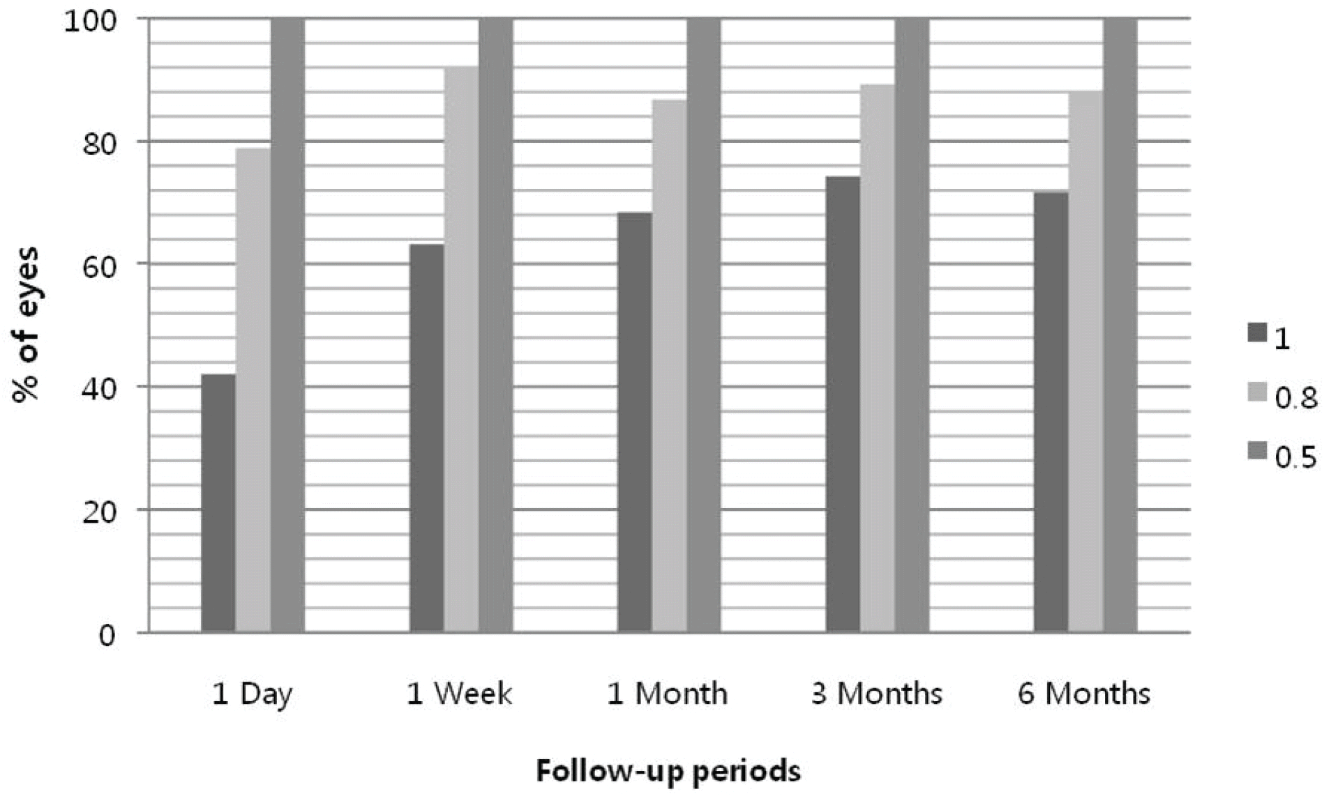 | Figure 1.Uncorrected visual acuity (UCVA) after surgery. UCVAover 20/20 was presented in 63% at postoperative 1 week and 74% at postoperative 3 and 6 months. Uncorrected visual acuity over 20/25 was presented in about 90 % after postoperative 1 week. |
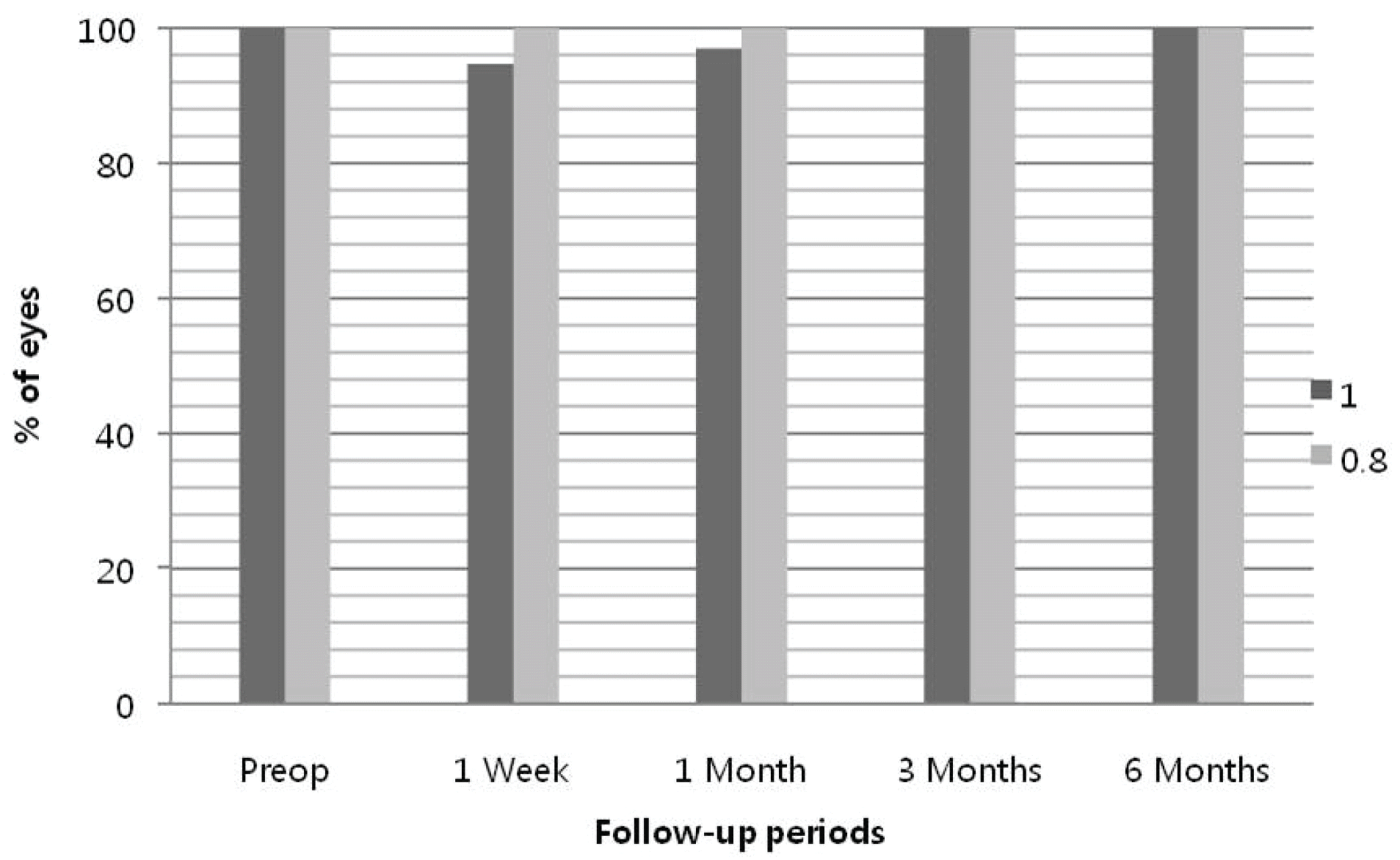 | Figure 2.Best corrected visual acuity (BCVA). BCVA over 20/25 was presented in all patients after post-operative 1 week. And there was no loss in BCVA compared with preoperative visual acuity. |
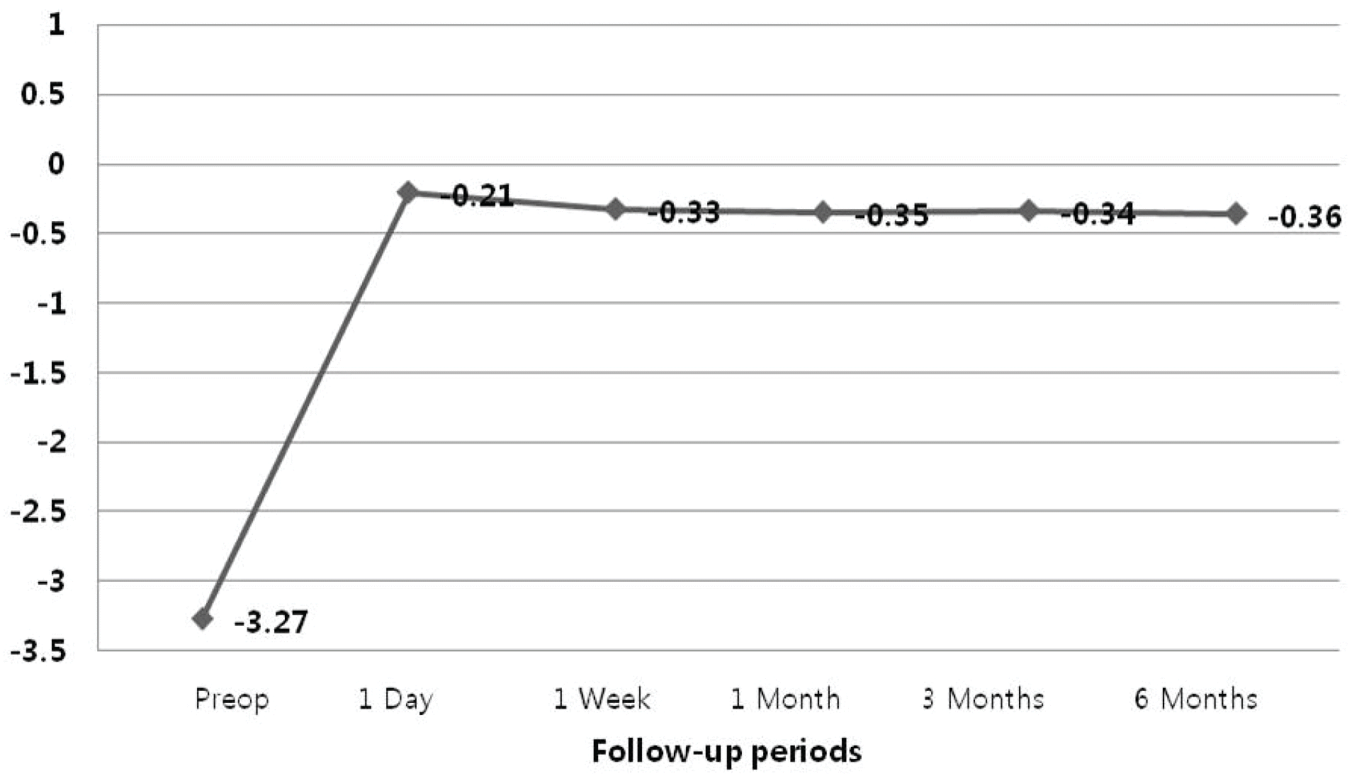 | Figure 3.Changes of spherical error. Changes of post-operative refractive error were minimal after post-operative 1 week. |
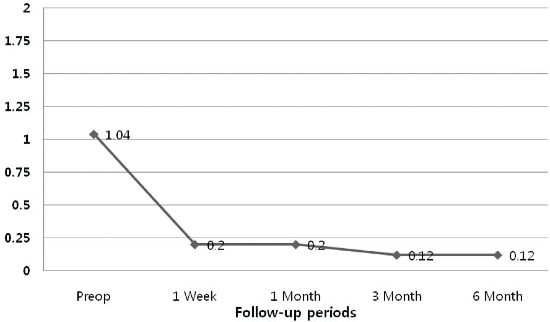 | Figure 4.Changes of cylindrical error. There was little change of astigmatism after operation. |
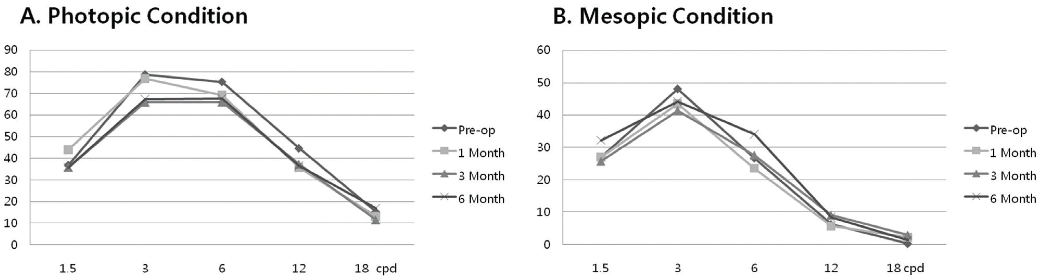 | Figure 5.Changes of contrast sensitivity. (A) Under photopic conditions although the chart shows slight decrease of postoperative contrast sensitivity values in all cpds, there was no significance. (B) There was no significant difference in preoperative and postoperative contrast sensitivity values under mesopic conditions. |
Table 1.
Patient demographics and refractive data
| M: F | 10: 9 |
|---|---|
| Age (year) | 30.0±5.20 (23∼42) |
| Mean Spherical error (Diopter) | −3.27±0.85 (−1.5∼-4.75) |
| Mean Cylindrical error (Diopter) | 1.04±0.69 (0∼1.75) |
| Mean corneal thickness (um) | 533.36±34.30 (447∼570) |
 XML Download