

 PDF
PDF ePub
ePub Citation
 Print
Print


Abstract
Purpose
To report the clinical features and treatment of canaliculitis associated with SmartPlug punctal plug insertion.
Methods
Case selection criteria included patients with canaliculitis, who were managed at Seoul National University Hospital from January 2006 to October 2008, presenting with a history of punctal plug insertion. The operation reports were reviewed to identify patients in whom SmartPlug was discovered during the operation. Six patients (8 eyes) were identified, and a retrospective chart review was performed for all the patients.
Results
The mean age of the patients was 34.3±8.6 years, and there were 1 men and 5 women. Common symptoms were mucous discharge (6 eyes) and conjunctival injection (2 eyes). The mean time from insertion of the plug to onset of symptoms was 27.0±27.0 months (range 4 to 77 months). All patients underwent surgical removal of the punctal plug by one-snip punctoplasty, canalicular retrograde compression using 2 cotton-tipped applications (2 eyes), or canalicular curettage (6 eyes). All patients had resolution of symptoms after the procedure.
Go to : 
References
1. Tai MC, Cosar CB, Cohen EJ, et al. The clinical efficacy of silicone punctal plug therapy. Cornea. 2002; 21:135–9.

2. Freeman JM. The punctum plug: evaluation of a new treatment for the dry eye. Trans Am Acad Ophthalmol Otolaryngol. 1975; 79:874–9.
3. White WL, Bartley GB, Hawes MJ, et al. Iatrogenic complications related to the use of Herrick lacrimal plugs. Ophthalmology. 2001; 108:1835–7.
4. Lee J, Flanagan JC. Complications associated with silicone intracanalicular plugs. Ophthal Plast Reconst Surg. 2001; 17:465–9.
5. Mazow ML, McCall T, Prager TC. Lodged intracanalicular plugs as a cause of lacrimal obstruction. Ophthal Plast Reconst Surg. 2007; 23:138–42.
6. Kay KM, Woo KI, Chang HR. Tuberculous blepharitis following removal of intracanalicular lacrimal plug. J Korean Ophthalmol Soc. 2003; 44:1428–32.
7. Lim DK, Joo MJ, Kim JH. Acase of chronic granulomatous canaliculitis induced by Herrick silicone punctual plug. J Korean Ophthalmol Soc. 2005; 46:384–7.
8. Jang JH, Chae JK, Kim BJ, Lee HB. Cases of complications after the use of punctal plugs. J Korean Ophthalmol Soc. 2005; 46:547–53.
9. SmartPlug Study Group. Management of complications after insertion of the SmartPlug punctual plug: a study of 28 patients. Ophthalmology. 2006; 113:1859e1–6.
10. Scheepers M, Pearson A, Michaelides M. Bilateral canaliculitis following SmartPLUG insertion for dry eye syndrome post LASIK surgery. Graefes Arch Clin Exp Ophthalmol. 2007; 245:895–7.
11. Burgess PI, Koay P, Clark P. SmartPlug versus silicone punctal plug therapy for dry eye. Cornea. 2008; 27:391–4.
12. Vesci VP, Huber-Spitzy V, Arocker-Mettinger E, Steinkogler FJ. Canaliculitis: Difficulties in diagnosis, differential diagnosis and comparison between conservative and surgical treatment. Ophthalmologica. 1994; 208:314–7.
13. Fowler AM, Dutton JJ, Fowler WC, Gilliqan P. Mycobacterium chelonae canaliculitis associated with SmartPlug use. Ophthal Plast Reconstr Surg. 2008; 24:241–3.
14. Hussain I, Bonshek RE, Loudon K, et al. Canalicular infection caused by Actinomyces. Eye. 1993; 7:542–4.
15. Demant E, Hurwitz JJ. Canaliculitis: review of 12 cases. Can J Ophthalmol. 1980; 15:73–5.
16. Lemp MA, Weiler HH. How do tears exit? Invest Ophthalmol Vis Sci. 1983; 24:619–22.
17. Zhu H, Bhatia S, Chauhan A. Dynamic mechanical properties of porcine lacrimal canaliculus. Curr Eye Res. 2007; 32:829–35.
18. Briscoe D, Edelstein E, Zacharopoulos I, et al. Actinomyces canaliculitis: diagnosis of a masquerading disease. Graefes Arch Clin Exp Ophthalmol. 2004; 242:682–6.
19. Pavilack MA, Frueh BR. Through curettage in the treatment of chronic canaliculitis. Arch Ophthalmol. 1992; 110:200–2.
Go to :
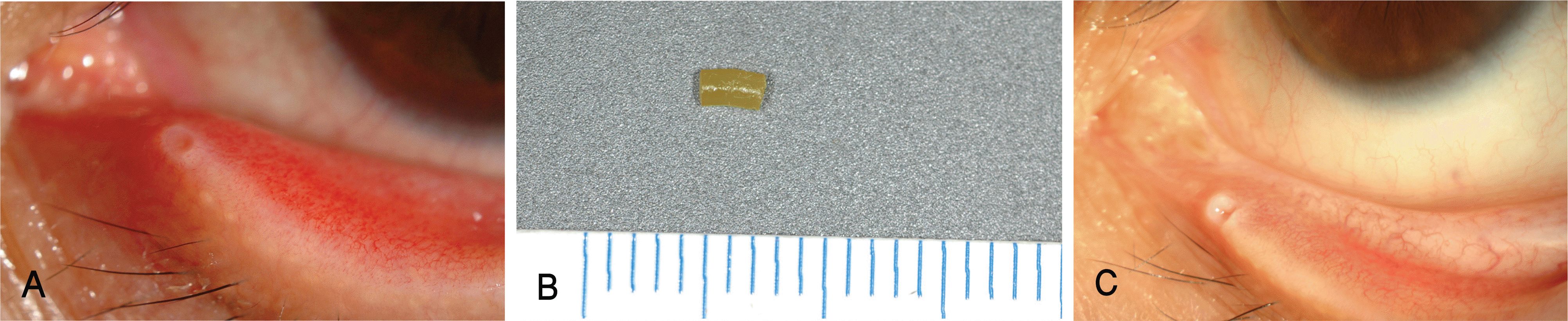 | Figure 1.(A) Preoperative appearance of a punctum in a patient with canaliculitis (Case No. 5–1). Note punctal and canalicular swelling, discharge in a lumen, and conjunctival injection. (B) SmartPlug removed during one snip punctoplasty and canalicular curettage. (C) Photograph of the same case 14 months after the procedure. There is no sign of canaliculitis. |
Table 1.
Clinical presentation and treatment of patients with canaliculitis after SmartPlug insertion
| Case No. | Age (years) | Sex | Affected area | Time from plug insertion to onset of symptom | Duration of symptom | Presenting symptoms | Treatment |
|---|---|---|---|---|---|---|---|
| 1 | 30 | F* | LLL‡ | 12 | 4 | Mucous discharge, lower lid swelling | One-snip punctoplasty, canalicular curettage |
| 2 | 36 | F | LLL | 18 | 4 | Mucous discharge | One-snip punctoplasty, canalicular curettage |
| 3 | 50 | M† | RLL§ | 4 | 1 | Mucous discharge | One-snip punctoplasty, canalicular curettage |
| 4 | 35 | F | LLL | 24 | 3 | Pus from punctum | One-snip punctoplasty, canalicular curettage |
| 5–1 | 25 | F | LLL | 4 | 1 | Mucous discharge, tearing | One-snip punctoplasty, canalicular curettage |
| 6–1 | 30 | F | LLL | 60 | 5 | Conjunctival injection, discharge | One-snip punctoplasty, canalicular curettage |
| 5–2 | 25 | F | RLL | 19 | 2 | Bloody discharge | One-snip punctoplasty, canalicular curettage & squeezing |
| 6–2 | 30 | F | RLL | 77 | 3 | Conjunctival injection, discharge | One-snip punctoplasty, canalicular squeezing |
 XML Download