Abstract
Purpose
To present the clinical feature of retinal toxicity of intravitreal tissue plasminogen activator which was used for treatment of submacular hemorrhage.
Case summary
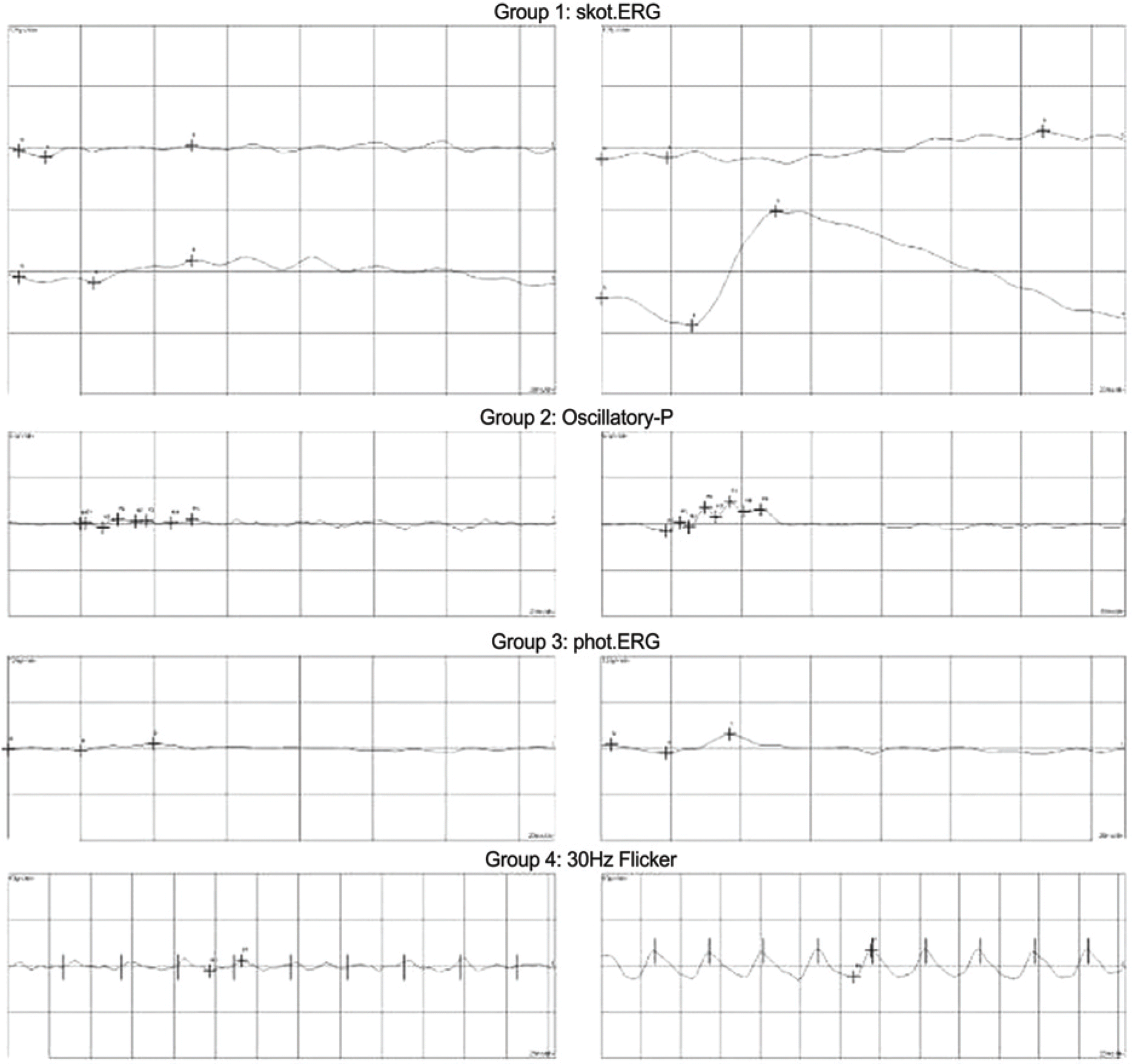
An intravitreal injection of tPA (100 μ g) with C3 F8 gas tamponade (0.2 cc) was given to treat the submacular hemorrhage in a patient with ARMD. The therapeutic effect was measured by visual acuity, slit lamp examination, indirect funduscopy and fluorescein angiogram. Three months after the operation, the hemorrhage was decreased but a pigmentary change was observed on the peripheral retina. After 8 months, the submacular hemorrhage completely reabsorbed but the peripheral pigmentary change had increased. Ten months later, the retinal pigmentary change was observed on the entire retina except the posterior pole. The fluorescein angiogram showed peripheral hyperfluorescene of the retina due to window defect from the pigmentary change but no leakage was detected. The electroretinogram showed reduced amplitude in the right eye.
Conclusions
Intravitreal tPA injection of 25 to 100 μ g with pneumatic displacement is typically used for the treatment of submacular hemorrhage. However, there is no established safety dose of tPA for use in human eyes. In the present study, 100 μ g of tPA was used and retinal toxicity was noted. Establishing a safety dose of tPA to prevent dosage dependent complications is necessary.
References
1. Heriot WJ. Management of submacular hemorrhage with intravitreal injection of tissue plasminogen activator and expansile gas. Retina. 1997; 11:211–2.
2. MacCumber MW, McCuen BW 2nd, Toth CA, et al. Tissue plasminogen activator for preserving inferior peripheral iridectomy patency in eyes with silicone oil. Ophthalmology. 1996; 103:269–73.
3. Johnson MW, Oslen KR, Hernandez E. Tissue plasminogen activator thrombolysis during surgical evacuation of experimental subretinal hemorrhage. Ophthalmology. 1992; 99:515–21.

4. Lewis H. Intraoperative fibrinolysis of submacular hemorrhage with tissue plasminogen activator and surgical drainage. Am J Ophthalmol. 1994; 118:559–68.

5. Gilbert H. Pigmentary retinopathy following intravitreal injection of tissue plasminogen activator. New Orleans; Vitreous Society Annual Meeting. September 20, 1997.
6. Kim KS, Rhee K, Huh K. Retnal Toxicty of Intravitreal Tissue Plasminogen Activator with C3 F8 Injection in Rabbit Eyes. J Korean Ophthalmol Soc. 2004; 45:1181–8.
7. Handwerger BA, Blodi BA, Chandra SR, et al. Treatment of Submacular Hemorrhage With Low-Dose Intravitreal Tissue Plasminogen Activator Injection and Pneumatic displacement. Arch Ophalmol. 2001; 119:28–32.
8. Heriot WJ. Further experience in management of submacular hemorrhage with intravitreal tPA. American Academy of Ophthalmology Vitreoretinal Update;October 24, 1997.
9. Hassan AS, Johnson MW, Schneiderman TE, et al. Management of submacular hemorrhage with intravitreal tPA injection and pneumatic displacement. Ophthalmology. 1999; 106:1900–6.
10. Lee J-Y, Kim HC. Intravitreal Injection of tPA and C3 F8 Gas in Submacular Hemorrhage. J Korean Ophthalmol Soc. 2002; 43:1928–37.
11. Chen SN, Yang TC, Ho CL, et al. Retinal toxicity of intravitreal tissue plasminogen activator. Case report and literature review. Ophthalmology. 2003; 110:704–8.
12. Kumada M, Niwa M, Wang X, et al. Endogenous tissue type plasminogen activator facilitates NMDA-induced retinal damage. Toxicol Appl Pharmacol. 2004; 200:48–53.

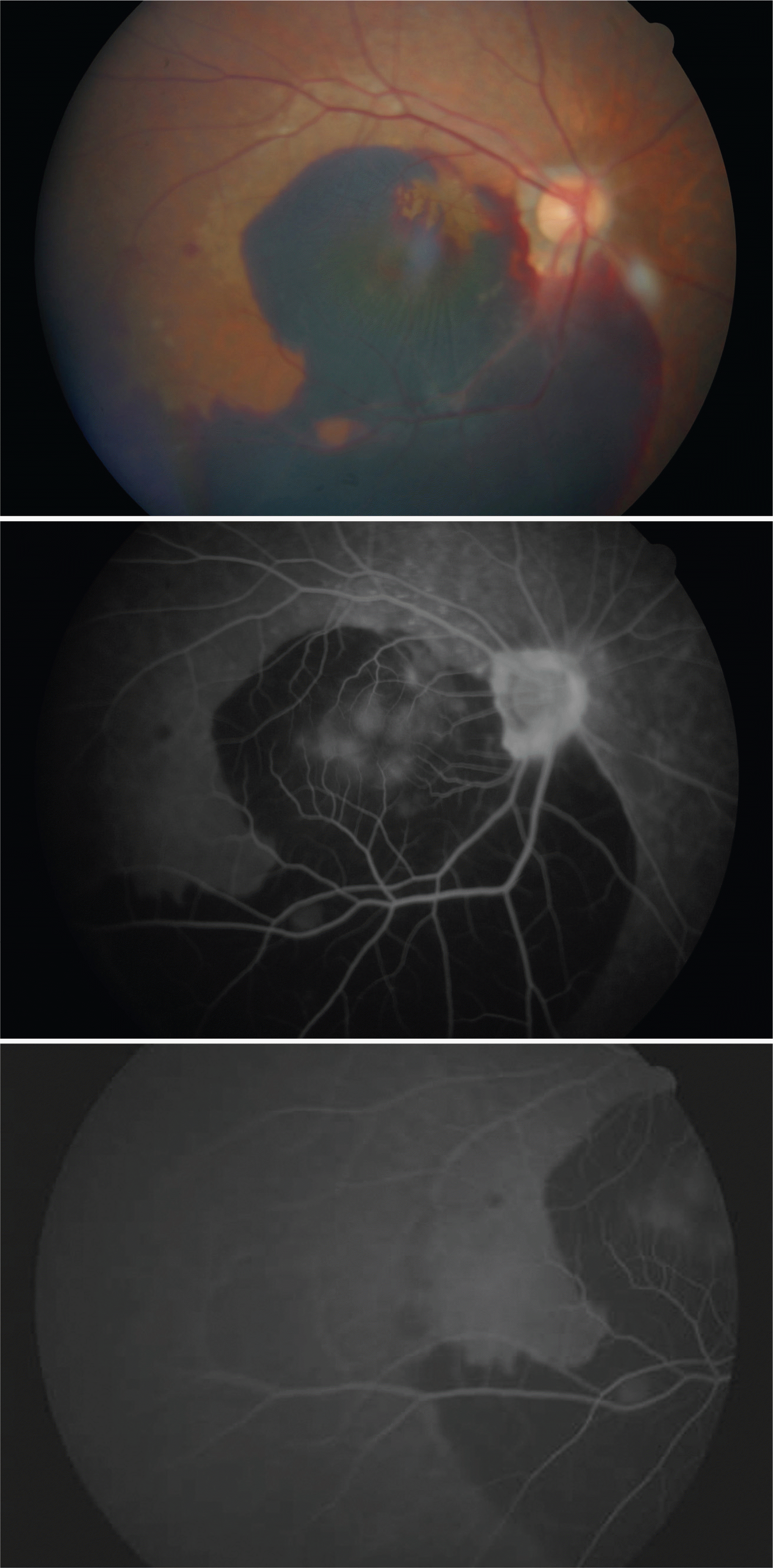
Figure 1.
Preoperative fundus photograph and fluorescein angiogram show large submacular hemorrhage (about 4.5DD) occupying the posterior pole extending inferiorly outside the major arcade.

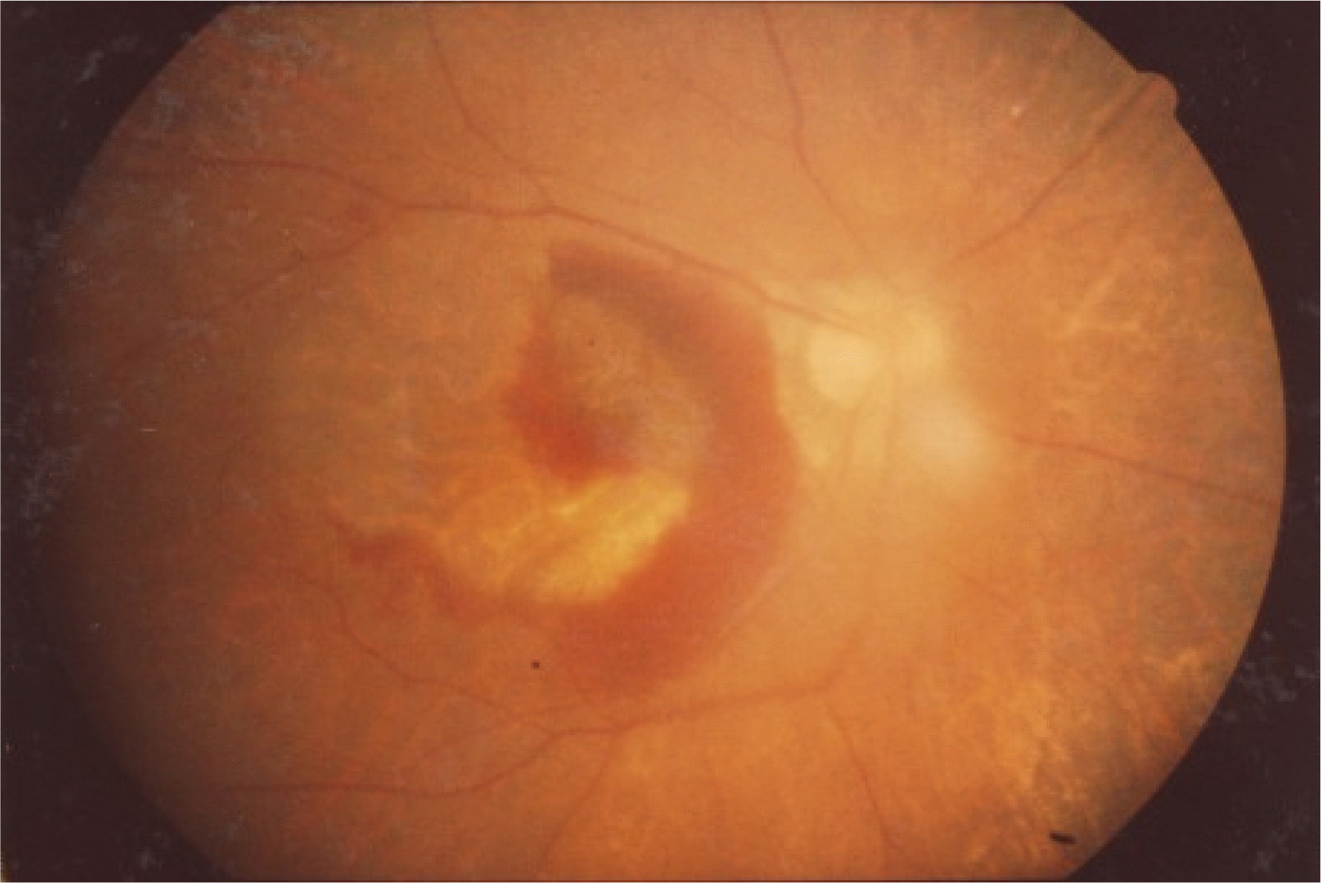
Figure 2.
Three months after intravitreal tPA and gas injection. The submacular hemorrhage was largely absorbed and peripheral pigmentary change was noted.


 PDF
PDF ePub
ePub Citation
 Print
Print


 XML Download