

 PDF
PDF ePub
ePub Citation
 Print
Print


Abstract
Purpose
To investigate the clinical features and prognosis of primary intraocular lymphoma (PIOL). Methods: A retrospective review of medical records was performed in 9 patients who were diagnosed and treated as PIOL in the Department of Ophthalmology, Seoul National University Hospital.
Results
Among patients who were enrolled in the study, 14 eyes were examined. Thirteen eyes (92.9%) showed yellowish subretinal or choroidal infiltrates which is a characteristic finding of PIOL in fundus examination and fluorescein angiography. Three patients presented with ocular symptoms initially, and 5 patients later presented with central nerve system (CNS) involvement. Only 1 patient showed PIOL without CNS involvement. Among 6 patients (9 eyes) that received systemic chemotherapy or ocular irradiation, 5 patients (7 eyes, 77.8%) responded. Among those patients, 3 patients (4 eyes) showed relapse of PIOL. Five patients died during the mean follow-up period of 43.3 months, and the median survival time was 47 months.
References
1. Chan CC, Buggage RR, Nussenblatt RB. Intraocular lymp-homa. Curr Opin Ophthalmol. 2002; 13:411–8.

2. Whitcup SM, de Smet MD, Rubin BI. . Intraocular lymphoma. Clinical and histopathologic diagnosis. Ophthal-mology. 1993; 100:1399–406.
3. Akpek EK, Ahmed I, Hochberg FH. . Intraocular-central nervous system lymphoma: clinical features, diagnosis, and outcomes. Ophthalmology. 1999; 106:1805–10.
4. Lee SH, Kim DJ, Kim IT. A case of primary central nervous system lymphoma with ocular involvement. J Korean Ophthal-mol Soc. 2005; 46:565–71.
5. Char DH, Ljung BM, Miller T, Phillips T. Primary intra-ocular lymphoma (ocular reticulum cell sarcoma) diagnosis and management. Ophthalmology. 1988; 95:625–30.
6. Peterson K, Gordon KB, Heinemann MH, DeAngelis LM. The clinical spectrum of ocular lymphoma. Cancer. 1993; 72:843–9.
7. Augustein WG, Buggage RR, Smith JA. . Fluorescein angiogram interpretation in the diagnosis of primary intra-ocular lymphoma. Invest Ophthalmol Vis Sci. 2001; 42:S462.
8. Gill MK, Jampol LM. Variations in the presentation of primary intraocular lymphoma: Case reports and a review. Surv Ophthalmol. 2001; 45:463–71.
9. Levy-Clarke GA, Chan CC, Nussenblatt RB. Diagnosis and management of primary intraocular lymphoma. Hematol Oncol Clin North Am. 2005; 19:739–49.
10. Karma A, von Willebrand EO, Tommila PV. . Primary intraocular lymphoma: improving the diagnostic procedure. Ophthalmology. 2007; 114:1372–7.
11. Jahnke K, Korfel A, Komm J. . Intraocular lymphoma 2000-2005: results of a retrospective multicentre trial. Graefes Arch Clin Exp Ophthalmol. 2006; 244:663–9.
12. Zaldivar RA, Martin DF, Holden JT, Grossniklaus HE. Primary intraocular lymphoma: clinical, cytologic, and flow cytometric analysis. Ophthalmology. 2004; 111:1762–7.
13. Tuaillon N, Chan CC. Molecular analysis of primary central nervous system and primary intraocular lymphomas. Curr Mol Med. 2001; 1:259–72.
14. Hoffman PM, McKelvie P, Hall AJ, Stawell RJ, Santamaria JD. Intraocular lymphoma: a series of 14 patients with clinicopathological features and treatment outcomes. Eye 2003; 17. 513–21.
15. Coupland SE, Bechrakis NE, Anastassiou G. . Evaluation of vitrectomy specimens and chorioretinal biopsies in the diagnosis of primary intraocular lymphoma in patients with masquerade syndrome. Graefes Arch Clin Exp Ophthalmol. 2003; 241:860–70.
16. Batchelor TT, Kolak G, Ciordia R. . High-dose methotrexate for intraocular lymphoma. Clin Cancer Res. 2003; 9:711–5.
17. Batchelor T, Carson K, O'Neill A. . Treatment of primary CNS lymphoma with methotrexate and deferred radiotherapy: a report of NABTT 96–07. J Clin Oncol. 2003; 21:1044–9.
18. Hayabuchi N, Shibamoto Y, Onizuka Y. Primary central nervous system lymphoma in Japan: a national survey. Int J Radiat Oncol Biol Phys. 1999; 44:265–72.

Figure 2.
Multiple dots of yellowish chorioretinal infiltration (A) and multiple hypofluoresecent dots intermingling with hyperfluorescent dots at the level of the retinal pigment epithelium, suggestive of an aggregate of tumor cells at fluorescein angiogram (B).

Figure 3.
Pretreatment fundus photograph demonstrates large infiltrative subretinal mass involving inferior retina and diffuse vitreous infiltration (A). Subretinal mass and vitreous infiltrationdisappeared 4 months after combined chemo-radiation therapy, and atrophy of optic disc and retina remained as a sequaele (B).

Figure 4.
Case 1 right eye. Wright-Giemsa stain of vitreous specimen shows immature pleomorphic lymphocytes with microcystic nucleus and ill-defined nucleus membrane (black arrows). Nectrotic atypical lymphocytes cells were also found (red arrows)(×200).
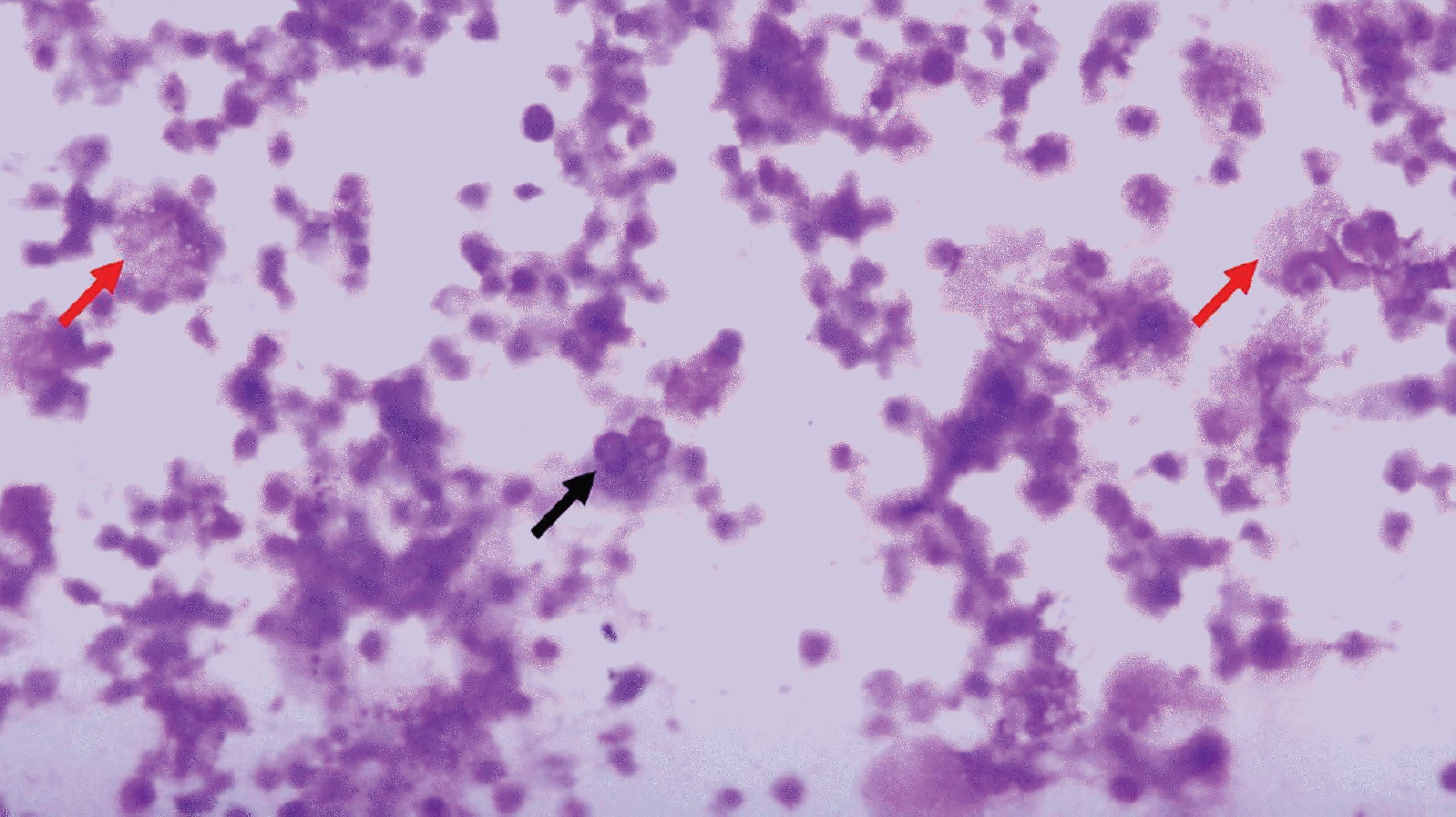
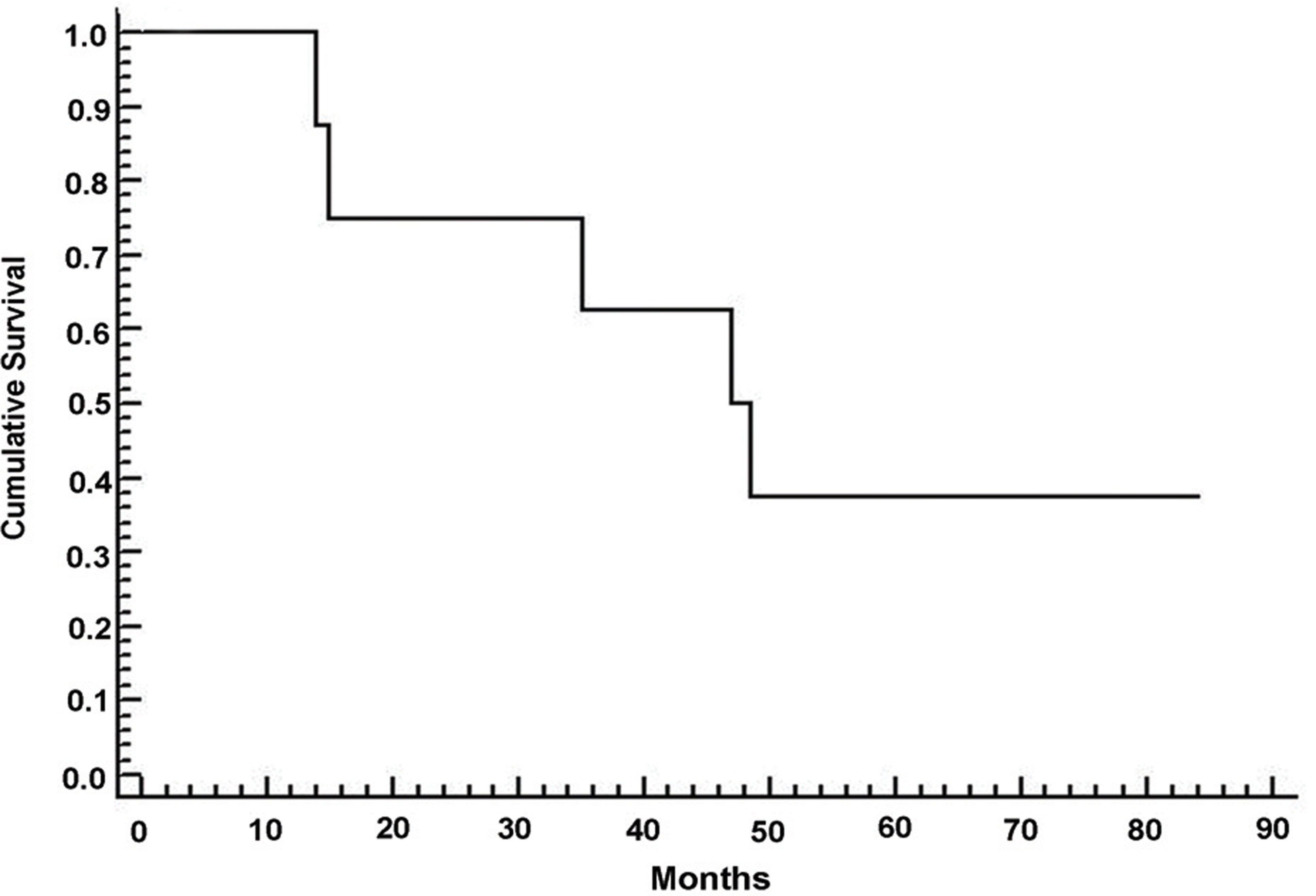
Table 1.
Demographics
| No | Age/Sex | later ality | Ocular features | Sympto m-Diag nosis1) | PIOL -PCN SL2) | Involve ment3) | No. & Result of ‡PPVs | f Diagnosis based on | Patho-logy | -Treatment in chronological order (treatment on PCNSL) T R | Treatme nt Repons e4) | Outcome (months after diagnosis) | Ref 5) |
|---|---|---|---|---|---|---|---|---|---|---|---|---|---|
| 1 | M/56 | ∗ R | panuveitis | 10 | -3 | 1,2 | 1, PIOL | PPV, Brain § MRI | П NA | (‗‗ brRT)-PPV-†† o cRT | Yes, Yes | ПП AND 35 | 3 |
| † L | panuveitis | 10 | -3 | 1,2 | 0 | Brain MRI | NA | (brRT)-ocRTx-P PV | Yes, Yes | AWD (ocular) 35 | 3 | ||
| 2 | F/57 | R | Vitreitis with chorioretinal lesion | 7 | 0 | 1,2,1 | 0 | Brain biopsy | # DLBL | L ‡‡HDMTX, brRT | Yes, Yes | ## AWD (ocular) 14 | 3 |
| L | Vitreitis with chorioretinal lesion | 7 | 0 | 1,2,1 | 0 | Brain biopsy | DLBL | HDMTX, brRT | Yes, Yes | AWD (ocular) 14 | 3 | ||
| 3 | M/63 | R | panuveitis | 7 | 0.5 | 1,2 | 0 | Brain biopsy | DLBL | HDMTX | NA6), No | Died 7.8 | 3 |
| L | panuveitis | 7 | 0.5 | 1,2 | 0 | Brain biopsy | DLBL | HDMTX | NA6), No | Died 7.8 | 3 | ||
| 4 | M /63 | R | panuveitis | 15 | NA | 11 | 1, Necroti atypical cells | ic PPV | NA | PPV-HDMTX | Yes, NA | AND 15 | 2 |
| L | panuveitis | 15 | NA | 1 1 | 1,Necrotic atypical cells | c PPV | NA | PPV-HDMTX | Yes, NA | AND 15 | 2 | ||
| 5 | F/64 | R | panuveitis | 9 | -12 | 1 2,1 | 1, Necroti atypical cells | ic Brain biopsy, PPV | DLBL | (HDMTx,brRT)-PPV-ocRT | Yes, Yes | AND 47 | 1 |
| L | panuveitis | 9 | -12 | 2,1 | 0 | Brain biopsy | DLBL | (HDMTx,brRT)-ocRT | Yes, Yes | AWD (ocular) 47 | 1 | ||
| 6 | F/64 | R | panuveitis | 1 | -57 | 2,1,2 | 0 | Brain biopsy | DLBL | (CHOP,brRT)-H DMTX,ocRT | Yes, No | Died 84 | 1 |
| 7 | F/71 | R | posterior chorioretinal lesion | 0.5 | -24.5 | 2,1 | 0 | Brain biopsy | DLBL | (HDMTx,brRT) | NA, Yes | AWD (ocular) 48.5 | 1 |
| 8 | M/57 | L | Vitreitis without chorioretinal lesion | NA | -48 | 2,1,2 | 0 | Brain biopsy | DLBL | (HDMTx,brRT) | NA, No | Died 55 | 1 |
| 9 | M/62 | R | panuveitis | 0.5 | -60.5 | 2,1,2 | 0 | Brain biopsy | DLBL | (§§ CHOP,brRT) | NA, No | Died 83.8 | 1 |
1) Time (mo) from onset of ocular symptoms to diagnosis.
2) Time from the diagnosis of primary intraocular lymphoma (PIOL) to that of primary central nervous system lymphoma (PCNSL).
3) Choronological relationship of ocular (1), CNS (2) involvement.
4) Best response to treatment (ocular /systemic).
5) Status of the referral: 1 represents previously diagnosed of PCNSL, 2 represents referred from first order medical service; 3, secondary or tertiary order medical service.
6) Died 0.5 month after the initial chemotherapy due to progression of PCNSL.
 XML Download