Abstract
Purpose
To analyze the clinical results of Goldmann applanation tonometry (GAT) and Pascal dynamic contour tonometry (PDCT) and the influences of central corneal thickness and keratometric power in eyes that underwent Epi-LASIK or penetrating keratoplasty.
Methods
Measurements of intraocular pressure by GAT and PDCT as well as keratometric power and central corneal thickness were measured in 45 eyes that underwent penetrating keratopasty and 63 eyes that underwent Epi-LASIK. These parameters were also measured in healthy eyes with no specific disorders to create a control group.
Results
In the keratoplasty group, the PDCT results were significantly higher than the GAT results by 1.22±2.84 mmHg (p=0.006), but neither method showed a significant correlation with CCT or keratometric power. In the Epi-LASIK group, PDCT was higher as 3.45±2.35 mmHg than GAT, and the corrected results of GAT were not different from the results of PDCT. In the control group, GAT was affected by central corneal thickness and keratometric power, but PDCT showed no significant relationship with these two factors.
References
1. Allingham RR, Damji KF, Freedman S, et al. Shield's Textbook of Glaucoma. 5th ed.Philadelphia: Lippincott;2005. p. 41–4.
2. Whitacre MM, Stein R. Sources of error with use of Goldmann type tonometers. Surv Ophthalmol. 1993; 38:1–30.
3. Mardelli PG, Piebenga LW, Whitacre MM, Siegmund KD. The effect of excimer laser photorefractive keratectomy on intraocular pressure measurements using the Goldmann applanation tonometer. Ophthalmology. 1997; 104:945–8.

4. Fournier AV, Podtetenev M, Lemire J, et al. Intraocular pressure change measured by Goldmann tonometry after laser in situ keratomileusis. J Cataract Refract Surg. 1998; 24:905–10.

5. Kniestedt C, Lin S, Choe J, et al. Clinical comparison of contour and applanation tonometry and their relationship to pachymetry. Arch Ophthalmol. 2005; 123:1532–7.

6. Ehlers N, Bramsen T, Sperling S. Applanation tonometry and central corneal thickness. Acta Ophthalmol (Copenh). 1975; 53:34–43.

7. Emara B, Probst LE, Tingey DP, et al. Correlation of intraocular pressure and central corneal thickness in normal myopic eyes and after laser in situ keratomileusis. J Cataract Refract Surg. 1998; 4:1320–5.

8. Kohlhaas M, Spoerl E, Boehm AG, Pollack K. A correction formula for the real intraocular pressure after LASIK for the correction of myopic astigmatism. J Refract Surg. 2006; 22:263–7.

9. Kanngiesser HE, Kniestedt C, Robert YC. Dynamic contour tonometry: presentation of a new tonometer. J Glaucoma. 2005; 14:344–50.
10. Kniestedt C, Nee M, Stamper RL. Accuracy of dynamic contour tonometry compared with applanation tonometry in human cadaver eyes of different hydration states. Graefes Arch Clin Exp Ophthalmol. 2005; 243:359–66.

11. Kao SF, Lichter PR, Bergstrom TJ, et al. Clinical comparison of the Oculab Tono-Pen to the Goldmann applanation tonometer. Ophthalmology. 1987; 94:1541–4.

12. Frenkel RE, Hong YJ, Shin DH. Comparison of the Tono-Pen to the Goldmann applanation tonometer. Arch Ophthalmol. 1988; 106:750–3.

13. Geyer C, Mayron Y, Lowenstein A, et al. Tono-Pen tonometry in normal and postkeratoplasty eyes. Br I Ophthalmol. 1992; 76:538–40.
14. Jiang B, Jiang Y. Clinical comparison of the Tono-Pen tonometer to the Goldmann applanation tonometer. Yan Ke Xue Bao. 2002; 18:226–9.
15. Frenkel RE, Hong YJ, Shin DH. Comparison of the Tono-Pen to the Goldmann applanation tonometer. Arch Ophthalmol. 1988; 106:750–3.

16. Ismail AR, Lamont M, Perera S, et al. Comparison of IOP measurement using GAT and DCT in patients with penetrating keratoplasties. Br J Ophthalmol. 2007; 91:980–1.

17. Kirkness CM, Ficker LA. Risk factors for the development of postkeratoplasty glaucoma. Cornea. 1992; 11:427–32.

18. Redbrake C, Arend O. Corneal transplantation and glaucoma. Ophthalmologe. 2000; 97:552–6.
19. Aldave AJ, Rudd JC, Cohen EJ, et al. The role of glaucoma therapy in the need for repeat penetrating keratoplasty. Cornea. 2000; 19:772–6.

20. Viestenz A, Langenbucher A, Seitz B, et al. Evaluation of dynamic contour tonometry in penetrating keratoplasties. Ophthalmologe. 2006; 103:773–6.
21. Meyenberg A, Iliev ME, Eschmann R, et al. Dynamic contour tonometry in keratoconus and postkeratoplasty eyes. Cornea. 2008; 27:305–10.

22. Kniestedt C, Nee M, Stamper RL. Dynamic contour tonometry: a comparative study on human cadaver eyes. Arch Ophthalmol. 2004; 122:1287–93.
23. Madjlessi F, Marx W, Reinhard T, et al. Impression and applanation tonometry in irregular corneas. Comparison with intraocular needle tonometry. Ophthalmologe. 2000; 97:478–81.
24. Feltgen N, Leifert D, Funk J. Correlation between central corneal thickness, applanation tonometry, and direct intracameral IOP reading. Br J Ophthalmol. 2001; 85:85–7.
25. Kaufmann C, Bachmann LM, Thiel MA. Intraocular pressure measurements using dynamic contour tonometry after laser in situ keratomileusis. Invest Ophthalmol Vis Sci. 2003; 44:3790–4.

26. Liu L, Lei C, Li X, Dong J. Measurement of intraocular pressure after LASIK by dynamic contour tonometry. J Huazhong Univ Sci Technolog Med Sci. 2006; 26:372–3. 377.
27. Siganos DS, Papastergioiu GI, Moedas C. Assessment of the Pascal dynamic contour tonometer in monitoring intraocular pressure in unoperated eyes and eyes after LASIK. J Cataract Refract Surg. 2004; 30:746–51.

28. Choi HJ, Kim SW, Kim TI, Kim EK. Intraocular Pressure Measurements Using Dynamic Contour Tonometer After Photorefractive Keratectomy. J Korean Ophthalmol Soc. 2008; 49:577–82.

29. Emara B, Probst LE, Tingey DP, et al. Correlation of intraocular pressure and central corneal thickness in normal myopic eyes and after laser in situ keratomileusis. J Cataract Refract Surg. 1998; 24:1320–5.

30. Mills RP. If intraocular pressure measurement is only an estimate then what? Ophthalmology. 2000; 107:1807–8.
32. Gimeno JA, Munoz LA, Valenzuela LA, et al. Influence of refraction on tonometric readings after photorefractive keratectomy and laser assisted in situ keratomileusis. Cornea. 2000; 19:512–6.

33. Park HJ, Uhm KB, Hong C. Reduction in intraocular pressure after laser in situ keratomileusis. J Cataract Refract Surg. 2001; 27:303–9.

34. Montes-Mico R, Charman WN. Intraocular pressure after excimer laser myopic refractive surgery. Ophthalmic Physiol Opt. 2001; 21:228–35.

35. Garzozi HJ, Chung HS, Lang Y, et al. Intraocular pressure and photorefractive keratectomy: a comparison of three different tonometers. Cornea. 2001; 20:33–6.
36. Liu J, Roberts CJ. Influence of corneal biomechanical properties on intraocular pressure measurement: quantitative analysis. J Cataract Refract Surg. 2005; 31:146–55.
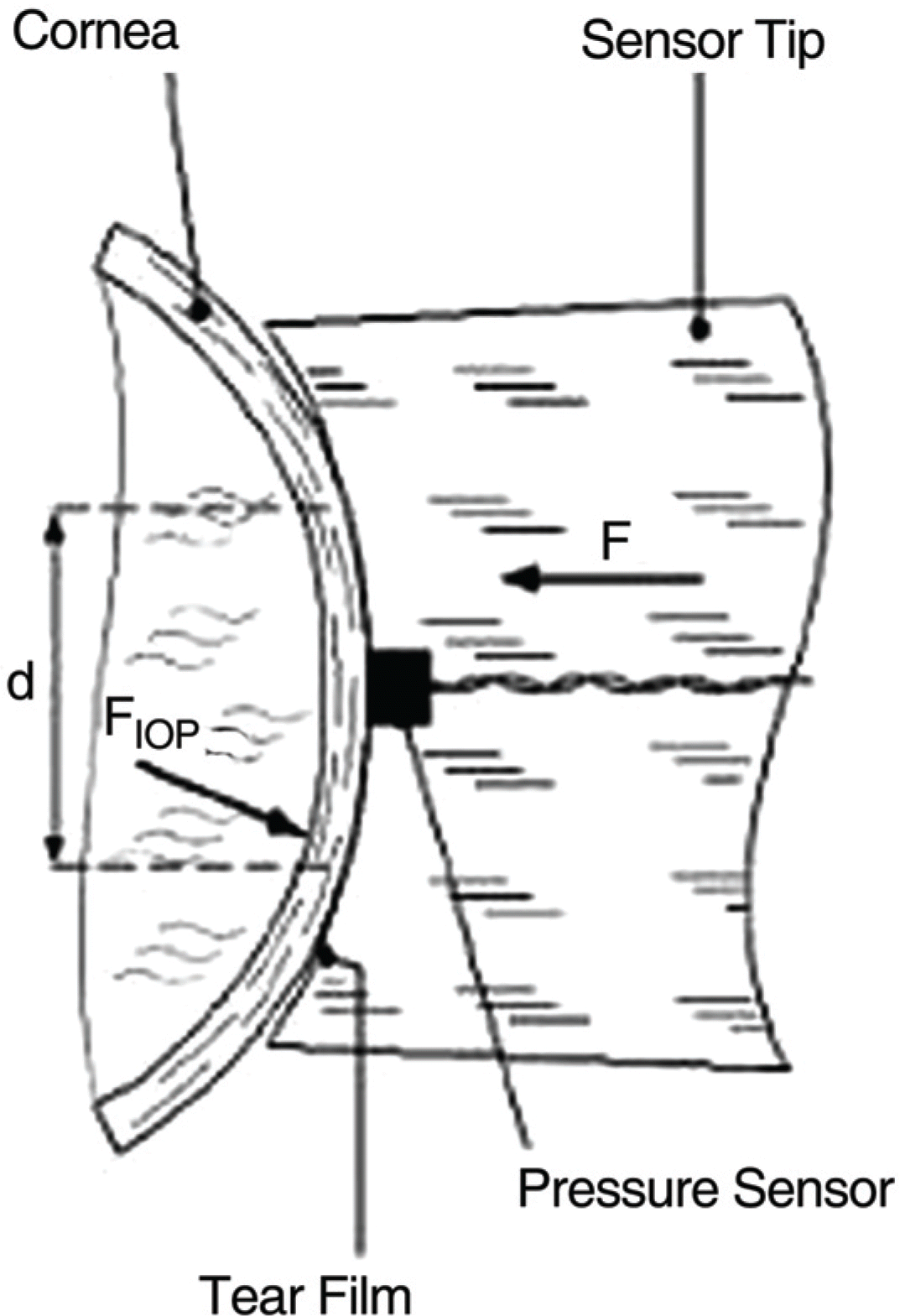
Figure 1.
Schematic diagram of Non-applanating contour-matched tip of the Pascal tonometer. When the match is achieved, the sensor tip embeddd in tip measures38

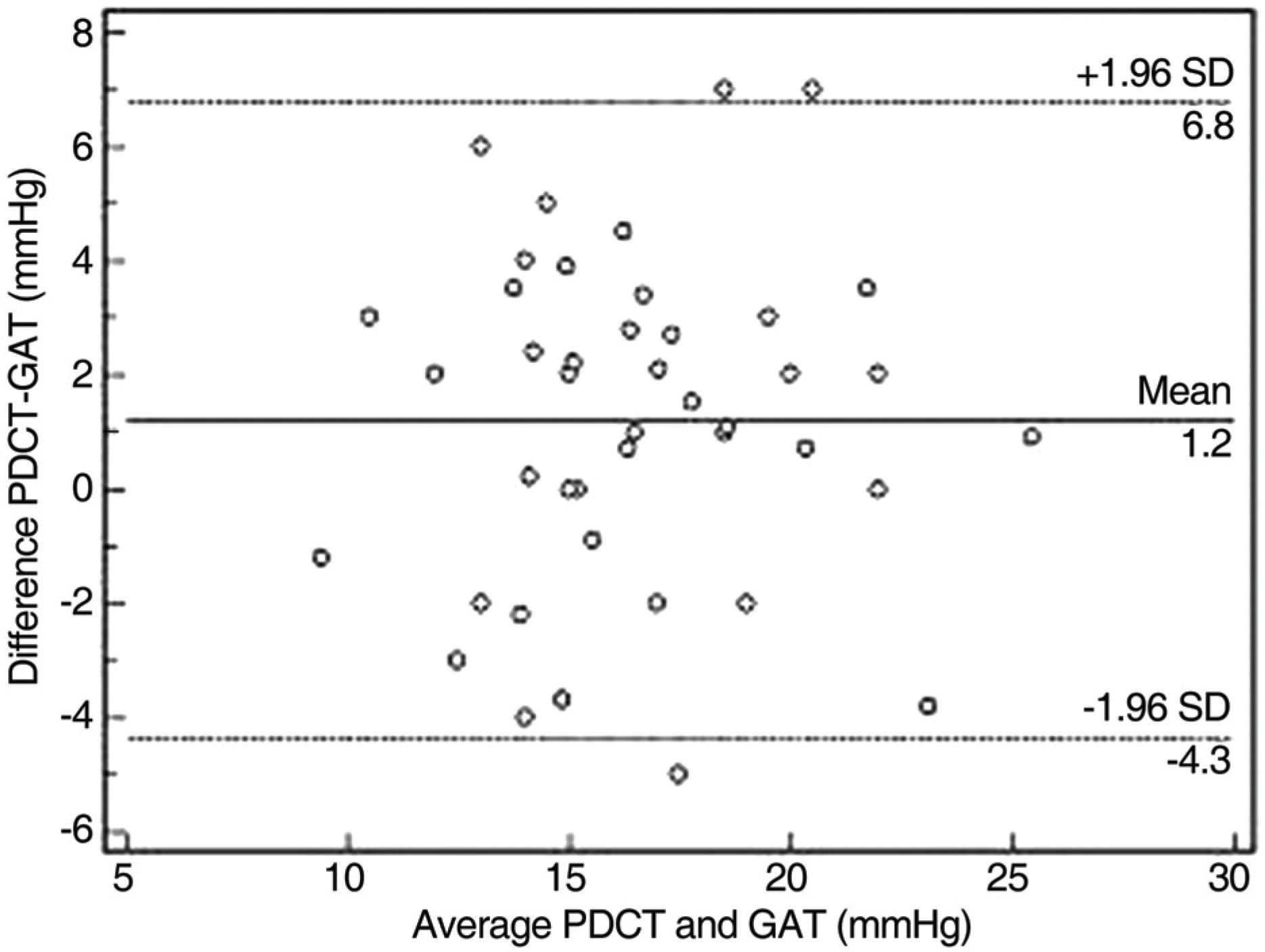
Figure 2.
Difference between PDCT and GAT IOP measurements, plotted against the mean of the two measurements. The measurements obtained by these two techniques were found to be within ±5.61 mm Hg of each other in 95% of cases (PPKP group).

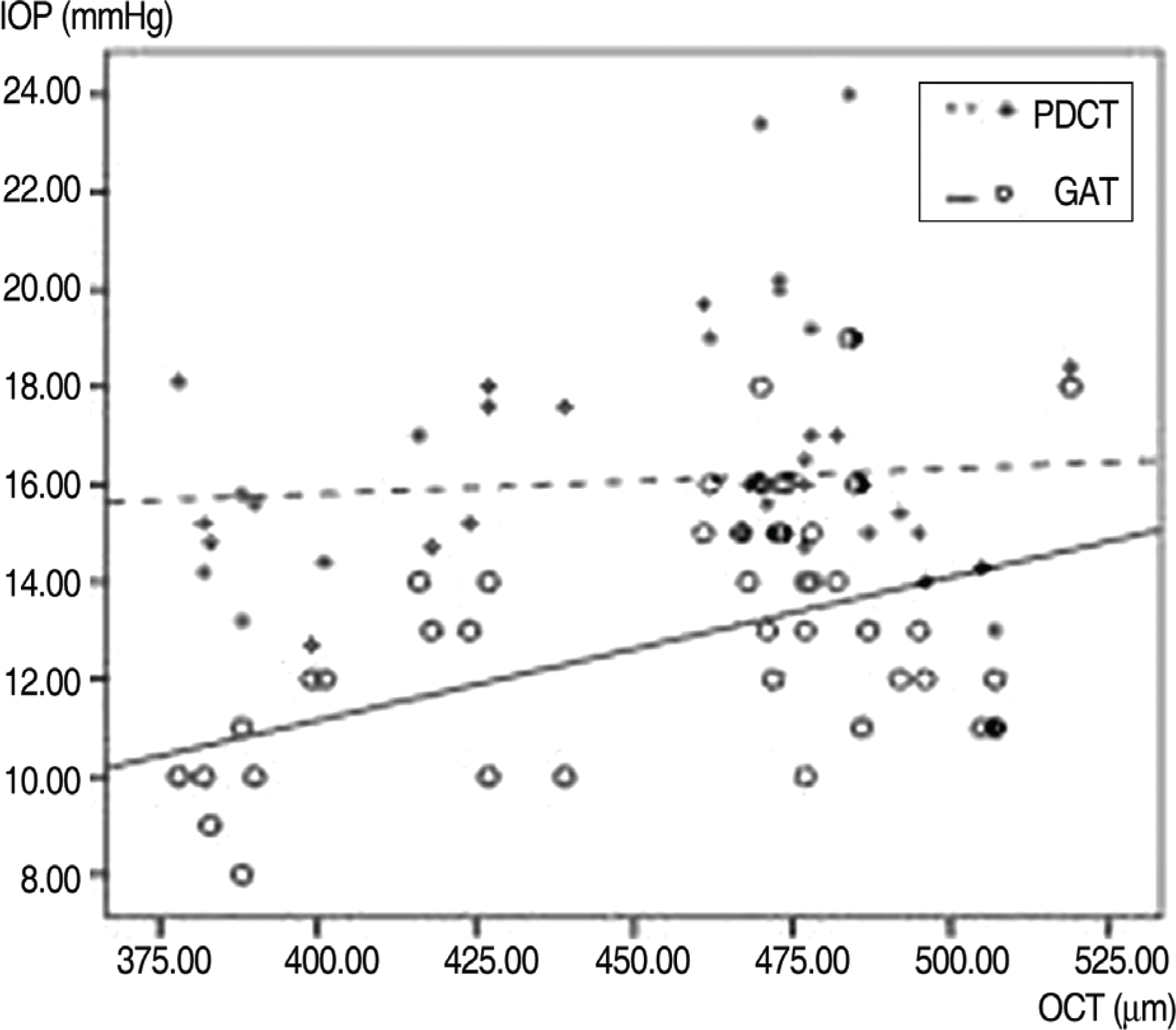
Figure 3.
Effect of CCT on PDCT and GAT in Epi-LASIK group Cross and dotted line represents PDCT result and its estimated regression line according to CCT. PDCT does not correlate with CCT in contrast to GAT. Dotted line (PDCT); IOP=0.005× CCT+13.725 (R2=0.009, p-value=2.479), Solid line (GAT); IOP= 0.029× CCT–0.597 (R2=0.266, p-value=0.000).

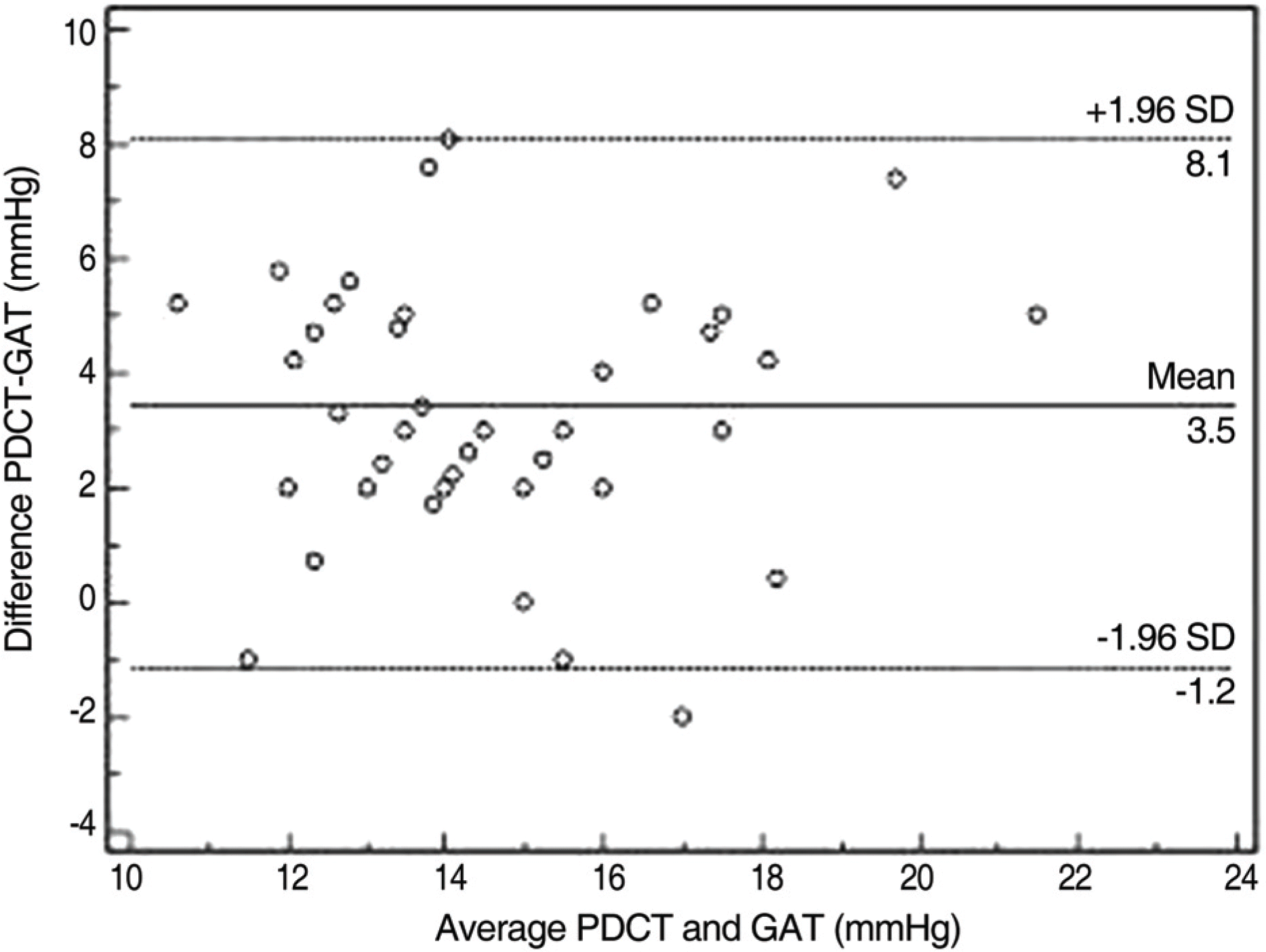
Figure 4.
Difference between PDCT and GAT IOP measurements, plotted against the mean of the two measurements. The measurements obtained by these two techniques were found to be within ±4.62 mmHg of each other in 95% of cases (EpiLasik group).

Table 1.
Subject characteristics
Table 2.
Methods of IOP measurements in PPKP group
| Method | GAT | PDCT | Corrected GAT 5 mmHg/70 μm | Corrected GAT 0.027 mmHg/1 μm | Corrected GAT Equation† |
|---|---|---|---|---|---|
| Mean (mm Hg) | 15.99±3.59 | 17.22±3.73 | 16.36±4.89 | 16.13±3.81 | 16.65±3.56 |
| t-test with GAT (p-value) | | 0.006* | 0.117 | 0.010* | 0.177 |
Table 3.
Correlation of methods and Keratometry or CCT (Pearson correlation)
| | GAT coefficient (p-value) | PDCT coefficient (p-value) |
|---|---|---|
| Keratometry (p-value) | | |
| PPKP | 0.260 (0.084) | 0.201 (0.186) |
| Epi-Lasik | 0.488 (0.000)* | 0.158 (0.216) |
| Control | −0.026 (0.875) | −0.100 (0.550) |
| CCT(p-value) | | |
| PPKP | −0.008 (0.958) | −0.249 (0.098) |
| Epi-Lasik | 0.515 (0.000)* | 0.093 (0.469) |
| Control | 0.376 (0.020) | 0.185 (0.265) |


 PDF
PDF ePub
ePub Citation
 Print
Print


 XML Download