Abstract
Purpose
To evaluate the clinical outcomes of macular laser photocoagulation after intravitreal injection of triamcinolone acetonide (IVTA) for macular edema in branch retinal vein occlusion (BRVO).
Methods
In this retrospective study we reviewed the medical records of patients who had been treated with an intravitreal injection of 4 mg triamcinolone acetonide for macular edema due to BRVO and followed up for more than six months. We divided the eyes into two groups, namely, the IVTA-only group and the additional grid laser group after IVTA. Visual acuity, macular thickness, intraocular pressure, and abundance of IVTA were compared.
Results
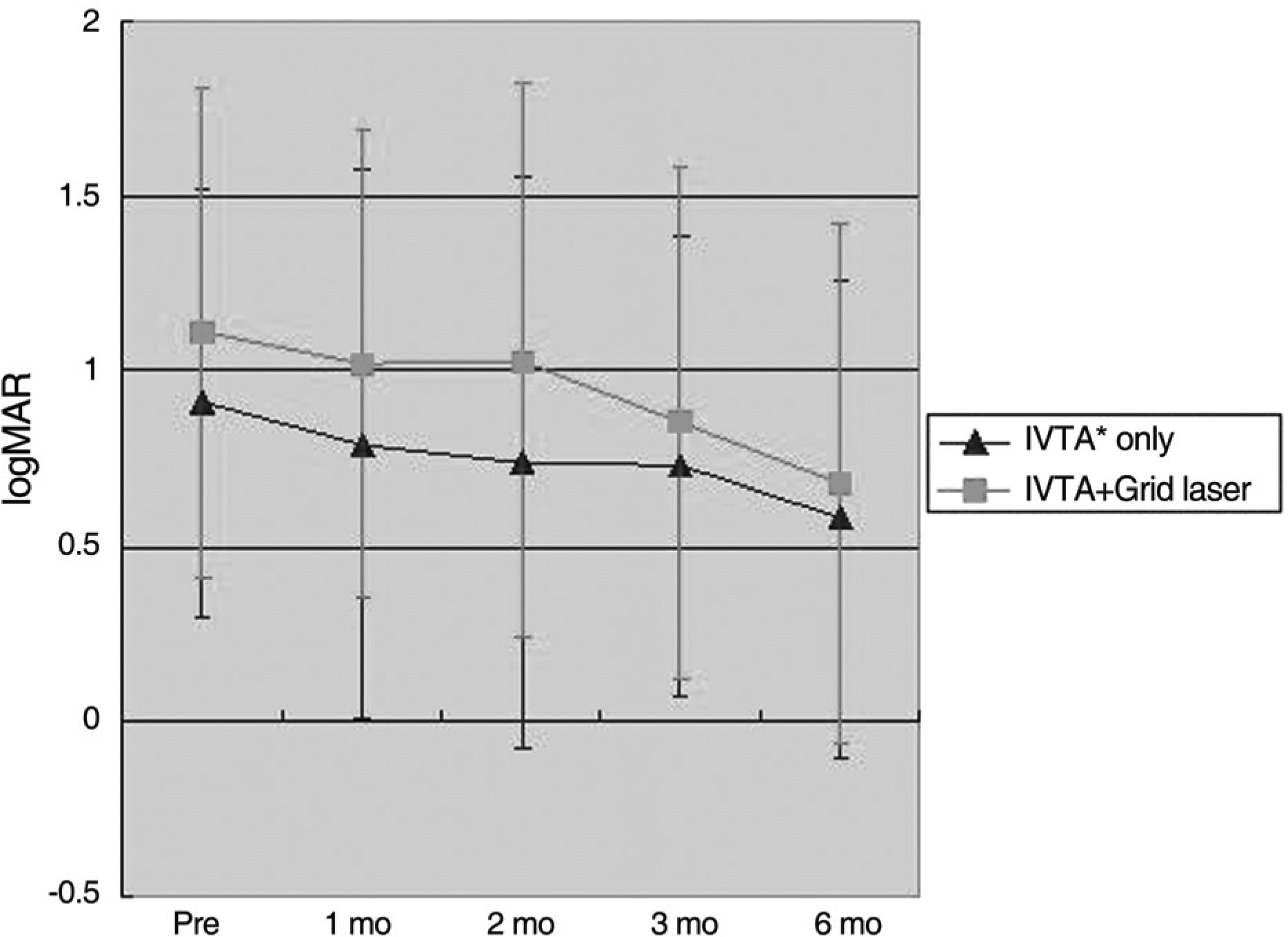
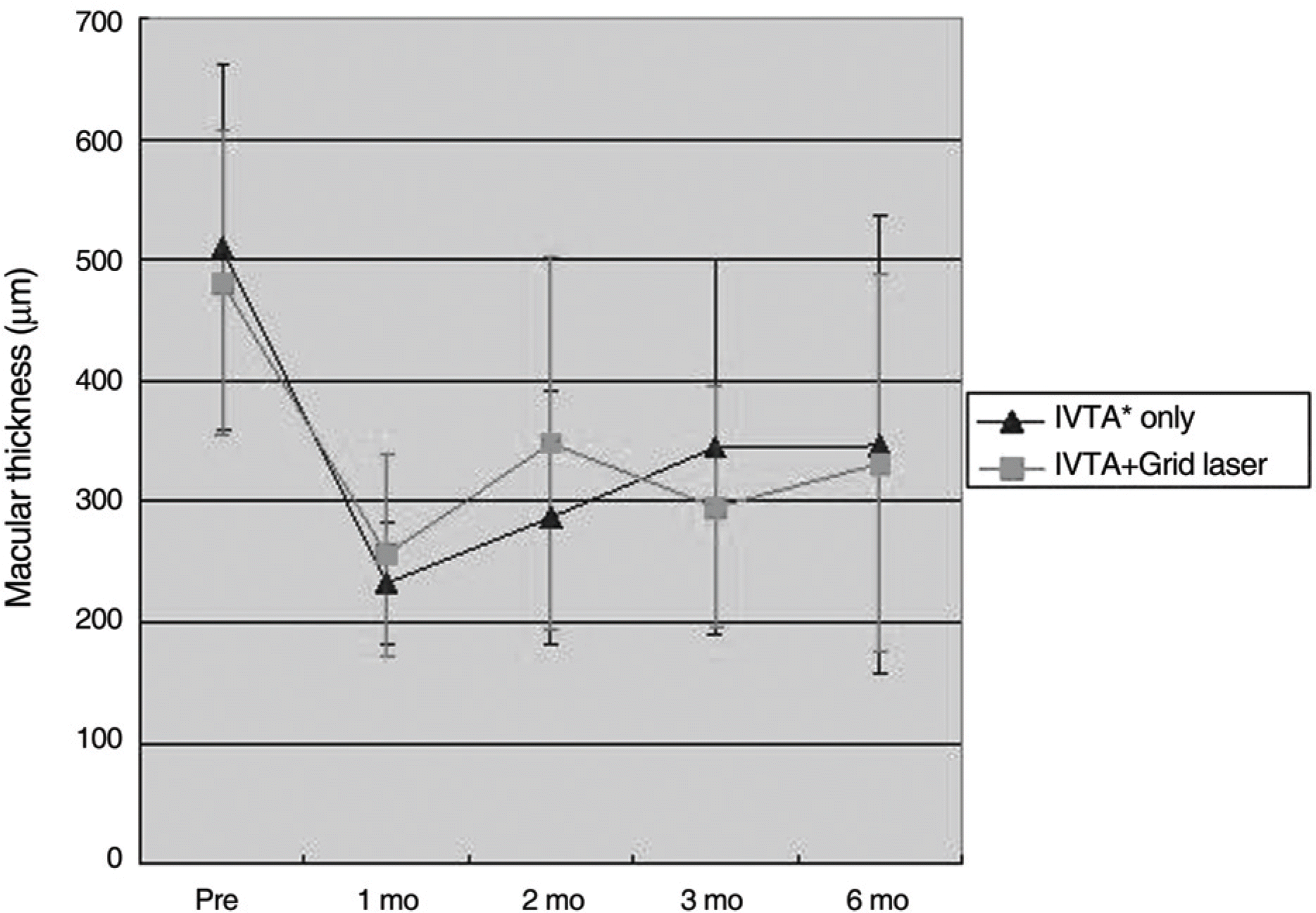
A total of 41 eyes underwent IVTA, 21 of which were treated with additional macular grid photocoagulation. The difference in best corrected visual acuity before IVTA between the two groups was not statistically significant. Laser photocoagulation was performed at an average of 2.8 months after IVTA. Final VA and foveal thickness improved significantly after IVTA in both groups and showed no significant difference between the two groups.
After six months a second injection was required in six eyes in the IVTA group and one eye in the photocoagulation group due to recurrence of macular edema, the difference of which was significant (p=0.04).
Go to : 
References
1. Finkelstein D, Clarkson JG. Branch Vein Occlusion Study Group. Branch and central vein occlusions. Focal points 1987: Clinical modules for ophthalmologists. Am Acad Ophthalmol. 1987; 5:1–11.
2. Branch Vein Occlusion Study Group. Argon laser photocoagulation for macular edema in branch vein occlusion. Am J Ophthalmol. 1984; 98:271–82.
3. Wallow IH, Danis RP, Bindley C, Neider M. Cystoid degeneration in experimental branch vein occlusion. Ophthalmology. 1988; 95:1371–9.
4. Early Treatment Diabetic Retinopathy Study Research Group. Photocoagulation for diabetic macular edema: Early Treatment Diabetic Retinopathy Study Report number 1. Arch Ophthalmol. 1985; 103:1796–806.
5. Lee H, Shah GK. Intravitreal triamconoloneas primary treatment of cystoid macular edema secondary to branch retinal vein occlusion. Retina. 2005; 25:551–5.
6. Jonas JB, Söfker A. Intraocular injection of crystalline cortisone as adjunctive treatment of diabetic macular edema. Am J Ophthalmol. 2001; 132:425–7.

7. Degenring RF, Kamppeter B, Kreissig I, Jonas JB. Morphological and functional changes after intravitreal triamcinolone acetonide for retinal vein occlusion. Acta Ophtalmol Scand. 2003; 81:548–50.

8. Beer PM, Bakri SJ, Singh RJ, et al. Intraocular concentration and pharmacokinetics of triamcinolone acetonide after a single intravitreal injection. Ophthalmology. 2003; 110:681–6.

9. Chen SD, Sundaram V, Lochhead J, Patel CK. Intravitreal triamcinolone for the treatment of ischemic macular edema associated with branch retinal vein occlusion. Am J Ophthalmol. 2006; 141:876–83.

10. Ozdek S, Deren YT, Gurelik G, Hasanreisoglu B. Posterior subtenon triamcinolon, intravitreal triamcinolone and grid laser photocoagulation for the treatment of macular edema in branch retinal vein occlusion. Ophthalmic Res. 2008; 40:26–31.
11. Lee DG, Cho NC. Combination of laser treatment and intravitreal triamcinolone injection for macular edema with branch retinal vein occlusion. J Korean Ophthalmol Soc. 2005; 46:287–96.
12. Yang HN, Kim YJ, Kim JC, Shyn KH. Clinical evaluation for branch retinal vein occlusion. J Korean Ophthalmol Soc. 1992; 33:593–9.
13. Roseman RL, Olk RJ. Krypton red laser photocoagulation for branch retinal vein occlusion. Ophthalmology. 1987; 94:1120–5.

14. Kang SJ, Chin HS, Moon YS. Visual prognosis of macular edema associated with macular ischemia in branch retinal vein occlusion. J Korean Ophthalmol Soc. 2002; 43:1621–8.
15. Rehak J, Rehak M. Branch retinal vein occlusion: pathogenesis, visual prognosis, and treatment modalities. Curr Eye Res. 2008; 33:11–31.

16. Duff IF, Fall HF, Linman JW. Anticoagulant therapy in occlusive vascular disease of retina. AMA Arch Ophthalmol. 1951; 46:601–17.
17. Michel RG, Gass JD. The natural course of retinal branch vein obstruction. Trans Am Acad Ophthalmol Otolaryngol. 1974; 78:166–77.
18. Weiter JJ, Zuckerman R. The influence of photoreceptor-RPE complex on the inner retina: explanation for beneficial effect of photocoagulation. Ophthalmology. 1980; 87:1133–9.
19. Gottfredottir MS, Stefansson E, Jonasson F, Gíslason I. Retinal vasoconstriction after laser treatment for diabetic macular edema. Am J Ophthalmol. 1993; 115:64–7.
20. Clover GM. The effects of argon and krypton photocoagulation on the retina: implications for the inner and outer blood retinal barrier. Gitter KA, Schatz H, Yannuzzi LA, McDinald HR, editors. Laser photocoagulation of retinal disease. San Francisco, Calif: Pacific medical Press;1988. v. 11. chap.p. 17.
21. Amarsson A, Stefansson E. Laser treatment and the mechanism of edema reduction in branch retinal vein occlusion. Invest Ophthalmol Vis Sci. 2000; 41:877–9.
22. Kim HC, Kim PS, Kim HK. The macular circulatory change in branch retinal vein occlusion. J Korean Ophthalmol Soc. 1999; 40:1911–7.
23. Oh J-Y, Seo JH, Ahn JK, et al. Early versus late intravitreal triamcinolone acetonide for macular edema associated with branch retinal vein occlusion. Korean J Ophthalmol. 2007; 21:18–20.

24. Yepremyan M, Wertz FD, Tivnan T, et al. Early treatment of cystoid macular edema secondary to branch retinal vein occlusion with intravitreal triamcinolone acetonide. Ophthalmic Surg Lasers Imaging. 2005; 36:30–6.

25. Lee J, Lee JH. The effect of intravitreal triamcinolone acetonide on cytoid macular edema. J Korean Ophthalmol Soc. 2004; 45:408–13.
Go to : 
 | Figure 1.The changes of mean visual acuity (mean± SD, LogMAR) of the intravitreal triamcinolone (IVTA) only group and the combined IVTA and grid laser photocoagulation group in patients with macular edema associated with branch retinal vein occlusion (* intravitreal triamcinolone injection). |
 | Figure 2.The changes of central macular thickness (mean± SD, μm) on optical coherence tomography after the intravitreal triamcinolone injection (IVTA) only and combined IVTA and grid laser photocoagulation in patients with macular edema of branch retinal vein occlusion (* intravitreal triamcinolone injection). |
Table 1.
Clinical outcomes of intravitreal triamcinolone injection (IVTA) only and combined IVTA and grid laser photocoagulations in patients with diffuse macular edema of branched retinal vein occlusion
| | | IVTA* only | IVTA* and Grid laser | p value |
|---|---|---|---|---|
| VA† | Baseline | 0.9±0.6 | 1.1±0.7 | 0.39§ |
| (mean± SD, logMAR) | 6 months | 0.58±0.37 | 0.68±0.43 | 0.20§ |
| Central macular thickness | Baseline | 509.8±150.8 | 481.1±125.4 | 0.54‡ |
| (mean± SD, μm) | 6 months | 285±120 | 252±113 | 0.18‡ |
| VA†<0.3 | Baseline | 3 (15.0%) | 3 (14.2%) | 0.34§ |
| (No. of eyes) | 6 months | 6 (30.0%) | 6 (28.6%) | 0.266§ |
| VA† increased > 2 lines at 6 months | | 12 (60.0%) | 17 (81.0%) | 0.095§ |
| (No. of eyes) | | | | |
| Re‐injection of IVTA (No. of eyes) | | 4 (30%) | 1 (4.8%) | 0.046§ |


 PDF
PDF ePub
ePub Citation
 Print
Print


 XML Download