

 PDF
PDF ePub
ePub Citation
 Print
Print


Abstract
Purpose
We compared the measurements of corneal thickness and anterior chamber depth (ACD) using three different methods Orbscan, Pentacam and ultrasound pachymetry.
Methods
In healthy volunteers, central corneal thickness was measured with Orbscan, Pentacam and ultrasound pachymetry. Estimation of peripheral corneal thickness and ACD were done by Orbscan and Pentacam. All results were compared statistically.
Results
The mean central corneal thickness of 64 eyes measured by Orbscan, Pentacam and ultrasound pachymetry were 509.1±61.0 μ m, 539.2±51.7 μ m and 527.4±48.3 μ m, respectively. There were statistically significant differences in the results among the three methods (repeated-measures analysis of variance (ANOVA), p<0.05). There were significant correlations between the ultrasonic pachymetry, the Orbscan and the Pentacam (Pearson correlation, r >0.90, p<0.05). The temporal and nasal peripheral corneal thicknesses were thinner in the Pentacam than in the Orbscan (paired t-test, p<0.05). The superior and inferior corneal thickness and ACD were not significantly different.
Go to : 
References
1. Brennan NA, Coles ML. Extended wear in perspective. Optom Vis Sci. 1997; 74:609–23.

2. Kim JH, Kim HB. Cornea. 1st ed.Seoul: Ilchokak;2000. p. 25–34.
3. Machat JJ, Slade S, Probst LE. The art of LASIK. 2nd ed.Thoro-fare, NJ: SLACK Inc.;1999. p. 127–38.
4. Wang Z, Chen J, Yang B. Posterior corneal surface topographic change after LASIK are related to residual corneal bed thickness. Ophthalmology. 1999; 106:406–9.
5. Whitacre MM, Stein RA, Hassanein K. Theeffect of corneal thickness on applanation tonometry. Am J Ophthalmol. 1993; 115:592–6.
6. Kim DH, Kim MS, Kim JH. Early corneal thickness changes after penetrating keratoplasty. J Korean Ophthalmol Soc. 1997; 38:55–61.
7. Olsen T. Sources of error in intraocular lens power calculation. J Cataract Refract Surg. 1992; 18:125–9.
8. Holladay JT. Standardizing constants for ultrasonic biometry, keratometry, and intraocular lens power calculations. J Cataract Refract Surg. 1997; 23:1356–70.
9. Alio JL, de la Hoz F, Perez-Santonja JJ, et al. Phakic anterior chamber lenses for correction of myopia; a 7-year cumulative analysis of complications in 263 cases. Ophthalmology. 1999; 106:458–66.
10. Holladay JT. Refractive power calculations for intraocular lenses in the phakic eye. Am J Ophthalmol. 1993; 116:63–6.
11. Iskander NG, Anderson Penno E, Peters NT, et al. Accuracy of Orbscan Pachymetry measurements and DHG ultrasound pachymetry in primary laser in situ keratomileusis and LASIK enhancement procedure. J Cataract Refract Surg. 2001; 27:681–5.
12. Rosa N, Lanza M, Borrelli M, et al. Comparision of central corneal thickness measured with Orbscan and Pentacam. J Refract Surg. 2007; 23:895–9.
13. Yaylali V, Kaufman SC, Thompson HW. Corneal thickness measurements with Orbscan topography system and ultrasonic pachymeter. J Cataract Refract Surg. 1997; 23:1345–50.
14. Kang PS, Yang YS, Kim JD. Comparision of corneal thickness measurements with the Orbscan and Ultrasonic pachymetry. J Korean Ophthalmol Soc. 2000; 41:1697–703.
15. Gherghel D, Hosking SL, Mantry S, et al. Corneal pachymetry in normal and keratoconic eyes: Orbscan II versus ultrasound. J Cataract Refract Surg. 2004; 30:1272–7.
16. Suzuki S, Oshika T, Oki K, et al. Corneal thickness measurements: scanning-slit corneal topography and noncontact specular microscopy versus ultrasonic pachymetry. J Cataract Refract Surg. 2003; 29:1313–8.
17. Modis L. Jr, Langenbucher A, Seitz B. Scanning-slit and specular microscopic pachymetry in comparison with ultrasonic determination of corneal thickness. Cornea. 2001; 20:711–4.
18. Buehl W, Stojanac D, Sacu S, et al. Comparison of three methods of measuring corneal thickness and anterior chamber depth. Am J Ophthalmol. 2006; 141:7–12.
19. Kim SW, Byun YJ, Kim EK, et al. Central corneal thickness measurements in unoperated eyes and eyes after PRK for myopia using Pentacam, Orbscan II and ultrasonic pachymetry. J Refract Surg. 2007; 23:888–94.
20. Shin YJ, Kim NH, Kim DH. Comparison of Pentacam with Orbscan. J Korean Ophthalmol Soc. 2007; 48:637–41.
21. Ryu HW, Kim RK, Chung SK. Comparison of A-scan, Scheimpflug camera, and Orbscan for measurement of anterior chamber depth. J Korean Ophthalmol Soc. 2006; 47:1287–91.
Go to :
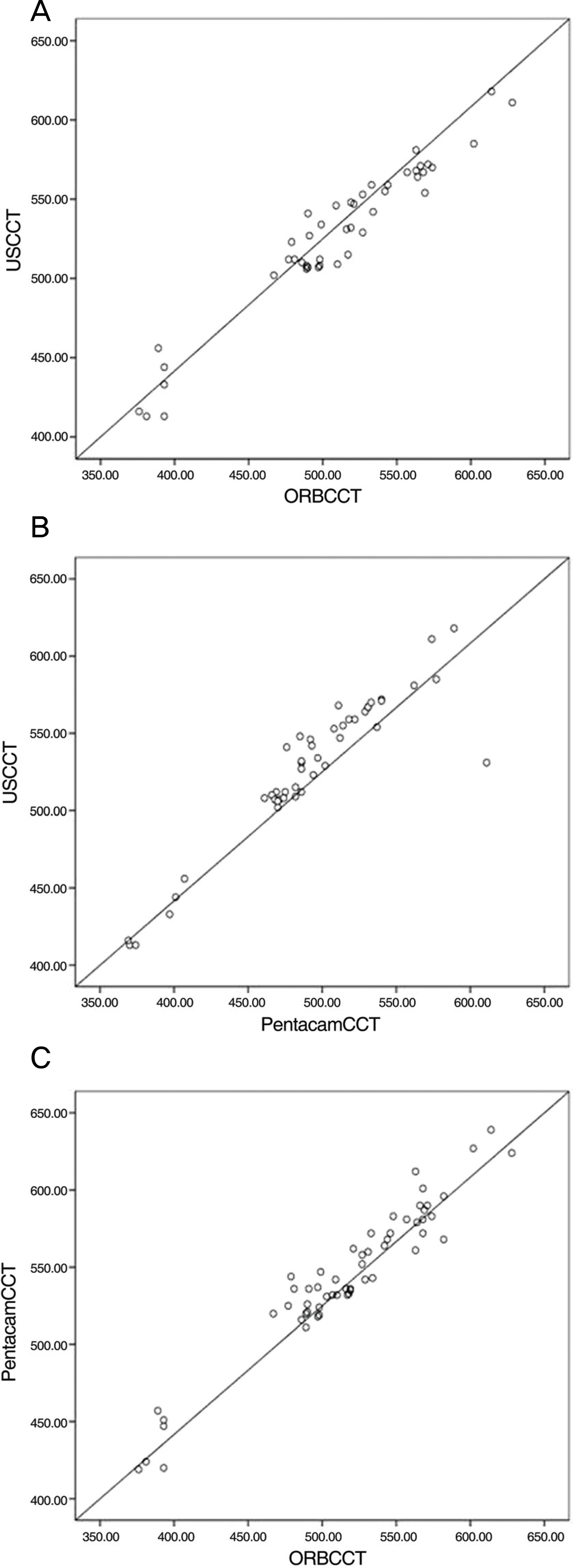 | Figure 1.The correlation plot of the central corneal thickness (CCT) measured by Ultrasound pachymetry (US), Orbscan (ORB) and Pentacam (in μm). (A) US CCT and ORB CCT (r=0.968, p=0.000), (B) US CCT and Pentacam CCT (r=0.927, p=0.000), (C) Pentacam CCT and ORB CCT (r=0.965, p=0.000). |
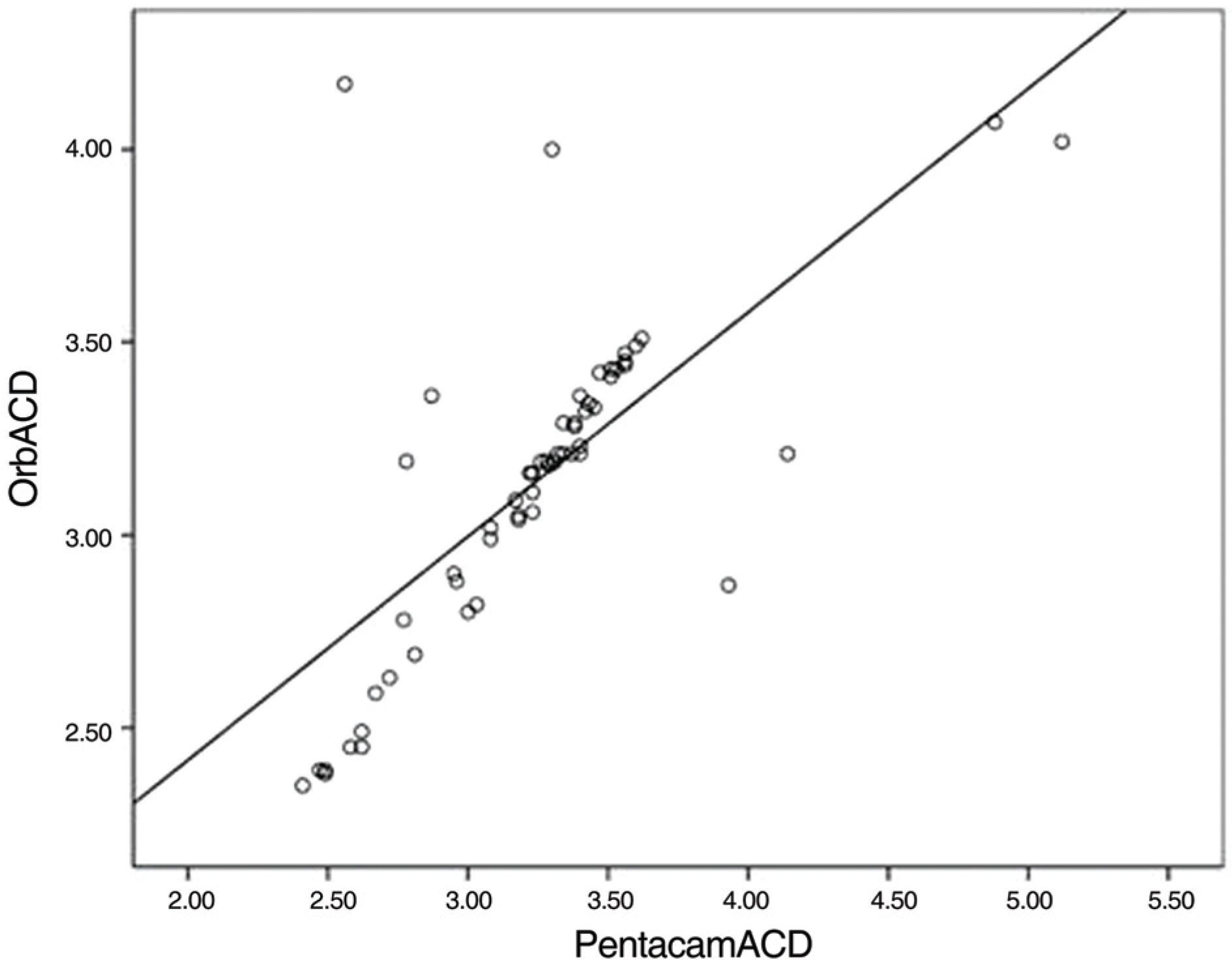 | Figure 2.The correlation plot of the anterior chamber depth (ACD, in mm) measured by Orbscan (Orb) and Pentacam (r=0.67, p<0.05) |
Table 1.
The central corneal thickness (m) measured by Ultrasound pachymetry (Ultrasound), Orbscan and Pentacam (Mean± standard deviation)
| | Ultrasound | Orbscan | Pentacam | p value* |
|---|---|---|---|---|
| CCT† (m) | 527.4±48.3 | 509.1±61.0 | 539.4±51.7 | 0.000 |
Table 2.
The comparison of corneal thickness (means± standard deviation) between Orbscan and Pentacam
| |
Corneal thickness |
||
|---|---|---|---|
| Orbscan (μm) | Pentacam (μm) | p value* | |
| Center | 515.94±56.75 | 544.51±47.89 | 0.00 |
| Thinnest | 510.16±58.07 | 540.69±49.54 | 0.00 |
| Temporal | 582.21±35.90 | 573.14±35.90 | 0.01 |
| Nasal | 613.43±43.46 | 593.90±49.75 | 0.00 |
| Superior | 613.67±40.84 | 586.96±41.24 | 0.93 |
| Inferior | 586.96±41.24 | 580.49±42.56 | 0.05 |
Table 3.
The comparison of anterior chamber depth (means± standard deviation) between Orbscan and Pentacam
| |
Anterior chamber depth |
||
|---|---|---|---|
| Orbscan (mm) | Pentacam (mm) | p value* | |
| Center | 3.15±0.40 | 3.24±0.51 | 0.09 |
| Deepest | 3.25±0.40 | 3.34±0.55 | 0.13 |
| Temporal | 2.93±0.32 | 2.90±0.33 | 0.30 |
| Nasal | 2.58±0.41 | 2.59±0.41 | 0.88 |
| Superior | 2.74±0.37 | 2.69±0.47 | 0.30 |
| Inferior | 2.93±0.39 | 3.02±0.33 | 0.13 |
 XML Download