Abstract
Purpose
To report the case of delayed suprachoroidal hemorrhage after Ahmed valve implantation in a neovascular glaucoma (NVG) patient.
Case summary
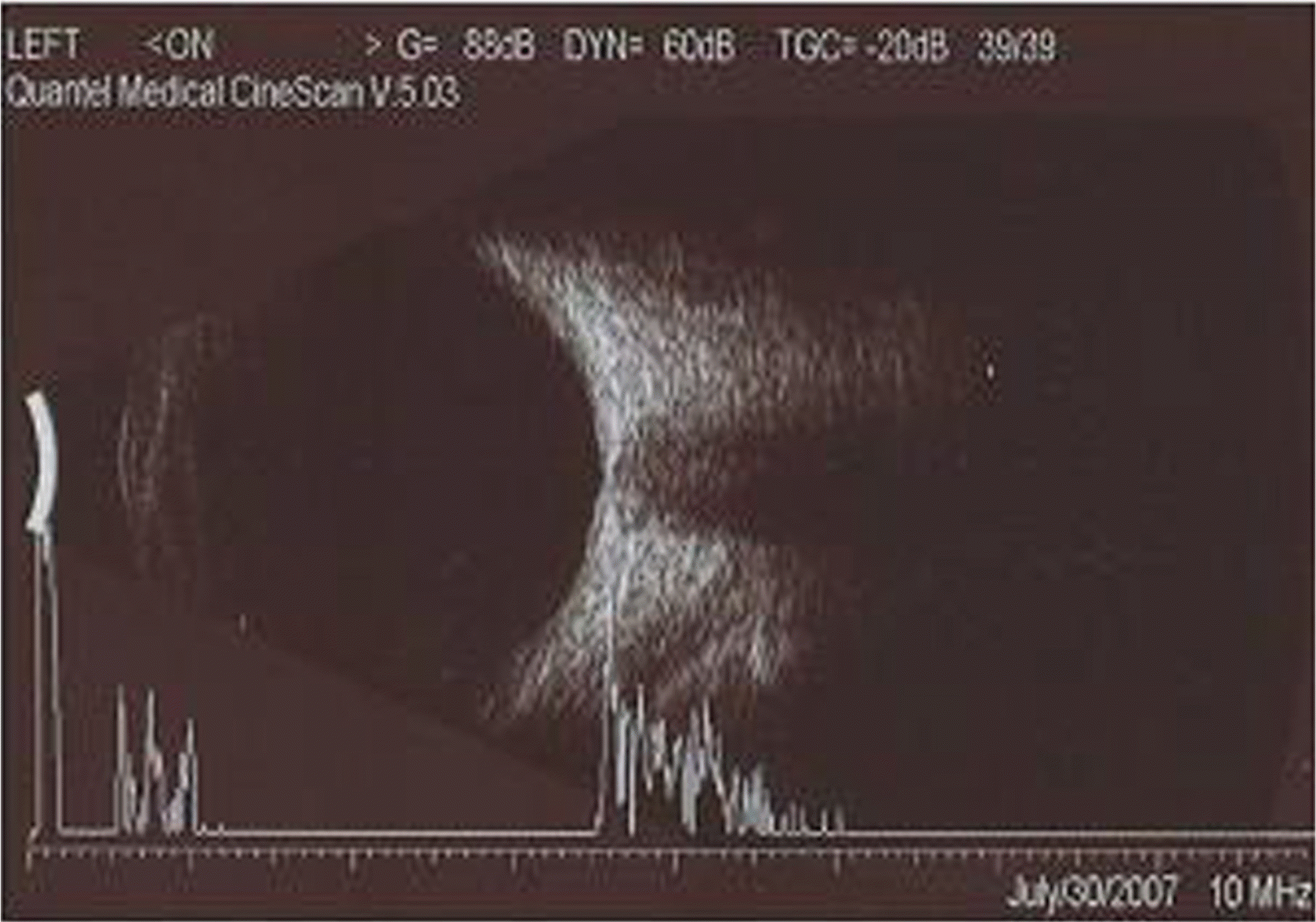
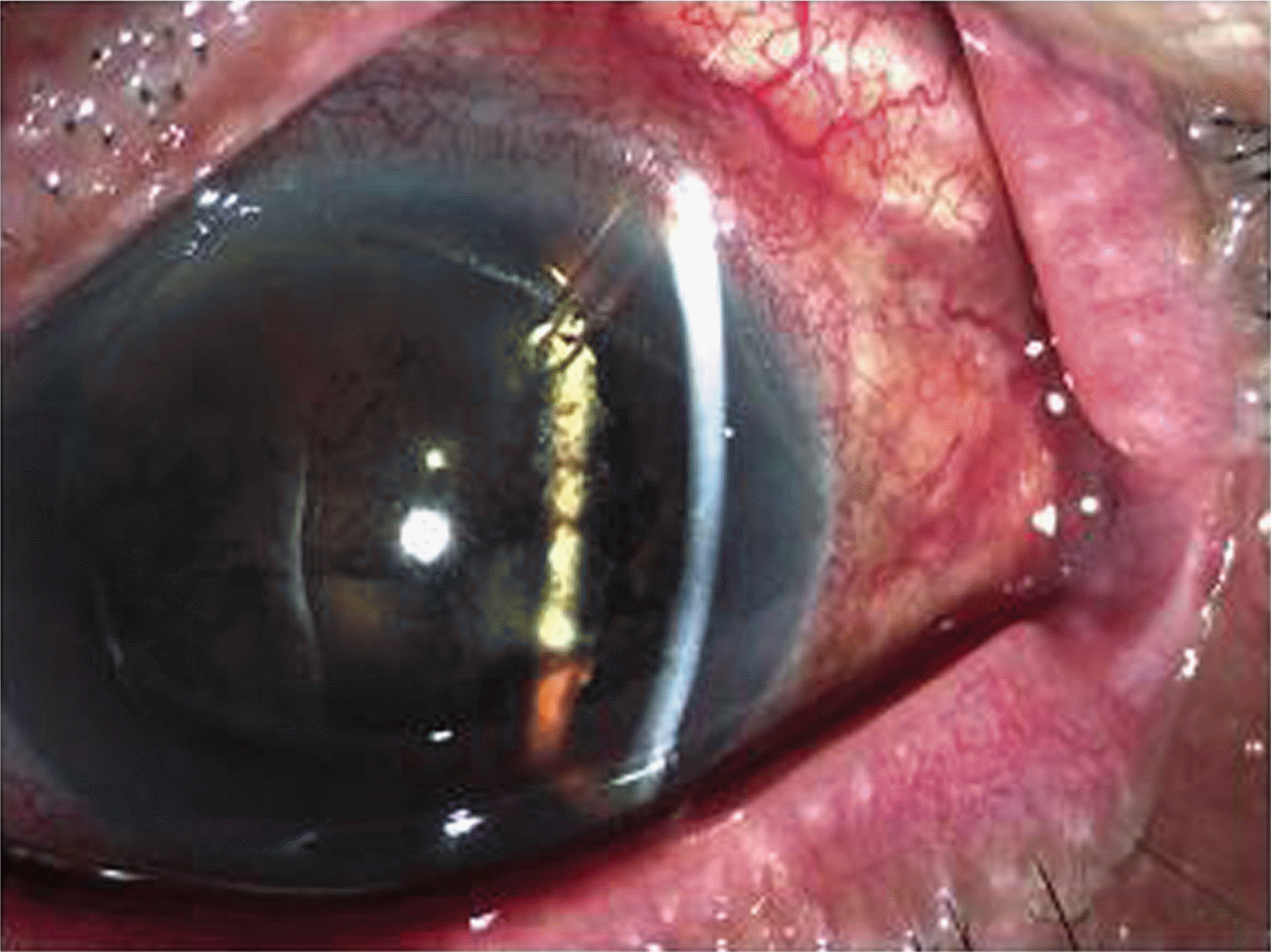
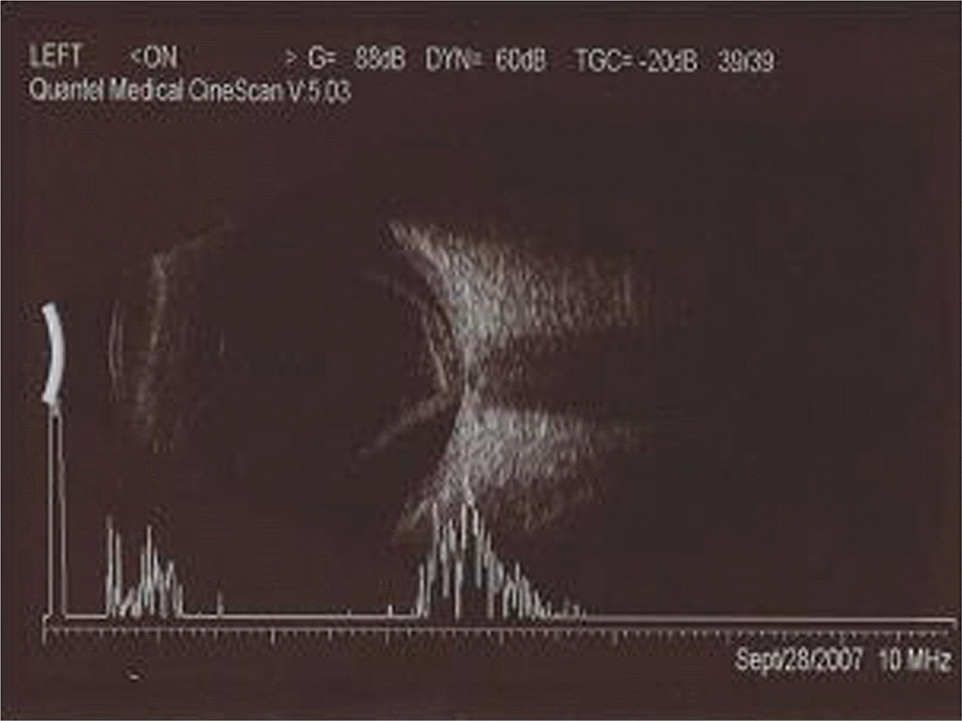
A 74-years-old male visited the hospital with ocular pain in the left eye. He had a history of vitrectomy and Intraocular lens (IOL) scleral fixation due to trauma in the left eye. NVG was diagnosed and Ahmed valve was implanted in his left eye. Three days later, hypotony occurred with all quadrant choroidal detachment. Next day, raised intraocular pressure (IOP) was checked and anterior chamber was flat on slit lamp examination. Vitreous hemorrhage and suprachoroidal hemorrhage were suspected. We performed anterior chamber formation with viscoelastics. The anterior chamber became deeper and hemorrhage gradually decreased. A month later, the patient visited us with severe ocular pain. Raised IOP and shallow anterior chamber due to moderate hyphema and anteriorly placed IOL were found. Retinal detachment was suspected on B-scan. Vitrectomy, IOL removal, silicone oil insertion, and Ahmed valve removal were performed.
Go to : 
References
1. Mueller H. Expulsive hemorrhage. Trans Ophthalmol Soc U K. 1959; 79:621–33.
2. Paysse E, Lee PP, Lloyd MA, et al. Suprachoroidal hemorrhage after Molteno implantation. J Glaucoma. 1996; 5:170–5.

3. Ruderman JM, Harbin TS, Campbell DG. Postoperative supra-chorodial hemorrhage following filtration procedures. Arch Ophthalmol. 1986; 104:201–5.
4. Givens K, Shields MB. Suprachoroidal hemorrhage after glaucoma filtering surgery. Am J Opthalmol. 1987; 103:689–94.

5. The Fluorouracil Filtering Surgery Study Group. Risk factors for suprachoroidal hemorrhage after filtering surgery. Am J Ophthalmol. 1992; 113:501–7.
7. Tuli SS, WuDunn D, Ciulla TA, Cantor LB. Delayed suprachoroidal hemorrhage after glaucoma filtration procedures. Ophthalmology. 2001; 108:1808–11.
8. Canning CR, Lavin M, McCartney AC, et al. Delayed suprachoroidal hemorrhage after glaucoma operation. Eye. 1989; 3:327–31.
9. Ayyala RS, Zurakowski D, Smith JA, et al. A clinical study of the Ahmed glaucoma valve implant in advanced glaucoma. Ophthalmology. 1998; 105:1968–76.
10. Coleman AL, Hill R, Wilson MR, et al. Initial clinical experience with the Ahmed glaucoma valve implant. Am J Ophthalmol. 1995; 120:23–31.

11. Frenkel RE, Shin DH. Prevention and management of delayed suprachoroidal hemorrhage after filtration surgery. Arch Ophthalmol. 1986; 104:1459–63.

12. Gressel MG, Parrish RK, Heuer DK. Delayed nonexpulsive suprachoroidal hemorrhage. Arch Ophthalmol. 1984; 102:1757–60.

13. Beyer CF, Peyman GA, Hill JM. Expulsive choroidal hemorrhage in rabbits. A histophathologic study. Arch Ophthalmol. 1989; 107:1648–53.
Go to : 
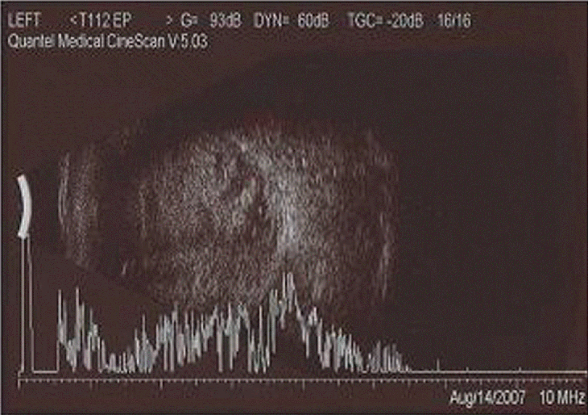 | Figure 2.Five days after Ahmed valve implantation, B-scan showed large amount of vitreous and suprachoroidal hemorrhage. |
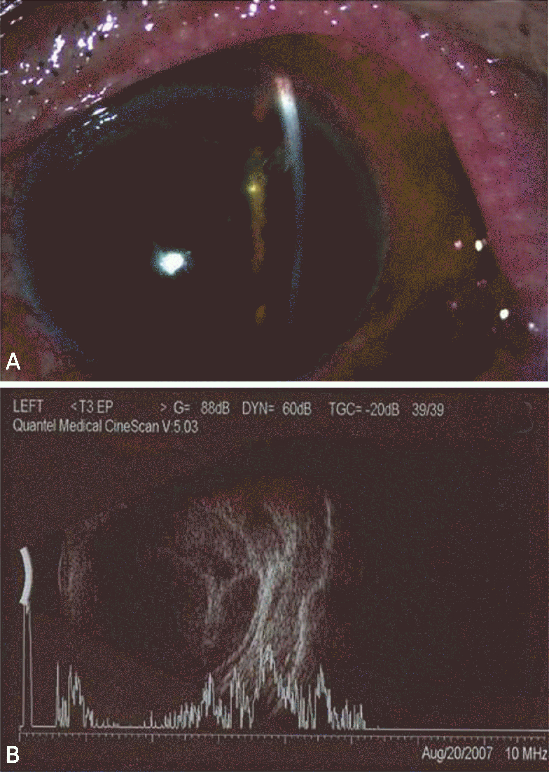 | Figure 3.After anterior chamber formation, anterior chamber became deep and Ahmed valve tip was in placed (A). On B-scan, vitreous and suprachoroidal hemorrhage decreased gradually (B). |


 PDF
PDF ePub
ePub Citation
 Print
Print


 XML Download