

 PDF
PDF ePub
ePub Citation
 Print
Print


Abstract
Purpose
We report a case of ophthalmic artery occlusion with third nerve paresis in the left eye due to acute occlusion of the left ICA.
Case summary
A 37-year-old man visited our emergency room with “black out” in the left eye, headache, and nausea. The corrected visual acuity was 20/25 in the right eye, and hand motion in the left eye. In the left eye, a relative afferent papillary defect was noted, with an intraocular pressure of six mmHg. Twenty prisms of exotropia in the primary position was observed, and ocular motor examination revealed limitations of supraduction, infraduction, and adduction in the left eye, suggesting third nerve palsy of the left eye. Fundus examination revealed a pale retina in the macula of the left eye. Brain MRI demonstrated multifocal faint low densities in the left caudate nucleus as well as the frontal and parietal lobes. CT angiography and four-vessel angiography demonstrated complete occlusion in the proximal part of the left internal carotid artery ICA.
After five months the corrected visual acuity of the left eye improved to 20/200. The 20 prism diopters of exotropia persisted, and ocular motor examination revealed only a mild limitation of adduction in the left eye.
References
1. Fisher M. Occlusion of the internal carotid artery. AMA Arch Neurol Psychiatry. 1951; 65:346–77.

2. Fisher M. Transient monocular blindness associated with hemiplegia. AMA Arch Ophthalmol. 1952; 47:167–203.
3. Appen RE, Wray SH, Cogan DG. Central retinal artery occlusion. Am J Ophthalmol. 1975; 79:374–81.
4. Brown GC, Magargal LE. The ocular ischemic syndrome. Clinical, fluorescein angiographic and carotid angiographic features. Int Ophthalmol. 1988; 11:239–51.
5. Guillon B, Levy C, Bousser MG. Internal carotid artery dissection: an update. J Neurol Sci. 1998; 153:146–58.
6. Biousse V, D'Anglejan-Chatillon J, Touboul PJ, et al. Time course of symptoms in extracranial carotid artery dissections. a series of 80 patients. Stroke. 1995; 26:235–9.
7. Biousse V, Touboul PJ, D'Anglejan-Chatillon J, et al. Ophthalmologic manifestations of internal carotid artery dissection. Am J Ophthalmol. 1998; 126:565–77.
8. Newman NJ, Kline LB, Leifer D, Lessell S. Ocular stroke and carotid artery dissection. Neurology. 1989; 39:1462–4.
9. Kline LB. The neuro-ophthalmologic manifestations of spontaneous dissection of the internal carotid artery. Semin Ophthalmol. 1992; 7:30–7.
10. Bogousslavsky J, Despland PA, Regli F. Spontaneous carotid dissection with acute stroke. Arch Neurol. 1987; 44:137–40.
11. Rao TH, Schneider LB, Patel M, Libman RB. Central retinal artery occlusion from carotid dissection diagnosed by cervical computed tomography. Stroke. 1994; 25:1271–2.
12. Cullom RD JR, Cullom ME, Kardon R, Digre K. Two neuro-ophthalmic episodes separated in time and space. Surv Ophthalmol. 1995; 40:217–24.
13. Koo HM, Kim JH. A case report of central retinal artery occlusion associated with internal artery occlusion. J Korean Ophthalmol Soc. 1986; 27:115–20.
14. Biousse V. Carotid disease and the eye. Curr Opin Ophthalmol. 1997; 8:16–26.
15. Klijn CJ, Kappelle LJ, Tulleken CA, van Gijn J. Symptomatic carotid artery occlusion: a reappraisal of hemodynamic factors. Stroke. 1997; 28:2084–93.
16. Mokri B, Silbert PL, Shievink WI, Piepgras DG. Cranial nerve palsy in spontaneous dissection of the extracranial internal carotid artery. Neurology. 1996; 46:356–9.
17. Wilson WB, Leavengood JM, Ringel SP, Bott AD. Transient ocular motor paresis associated with acute internal carotid artery occlusion. Ann Neurol. 1989; 25:286–90.
18. Maitland CG, Black JL, Smith WA. Abducens nerve palsy due to spontaneous dissection of the internal carotid artery. Arch Neurol. 1983; 40:448–9.
19. Vargas ME, Desrouleaux JR, Kupersmith MJ. Ophthalmoplegia as a presenting manifestation of internal carotid artery dissection. J Clin Neuroophthalmol. 1992; 12:268–71.
20. Lapresle J, Lasjaunias P. Cranial nerve ischemic arterial syndromes. a review. Brain. 1986; 109:207–16.
21. Duker JS, Belmont JB. Ocular ischemic syndrome secondary to carotid artery dissection. Am J Ophthalmol. 1988; 106:750–2.
22. Galetta SL, Leahey A, Nichols CW, Raps EC. Orbital ischemia, ophthalmoparesis, and carotid dissection. J Clin Neuroophthalmol. 1991; 11:284–7.
23. Asbury AK, Aldredge H, Hershberg R, Fisher CM. Oculomotor palsy in diabetes mellitus: a clinicopathological study. Brain. 1970; 93:555–66.
24. Parkinson D. A surgical approach to the cavernous portion of the carotid artery: anatomical studies and case report. J Neurosurg. 1965; 23:474–83.
25. Fisher CM. The headache and pain of spontaneous carotid artery dissection. Headache. 1982; 22:60–5.
26. Biousse V, D'Anglejan-Chatillon J, Massiou H, Bousser MG. Head pain in non traumatic artery dissection: a series of 65 patients. Cephalalgia. 1994; 14:33–6.
27. Silbert PL, Mokri B, Schievink WI. Headache and neck pain in spontaneous internal carotid and verterbral artery dissections. Neurology. 1995; 45:1517–22.
28. Biousse V, Woimant F, Amarenco P, et al. Pain as the only manifestation of internal carotid artery dissection. Cephalalgia. 1992; 12:314–7.
Figure 1.
On the 1st day of hospitalization, ocular motor examination revealed limitation of adduction, supraduction, and infraduction in the left eye, and in the primary position, 20 prism diopters of exotropia was seen.

Figure 2.
On the 1st day of hospitalization, fundus examination revealed pale retina in the macula of the left eye.
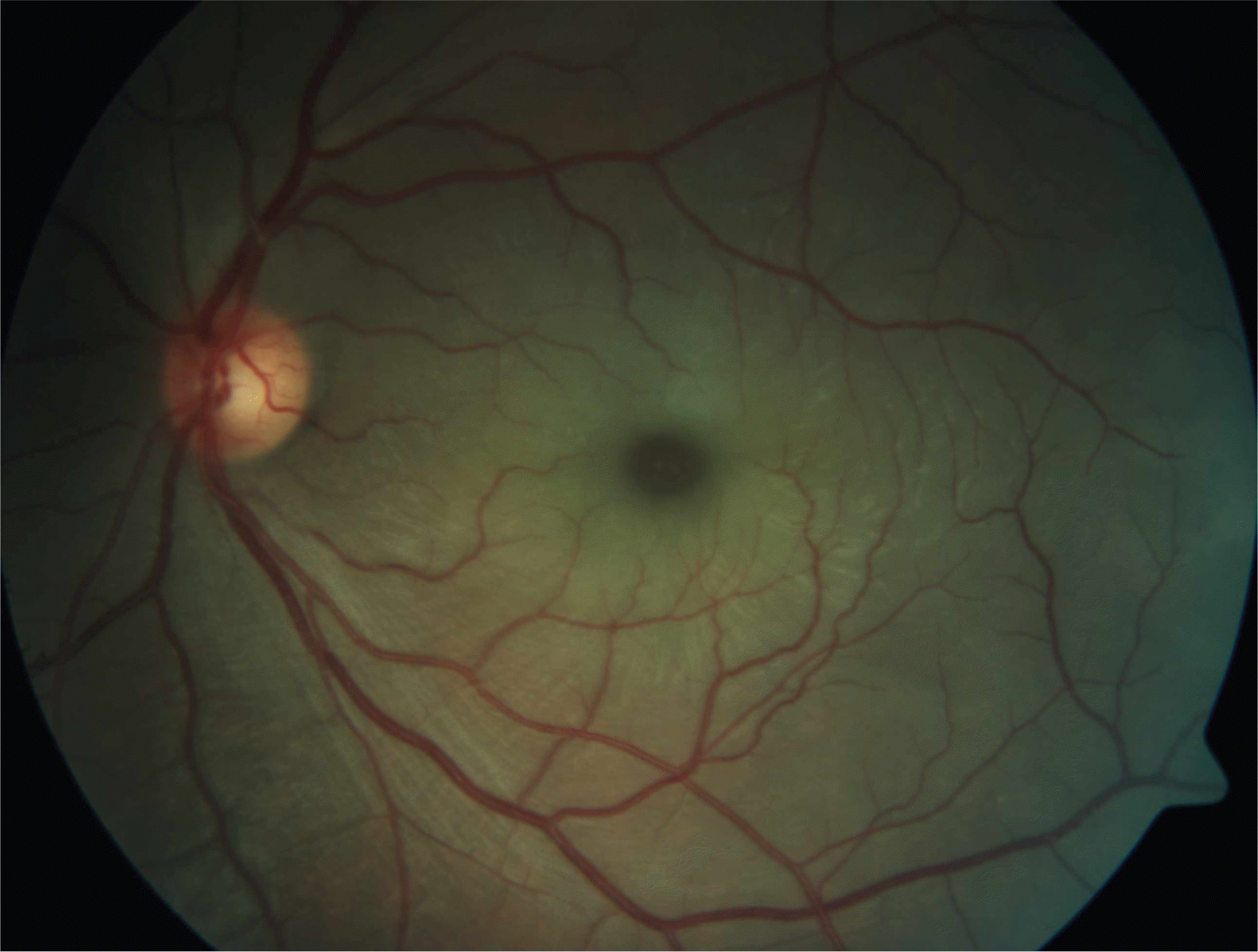
Figure 3.
Fluorescein angiography. There were choroidal filling delay and prolonged arteriovenous transit time.
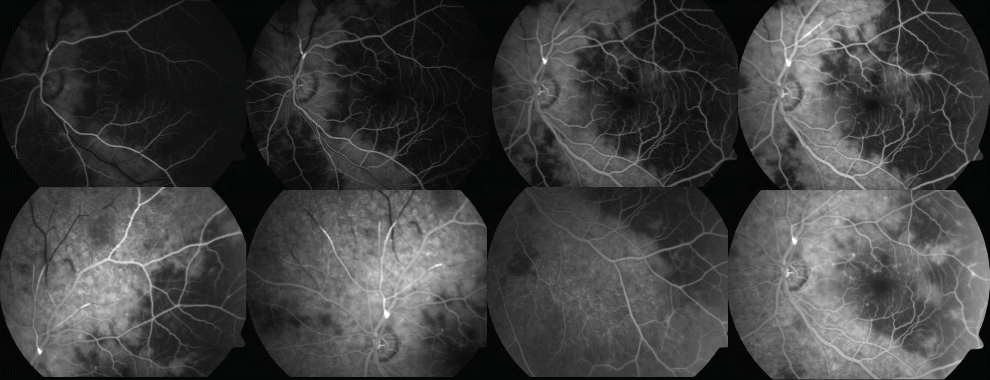
Figure 4.
Brain MRI. There are multifocal faint low densities in the left caudate nucleus, and frontal and parietal lobes.
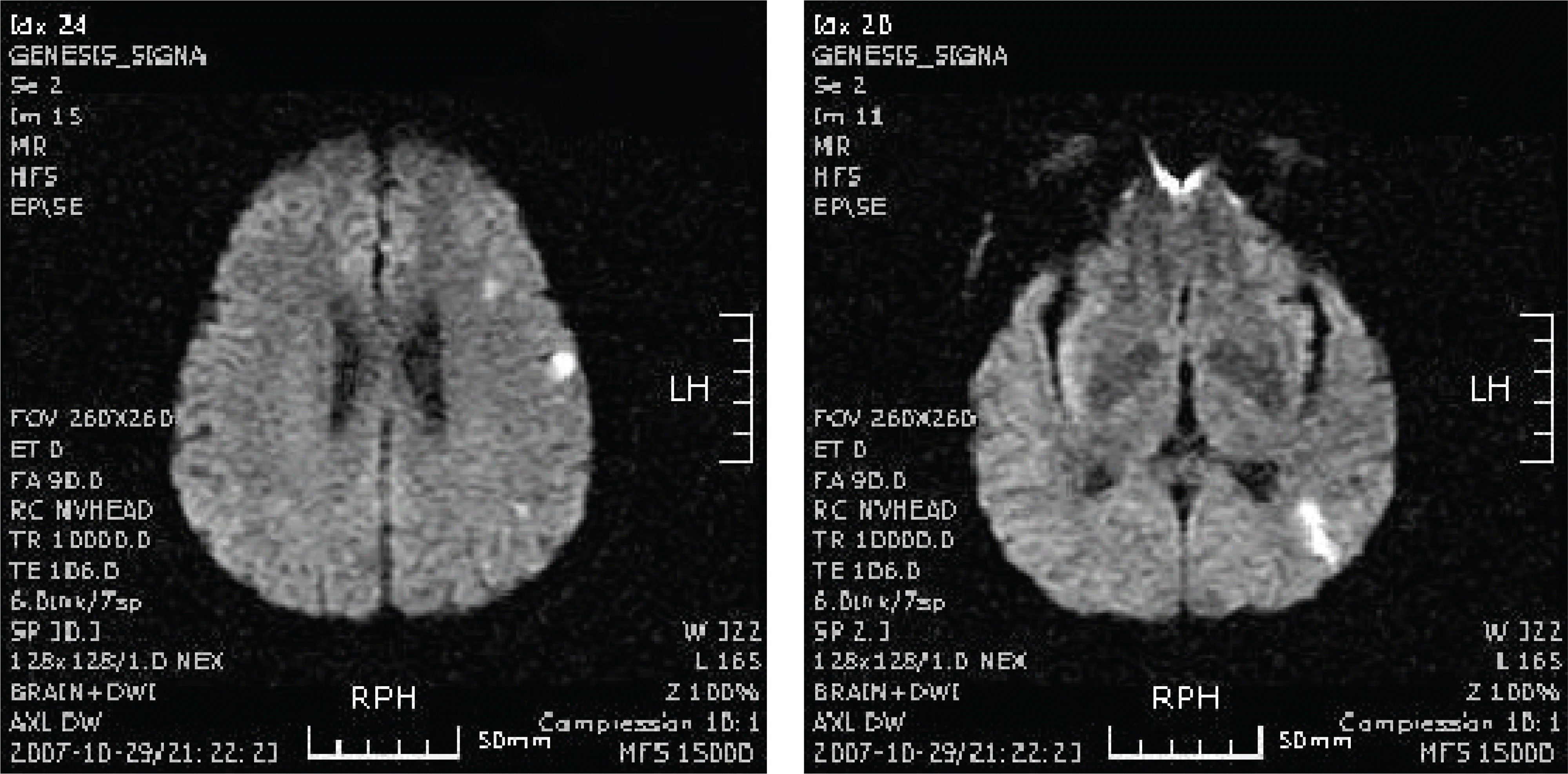
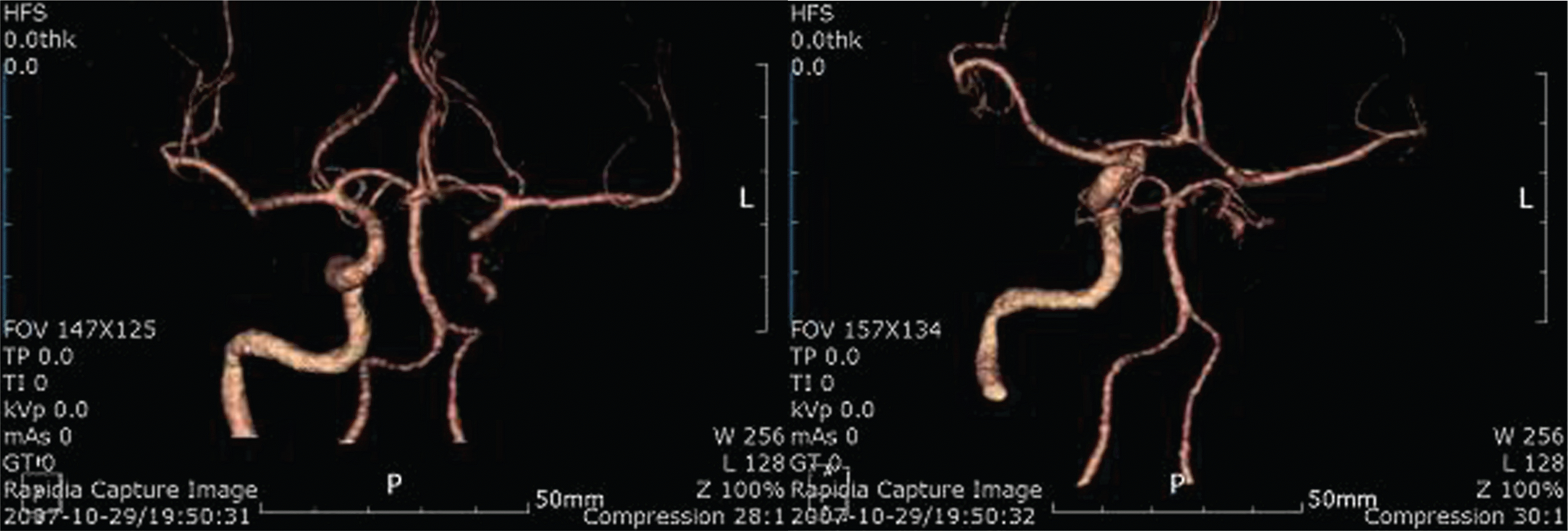
Figure 5.
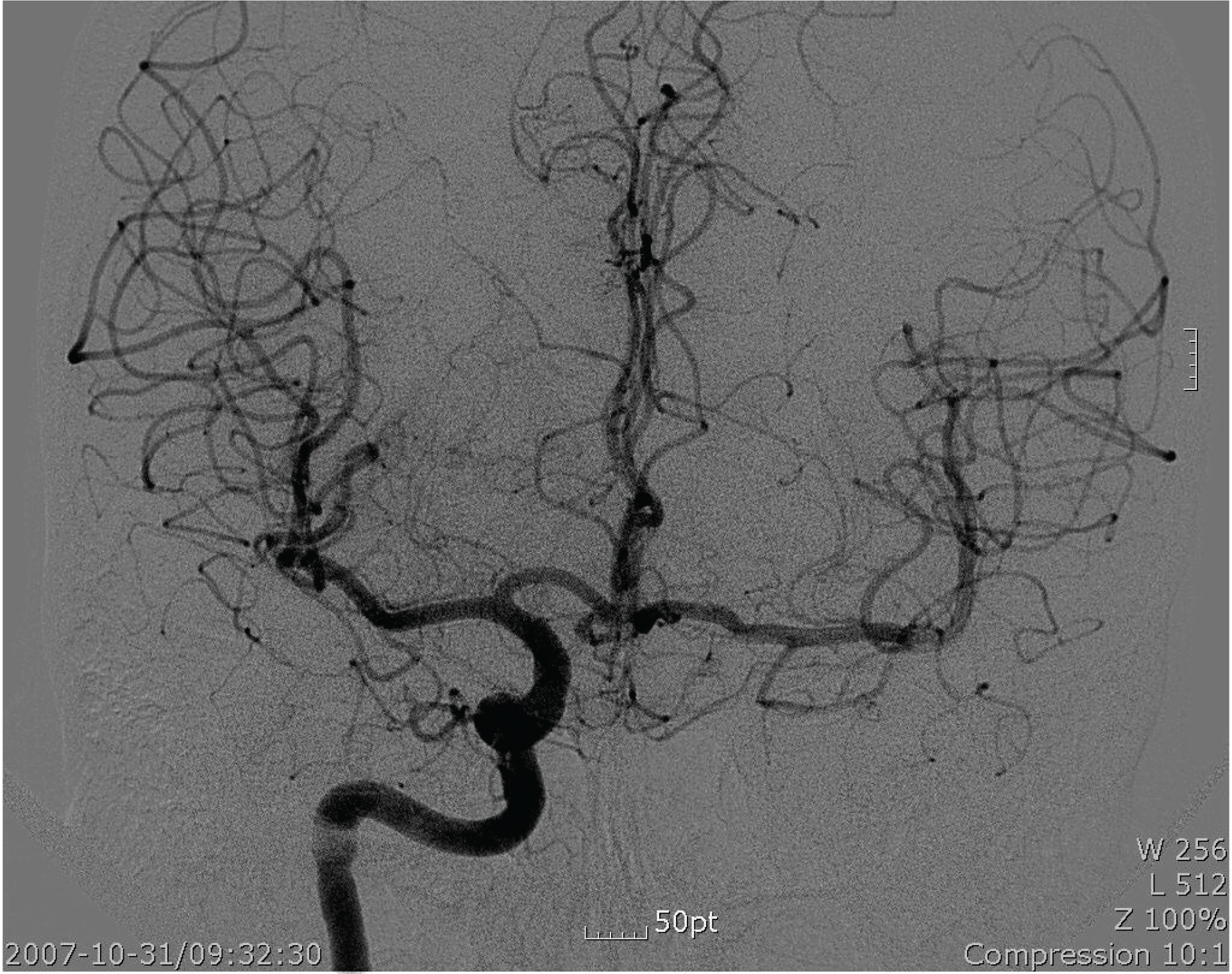
Four-vessel angiography. There was an irregular narrowing in the cavernous portion of the left ICA and the petrous and neck portion of the left ICA was not visualized.

 XML Download