Abstract
Purpose
We report two cases of mucocele formation after medial orbital wall fracture repair with an alloplastic implant.
Case summary
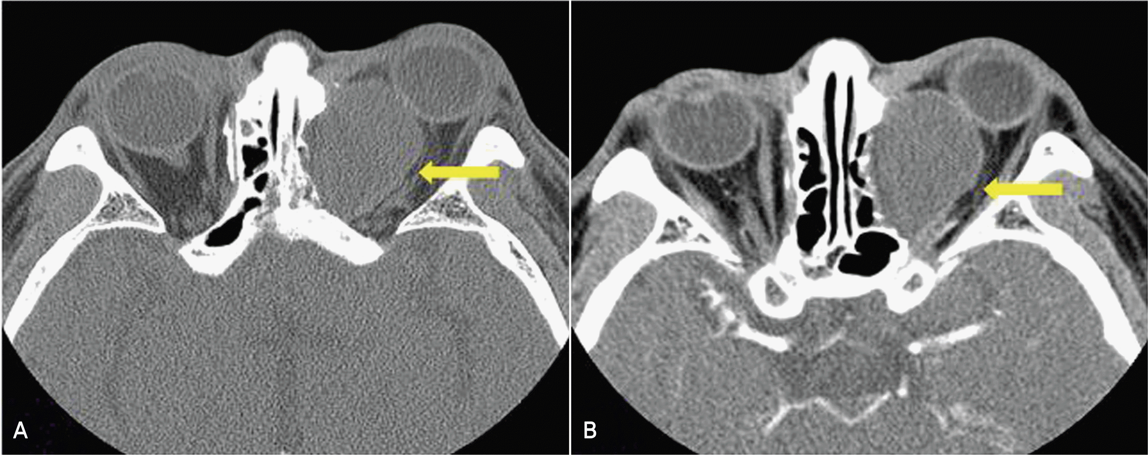
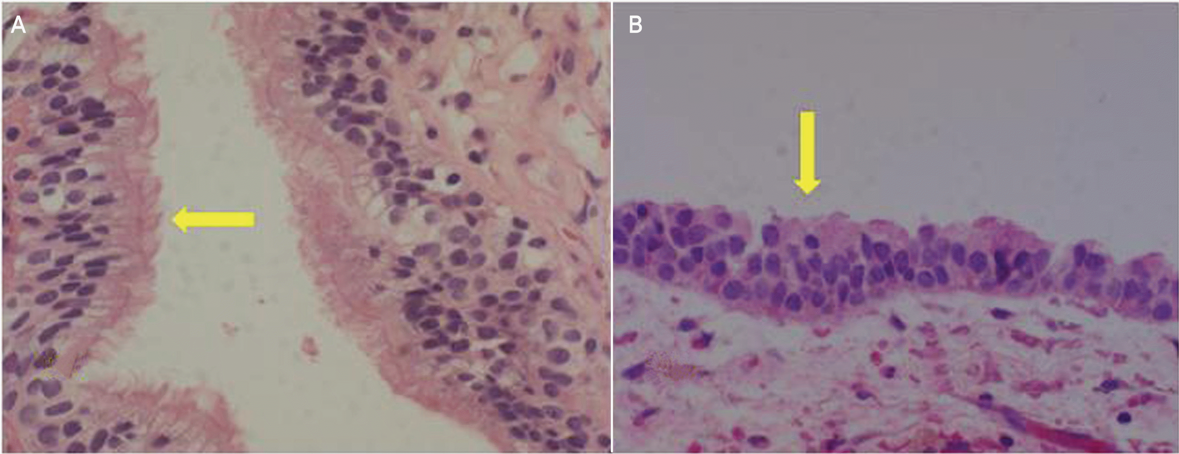
A 61‐ year‐ old man with a history of a medial orbital wall fracture repaired with an alloplastic implant five years earlier presented with a several‐ month history of left proptosis without diplopia, pain, or lid edema. A 55‐ year‐ old man with a history of a medial orbital wall fracture repaired with an alloplastic implant seven years prior, presented with a five‐ year history of left proptosis with diplopia. Computed tomography (CT) scans revealed a large cyst on the orbital medial wall, which surrounded the alloplastic implant and had no definite enhancement. The patients underwent orbital surgery to remove both the cyst and implant. Histologic examination of the cyst revealed a capsule lined with ciliated pseudostratified columnar epithelium. Both patients had an uncomplicated postoperative course with resolution of the proptosis.
Go to : 
References
1. Kim HK, Lim HS, Chung WS. Surgical effect of Medpor in the reconstruction of orbital wall fracture. J Korean Ophthalmol Soc. 1998; 39:623–30.
2. Jordan DR, St Onge P, Anderson RL, et al. Complications associated with alloplastic implants used in orbital fracture repair. Ophthalmology. 1992; 99:1600–8.

3. Cho BJ, Kim YD. Repair for orbital bone fracture with Supramid plate. J Korean Ophthalmol Soc. 1993; 34:925–35.
4. Stewart MG, Patrinely JR, Appling WD, Jordan DR. Late proptosis following orbital floor fracture repair. Arch Otolaryngol Head Neck Surg. 1995; 121:649–52.

5. Morrison AD, Sanderson RC, Moos KF. The use of silastic as an orbital implant for reconstruction of orbital wall defects: review of 311 cases treated over 20 years. J Oral Maxillofac Surg. 1995; 53:412–7.
6. Shapiro A, Tso MO, Putterman AM, Goldberg MF. A clinicopathologic study of hematic cysts of the orbit. Am J Ophthalmol. 1986; 102:237–41.

7. Gilhotra JS, McNab AA, McKelvie P, O'Donnell BA. Late orbital haemorrhage around alloplastic orbital floor implants: a case series and review. Clin Experiment Ophthalmol. 2002; 30:352–5.

8. Lee SB, Park KS, Kim YD. Orbital cyst after repair of blow– out fracture. J Korean Ophthalmol Soc. 1999; 40:273–7.
9. Wang TJ, Liao SL, Jou JR, Lin LL. Clinical manifestations and management of orbital mucoceles: the role of ophthalmologists. Jpn J Ophthalmol. 2005; 49:239–45.

10. Neves RB, Yeatts RP, Martin TJ. Pneumo– orbital cyst after orbital fracture repair. Am J Ophthalmol. 1998; 125:879–80.
11. Mauriello JA Jr., Flanagan JC, Peyster RG. An unusual late complication of orbital floor fracture repair. Ophthalmology. 1984; 91:102–7.

12. Tan CS, Ang LP, Choo CT, et al. Orbital cysts lined with both stratified squamous and columnar epithelia: a late complication of silicone implants. Ophthal Plast Reconstr Surg. 2006; 22:398–400.

13. Nkenke E, Amann K, Maier T, et al. Untreated ‘blow– in' fracture of the orbital floor causing a mucocele: report of an unusual late complication. J Craniomaxillofac Surg. 2005; 33:255–9.
14. Curtin HD, Rabinov JD. Extension to the orbit from paraorbital disease. The sinuses. Radiol Clin North Am. 1998; 36:1201–13.
Go to : 
 | Figure 1.A 61-year-old man with a history of a medial orbital wall fracture repaired with an alloplastic implant 5 years earlier, presented with a several‐month history of left proptosis. |
 | Figure 2.Axial (A) and coronal (B) orbit CT scans show a large cystic mass adjacent to the medial wall of the orbit. Axial (C) and coronal (D) MRI scans of the orbit show a large cystic mass that has high signal intensity in T1-, T2-weighted image. |
 | Figure 3.A 55-year-old man with a history of a medial orbital wall fracture repaired with an alloplastic implant 7 years before, presented with a 5-year history of proptosis in the left eye. |


 PDF
PDF ePub
ePub Citation
 Print
Print


 XML Download