

 PDF
PDF ePub
ePub Citation
 Print
Print


Abstract
Purpose
To investigate the effects of sutureless faden operation to eliminate suture-related perioperative risks in a rabbit model.
Methods
Twenty-eighty superior recti muscles of 14 rabbits were subjected to faden operation, at a distance of 6 mm from the insertion of the muscle. They were divided into four groups of 7 muscles each: group A, Beriplast-P; group B, Bard® mesh (12×2 mm); group C, Surgipro® mesh (6×2 mm); group D, Surgipro® mesh (6×6 mm). Rabbits were sacrificed and the eyes were enucleated. The operative field was examined upon dissection, 4 weeks after the surgery. Histopathologic sections were examined for the degree (grade 0 to 4) of inflammation, fibrosis, atrophy and/or degeneration of muscle fiber.
Results
Grossly, there was a mild adhesion at the myopexy site in group A, while there was greater adhesion at the myopexy site in groups B, C and D. Group B produced a pronounced adhesion, and the mesh firmly adhered to the sclera as well as to adjacent tissue. Upon histologic examination, group A showed a mild inflammatory reaction and fibrosis, whereas groups B and D showed a moderate inflammatory reaction and fibrosis. The degree of fibrosis in group D was more severe than that in group A (p<0.008), and there was no statistical significance among the other groups (p>0.05). There was atrophy and degeneration in the muscle fiber in all groups, with the exception of group D.
Go to : 
References
1. Shuckett EP, Hiles DA, Biglan AW, Evans DE. Posterior fixation suture operation (faden operation). Ophthalmic Surg. 1981; 12:578–85.
2. Alio JL, Faci A. Fundus changes following faden operation. Arch Ophthalmol. 1984; 102:211–3.

3. Tonelli E, de Almeida HC, Bambirra EA. Tissue adhesives for a sutureless fadenoperation: an experimental study in a rabbit model. Invest Ophthalmol Vis Sci. 2004; 45:4340–5.
4. Dunlap EA, Dunn M, Rossomondo R. Adhesives for sutureless muscle surgery. Arch Ophthalmol. 1969; 82:751–5.
5. Park KS, Min BM. Sutureless strabismus surgery with tissue adhesive in rabbit model. J Korean Ophthalmol Soc. 1995; 36:783–6.
6. Seong GJ, Kim HB, Koh HJ. Experimental strabismus surgery with Tissel in rabbit models. J Korean Ophthalmol Soc. 1991; 32:83–8.
7. Cho YA, Lee JY, Shin HY. Scleral attachment of extraocular muscle using a stitch combined with tissue adhesive in rabbits. J Korean Ophthalmol Soc. 2000; 41:411–6.
8. Kim SK, Lee JB. The usefulness of sutureless strabismus surgery with tissue adhesives in rabbit. J Korean Ophthalmol Soc. 1997; 38:1445–50.
9. Hadi HI, Maw A, Sarmah , Kumar P. Intraperitoneal tension-free repair of small midline ventral abdominal wall hernias with a Ventralex hernia patch: initial experience in 51 patient. Hernia. 2006; 10:409–13.
10. Quilici PJ, Greaney EM, Quilici J, Anderson S. Transabdominal preperitoneal laparoscopic inguinal herniorrhaphy: results of 509 repairs. Am Surg. 1996; 62:849–52.
11. Quilici PJ, Greaney EM, Quilici J, Anderson S. Laparascopic inguinal hernia repair: optimal technical variations and results in 1700 cases. Am Surg. 2000; 66:848–52.
12. Uen YH. Comparative laparascopic evaluation of the prolene polyprolene hernia system vs the perfix plug repair in a porcine groin hernia repair model. J Lapraendosc Adv Tech A. 2004; 14:368–73.
13. Prince JH, Eglitis I. Extraocular muscles. Prince JH, editor. The rabbit in eye research. 1964 ed.Springfield IL: Charles C. Thomas Publisher;1964. 1:chap 11.
14. Munton CG. Tissue adhesive in ocular surgery: a prospective study. Exp Eye Res. 1971; 11:1–6.
15. Ozkan SB, Kir E, Culhaci N, Dayanir V. The effect of Seprafilm on adhesion in strabismus surgery-An experimental study. J AAPOS. 2004; 8:46–9.
16. Berguer R, Staerkel RL, Moore EE, et al. Warning: fatal reaction to the use of fibrin glue in deep hepatic wounds. Case reports. J Trauma. 1991; 31:408–11.
17. Berruyer M, Amiral J, Ffrench P, et al. Immunization by bovine thrombin used with fibrin glue during cardiovascular operations. Development of thrombin and factor V inhibitors. J Thorac Cardiovasc Sur. 1993; 105:892–7.
18. Oswald AM, Joly LM, Gury C, et al. Fatal intraoperative anaphylaxis related to aprotinin after local application of fibrin glue. Anesthesiology. 2003; 99:762–3.
19. Uy HS, Reyes JM, Flores JD, Lim-Bon-Siong R. Comparison of fibrin glue and sutures for attaching conjunctival autografts after pterygium excision. Ophthalmology. 2005; 112:667–71.
20. Rabah DM, Begin LR, Zahran A, Corcos J. Tissue reactions of the rabbit urinary bladder to cadaveric human fascia lata and poly-propylene surgical mesh. Can J Urol. 2004; 11:2344–9.
21. Bringman S, Wollert S, Osterberg J, et al. One year results of a randomised controlled multi-centre study comparing Prolene and Vypro II-mesh in Lichtenstein hernioplasty. Hernia. 2005; 9:223–37.
22. Puccio F, Solazzo M, Marciano P. Comparison of three different mesh materials in tension-free inguinal hernia repair: prolene versus Vypro versus surgisis. Int Surg. 2005; 90:21–3.
23. Ingram RM. Rate at which muscle becomes joined to sclera after operations of recession and resection. Br J Ophthalmol. 1965; 49:235–45.
24. Alio JL, Chacon M, Faci A, et al. Muscular structural changes following faden operation. J Pediatr Ophthalmol strabismus. 1984; 21:102–9.
25. Lehman RA, Hayes GJ, Leonard F. Toxicity of alkyl 2-cyanoacrylates.Ⅰ. Peripheral nerve. Arch Surg. 1966; 93:441–6.
26. Goldstein JH, Kopelowitz N. Tissue response to the Fadenoperation. Br J Ophthalmol. 1979; 63:684–6.
Go to :
 | Figure 1.The methods of sutureless Fadenoperation in a rabbit model. Fadenoperation was performed at a distance of 6 mm from the insertion of superior rectus. The Beriplast-P® was gently applied with an injector between the superior rectus and sclera (A). Bard® mesh (12×2 mm) was encircled the superior rectus and the ends of the mesh were sutured with 6–0 Vicryl (B). Surgipro® mesh was inserted between the superior rectus and sclera (C,D). The size of Surgipro® mesh was 6×2 mm (C) and 6×6 mm (D). The dotted boxes mean the position of inserted mesh. |
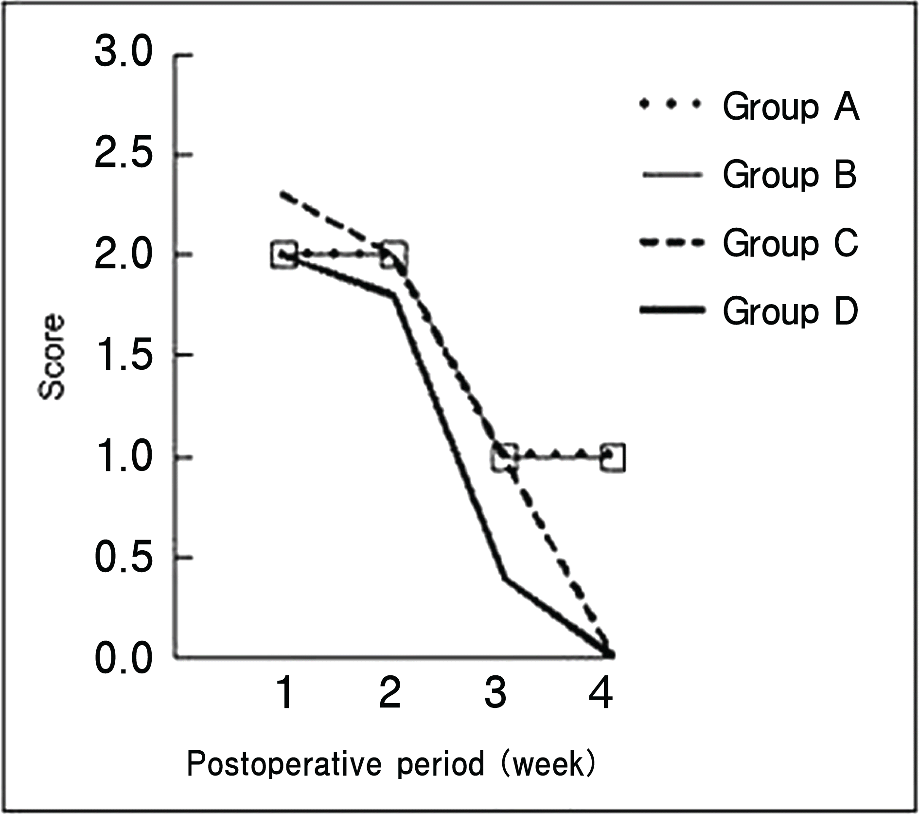 | Figure 2.The comparison of conjunctival injection and edema after surgery; Group A=Application of the Beriplast-P®; Group B=Bard® mesh (12×2 mm); Group C=Surgipro® mesh (6×2 mm); Group D=Surgipro® mesh (6×6 mm). |
 | Figure 3.Gross appearance at 4 weeks after surgery. (A) No remarkable finding in group A. (B) Irregular surface due to severe adhesion in group B. (C) Linear adhesion (arrow) in group C. (D) Diffuse adhesion in group D. Group A=Application of the Beriplast-P®; Group B=Bard® mesh (12×2 mm); Group C=Surgipro® mesh (6×2 mm); Group D=Surgipro® mesh (6×6 mm). |
 | Figure 4.Histologic findings of Fadenoperation site at 4 weeks after surgery (hematoxylin-eosin stain). (A) Mild inflammatory infiltrates (arrow) and perimuscular, fibrotic reaction (asterisk) in group A. (B) Moderate inflammatory infiltrate including lymphocytes and plasma cells (arrow) and thick fibrotic bands (asterisk) in group B. (C) Linear fibrosis (arrow) between the sclera and muscle in group C. (D) Vascular proliferation (arrow) into the mesh in group C. (E) Well-developed, dense bands of collagen (arrows) in group D. (F) Many neutrophils and mononuclear cells (arrow) in group D. S, sclera; M, muscle; magnification: a,×100; b,×40; c-f,×200. Group A=Application of the Beriplast-P®; Group B=Bard® mesh (12×2 mm); Group C=Surgipro® mesh (6×2 mm); Group D=Surgipro® mesh (6×6 mm). |
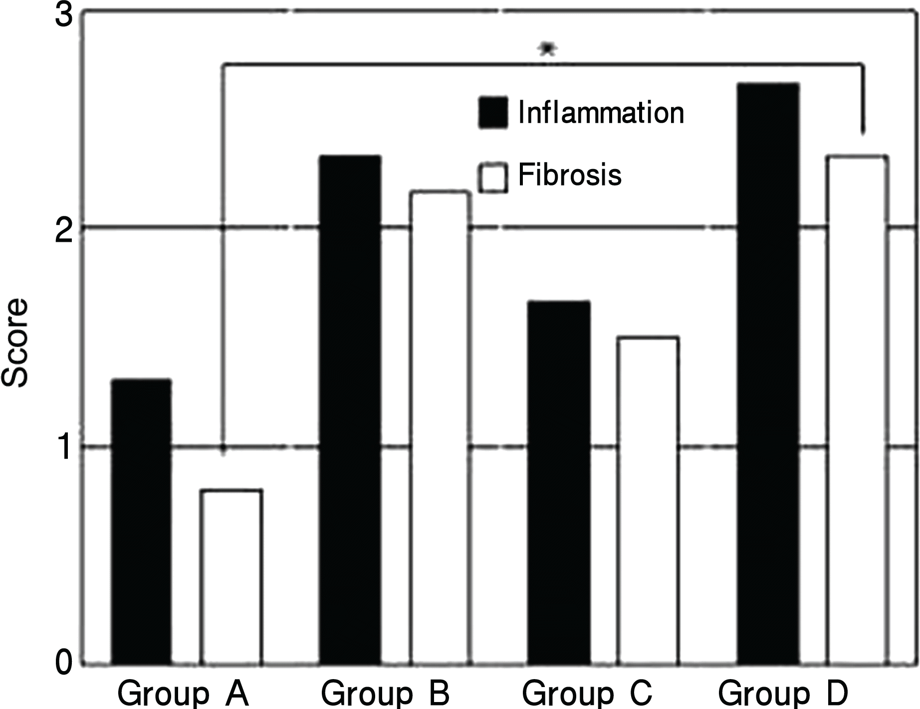 | Figure 5.The comparison of inflammation and fibrosis on histologic findings at 4 weeks after surgery. * The degree of fibrosis in group D was significantly greater than that in group A. Otherwise, there was no statistical significance of the degree of fibrosis or inflammation between the groups (Mann-Whitney test, p<0.008). Group A=Application of the Beriplast-P®; Group B=Bard® mesh (12×2 mm); Group C=Surgipro® mesh (6×2 mm); Group D=Surgipro® mesh (6×6 mm). |
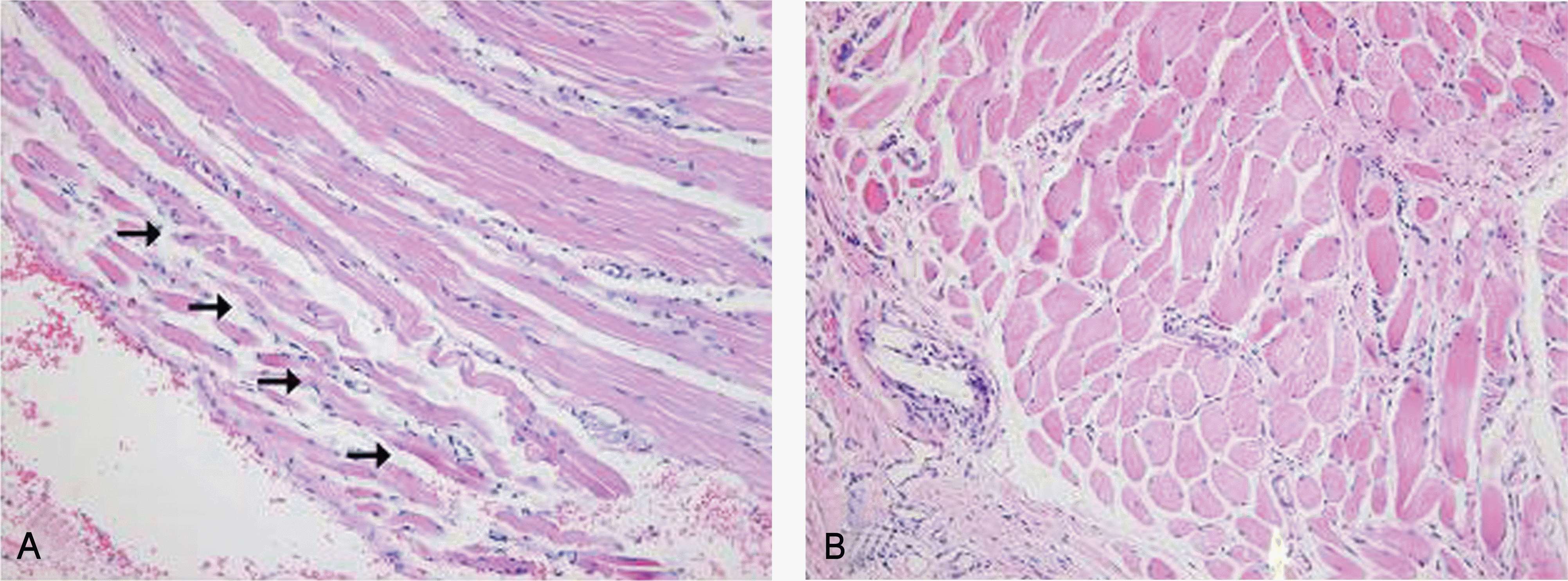 | Figure 6.Histological findings of muscular atrophy at 4 weeks after surgery (hematoxylin-eosin stain, ×200). (A) Focal atrophic changes (arrows) in the muscle of group A. These changes were noticed in 5 out of 6 eyes in group A, 2 out of 5 eyes in group B, and 1 eye among 7 eyes in group C. (B) There was no such atrophic change in the muscle of group D; Group A=Application of the Beriplast-P; Group B=Bard® mesh (12×2 mm); Group C=Surgipro® mesh (6×2 mm); Group D=Surgipro® mesh (6×6 mm). |
Table 1.
Score charts of conjunctival injection and edema14
| Score | Injection and edema |
|---|---|
| 0 | None |
| 1 | Injection only |
| 2 | A few blood vessels and mild edema |
| 3 | Moderate injection and edema |
| 4 | Severe injection and gross edema |
Table 2.
The grading score for inflammation15
 XML Download