Abstract
Purpose
To evaluate the effects of the Wnt protein on proliferation and stemness maintenance of cultured corneal limbal stem cells.
Methods
We examined the expression of Wnt proteins by Western blot analysis. We then evaluated the effects of Wnt on cell proliferation by colony forming efficiency. β-catenin activation using Wnt proteins was examined by immunocytochemistry. We also examined the effects of Wnt on proliferation and stemness maintenance by reverse transcriptase polymerase chain reaction of p63 and connexin43.
Results
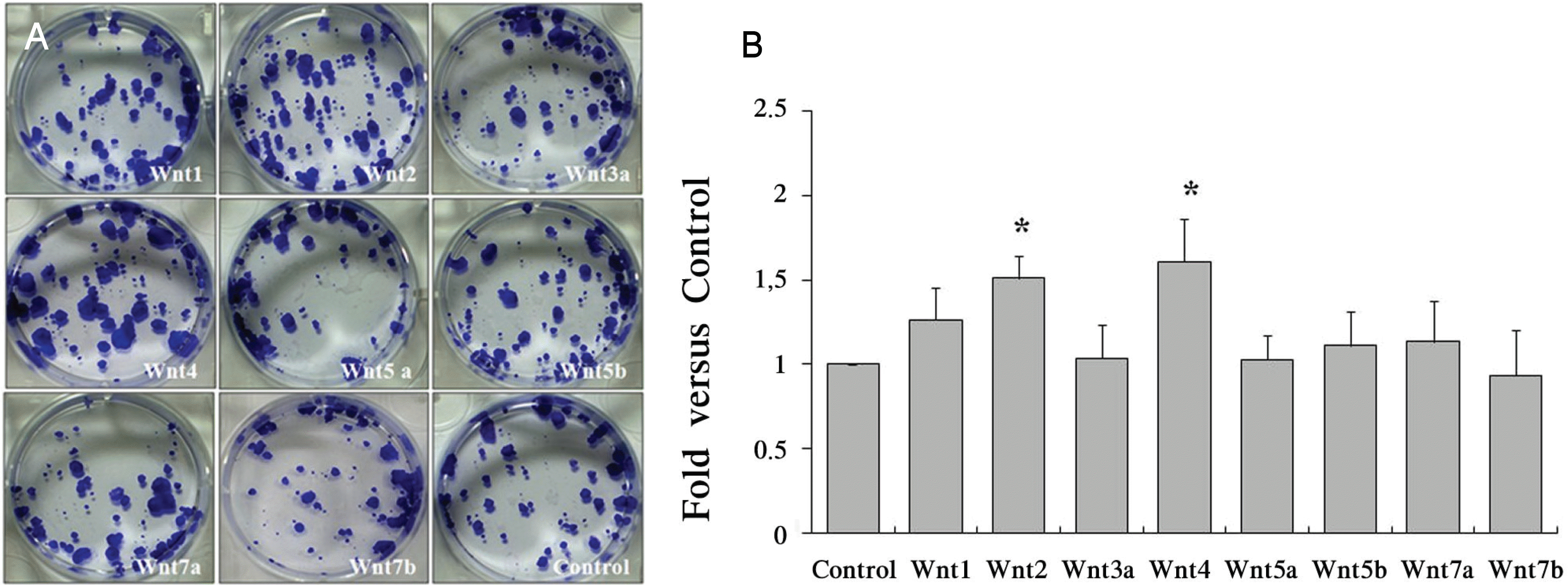
Wnt has a different effect on corneal epithelial stem cells. Colony forming efficiency was also significantly higher in treated Wnt2 and Wnt4 cells compared with controls. The Wnt2 and Wnt4 treated cells showed nuclear accumulation of β-catenin. In addition, the limbal stem cell marker p63 was strongly expressed in Wnt2, Wnt4 Wnt5a, and Wnt5b. Connexin43 mRNA was also strongly expressed in Wnt5a, Wnt5b and Wnt7b cells.
Go to : 
References
1. Davanger M, Evensen A. Role of the pericorneal papillary structure in renewal of corneal epithelium. Nature. 1971; 229:560–1.

2. Daniels JT, Dart JK, Tuft SJ, et al. Corneal stem cells in review. Wound Repair Regen. 2001; 9:483–94.

3. Revoltella RP, Papini S, Rosellini A, et al. Epithelial stem cells of the eye surface. Cell Prolif. 2007; 40:445–61.

4. Kenyon KR, Tseng SC. Limbal autograft transplantation for ocular surface disorders. Ophthalmology. 1989; 96:709–22.

5. Frucht-Pery J, Siganos CS, Solomon A. Limbal cell autograft transplantation for severe ocular surface disorders. Graefes Arch Clin Exp Ophthalmol. 1998; 236:582–7.

6. Kim MK, Lee JL, Shin KS, et al. Isolation of putative corneal epithelial stem cells from cultured limbal tissue. Korean J Ophthalmol. 2006; 20:55–61.

7. Park KS, Lim CH, Min BM, et al. The side population cells in the rabbit limbus sensitively increased in response to the central cornea wounding. Invest Ophthalmol Vis Sci. 2006; 47:892–900.

8. Dogru M, Tsubota K. Current concepts in ocular surface reconstruction. Semin Ophthalmol. 2005; 20:75–93.

9. Shortt AJ, Secker GA, Notara MD, et al. Transplantation of ex vivo cultured limbal epithelial stem cells: a review of techniques and clinical results. Surv Ophthalmol. 2007; 52:483–502.

10. Liu J, Song G, Wang Z, et al. Establishment of a corneal epithelial cell line spontaneously derived from human limbal cells. Exp Eye Res. 2007; 84:599–609.

11. Chee KY, Kicic A, Wiffen SJ. Limbal stem cells: the search for a marker. Clin Experiment Ophthalmol. 2006; 34:64–73.

12. Schlötzer-Schrehardt U, Kruse FE. Identification and characterization of limbal stem cells. Exp Eye Res. 2005; 81:247–64.

13. Ivekovic R, Tedeschi-Reiner E, Novak-Laus K, et al. Limbal graft and/or amniotic membrane transplantation in the treatment of ocular burns. Ophthalmologica. 2005; 219:297–302.

14. Vossmerbaeumer U, Kuehl S, Bieback K, et al. Cultivation and differentiation characteristics of human limbal progenitor cells. Tissue Cell. 2008; 40:83–8.

15. Zhao B, Allinson SL, Ma A, et al. Targeted cornea limbal stem/ progenitor cell transfection in an organ culture model. Invest Ophthalmol Vis Sci. 2008; 49:3395–401.
16. Moon RT, Brown JD, Torres M. Wnts modulate cell fate and behaviour during vertebrate development. Trends Genet. 1997; 13:157–62.
17. Logan CY, Nusse R. The Wnt signaling pathway in development and disease. Annu Rev Cell Dev Biol. 2004; 20:781–810.

19. Willert K, Nusse R. Beta-catenin: a key mediator of Wnt signaling. Curr Opin Genet Dev. 1998; 8:95–102.
Go to : 
 | Figure 1.Wnt gene expression in NIH3T3 cells. HA‐Wnt protein expression was determined by western blot analysis. The protein was extracted from cytoplasm and media. HA was expressed Wnt trasnfected. Cnt as an empty vector‐transfected control. |
 | Figure 2.Proliferation of limbal stem cells by Wnt. (A) Proliferation capacity of Wnt was evaluated by 0.1% crystal violet staining at 13 days. (B) The colony formed in Wnt2 and Wnt4 higher in treated Wnt2 and Wnt4 compared with control. In addition, CFE was calculated as the number of colonies/number of inoculated cells. Colony counting (holoclone): diameter of colony>1 mm. Values represent the mean± S.E.M of three independent experiments. Significant difference (* p<0.05) compared with vector‐transfected control are represented by* for Wnt-treated. Control as a vector-transfected control. Abbreviation: CFE, colony‐forming efficiency. |
 | Figure 3.Nuclear translocation of β‐catenin in the colony induced by Wnt2 and Wnt4. The localization of β‐catenin was examined by immunocytochemistry at 11 days. (A) β‐catenin is observed in the cytoplasm and plasma membrane. (B)(C) Wnt2 and Wnt4 treated cells show nuclear accumulation of β‐catenin. β‐catenin proteins were detected as a red color using anti‐rabbit alexa 546. Cell nuclei were stained with Hoechst 33342 Scale bar=10 μm. |
 | Figure 4.Expression of limbal stem cells and differentiation marker in cultured limbal stem cells. RT‐PCR profiles showing mRNA expression of Connexin43 and p63 with GAPDH as an internal control. Limbal stem cell marker p63 mRNA was strongly expressed in Wnt4 and Wnt5a. Otherwise, Differentiation marker Connexin43 mRNA strongly expressed in Wnt5a, Wnt5b, Wnt7b. |


 PDF
PDF ePub
ePub Citation
 Print
Print


 XML Download