

 PDF
PDF ePub
ePub Citation
 Print
Print


Abstract
Purpose
To obtain the average values of anterior segment structures of adult Koreans using Visante OCT in relation to age and sex, and to compare the central corneal thickness values with these measurements using an A-scan ultrasonic pachymeter.
Methods
Anterior segment images were obtained in 185 Korean (298 eyes) using Visante OCT. Four different parameters (central corneal thickness, anterior chamber depth, anterior chamber angle, and internal anterior chamber diameter) were measured at four meridians (vertical, horizontal, 45 degrees, and 135 degrees). Afterwards, the same examiner performed A-scan ultrasonic pachymetry to measure central corneal thickness.
Results
Central corneal thickness values measured using Visante OCT were significantly less than those obtained using an A-scan ultrasonic pachymeter (p=0.000). Central corneal thickness, anterior chamber angle, and internal anterior chamber diameters were all smaller in females than in males (p<0.05). Anterior chamber depth, anterior chamber angle, and internal anterior chamber diameter showed a significant decrease according to increasing age in both sexes (p<0.05).
Conclusions
Average values and changes of anterior segment structures in relation to age and sex were established using Visante OCT in Korean adults. These results may be useful as standard values in anterior segment surgeries as well as in the diagnosis and follow-up of certain anterior segment diseases.
Go to : 
References
1. Baïkoff G. Anterior segment OCT and phakic intraocular lenses: a perspective. J Cataract Refract Surg. 2006; 32:1827–35.

2. Memarzadeh F, Li Y, Chopra V, et al. Anterior segment optical coherence tomography for imaging the anterior chamber after laser peripheral iridotomy. Am J Ophthalmol. 2007; 143:877–9.
3. Nolan WP, See JL, Chew PT, et al. Detection of primary angle closure using anterior segment optical coherence tomography in Asian eyes. Ophthalmology. 2007; 114:33–9.
4. Müller M, Dahmen G, Pörksen E, et al. Anterior chamber angle measurement with optical coherence tomography: intraobserver and interobserver variability. J Cataract Refract Surg. 2006; 32:1803–8.
5. Fine IH, Hoffman RS, Packer M. Profile of clear corneal cataract incisions demonstrated by ocular coherence tomography. J Cataract Refract Surg. 2007; 33:94–7.
6. Huang D, Swanson EA, Lin C-P, et al. Optical coherence tomography. Science. 1991; 254:1178–81.
7. Puliafito CA, Hee MR, Schuman JS, Fujimoto JG. Optical Coherence Tomography of Ocular Diseases. Thorofare, NJ: Slack;1996.
8. Izatt JA, Hee MR, Swanson EA, et al. Micrometer-scale resolution imaging of the anterior eye in vivo with optical coherence tomography. Arch Ophthalmol. 1994; 112:1584–9.
9. Radhakrishnan S, Rollins AM, Roth JE, et al. Real-time optical coherence tomography of the anterior segment at 1310 nm. Arch Ophthalmol. 2001; 119:1179–85.
10. Dada T, Sihota R, Gadia R, et al. Comparison of anterior segment optical coherence tomography and ultrasound biomicroscopy for assessment of the anterior segment. J Cataract Refract Surg. 2007; 33:837–40.
11. Alsbirk PH. Corneal thickness. I. Age variation, sex, difference and oculometric correlations. Acta Ophthalmol (Copenh). 1978; 56:95–104.
12. Cho P, Lam C. Factors affecting the central corneal thickness of Hong-Kong Chinease. Curr Eye Res. 1999; 18:468–74.
13. Siu A. Herse P. The effect of age on human corneal thickness. Stastical implications of power analysis. Acta Ophthalmol (Copenh). 1993; 71:51–6.
14. Park SO, Cho BJ. Central Corneal Thickness Measured by Ultrasonic Pachymeter in Normal Koreans. J Korean Ophthalmol Soc. 2000; 41:2332–7.
15. Ho T, Cheng AC, Rao SK, et al. Central corneal thickness measurements using Orbscan II, Visante, ultrasound, and Pentacam pachymetry after laser in situ keratomileusis for myopia. J Cataract Refract Surg. 2007; 33:1177–82.
16. Zhao PS, Wong TY, Wong WL, et al. Comparison of central corneal thickness measurements by visante anterior segment optical coherence tomography with ultrasound pachymetry. Am J Ophthalmol. 2007; 143:1047–9.
17. Gao L, Fan H, Cheng AC, et al. The effects of eye drops on corneal thickness in adult myopia. Cornea. 2006; 25:404–7.
18. Nam SM, Lee HK, Kim EK, et al. Comparison of Corneal Thickness After the Instillation of Topical Anesthetics: Proparacaine Versus Oxybuprocaine. Cornea. 2006; 25:51–4.
19. Piñero DP, Plaza AB, Alió JL. Anterior segment biometry with 2 imaging technologies: very-high-frequency ultrasound scanning versus optical coherence tomography. J Cataract Refract Surg. 2008; 34:95–102.
20. Fishman GR, Pons ME, Seedor JA, et al. Assessment of central corneal thickness using optical coherence tomography. J Cataract Refract Surg. 2005; 31:707–11.
21. Wirbelauer C, Scholz C, Hoerauf H, et al. Noncontact corneal pachymetry with slit lamp-adapted optical coherence tomography. Am J Ophthalmol. 2002; 133:444–50.
22. Philips GI. Aetiology of angle closure glaucoma. Br J Ophthalmol. 1972; 56:248–53.
23. Fontana ST, Brubaker RF. Volume and depth of the anterior chamber in the normal aging human eye. Arch Ophthalmol. 1981; 98:1803–8.
24. Thomlison A, Leighton DA. Ocular dimensions and the heredity of open angle glaucoma. Br J Ophthalmol. 1974; 58:68–74.
25. Kim HK, Kim HB, Choi O. The Effects of the Mydriatics on the Anterior Chamber Depth. J Korean Ophthalmol Soc. 1984; 25:181–7.
26. Lee JH, Park WC, Rho SH. The Effects of Pilocarpine on the Anterior Chamber Depth and Angle. J Korean Ophthalmol Soc. 1994; 35:572–9.
27. Choi HH. A Consideration for Corneal Curvature, Its Thickness and Anterior Chamber Depth. J Korean Ophthalmol Soc. 1978; 19:417–22.
28. Lee HS, Kwon JY. The Differences of the Anterior Chamber Depth and the Corneal Thickness by the Refractive Error. J Korean Ophthalmol Soc. 1982; 23:395–400.
29. Oh DE, Jun RM, Choi KR. Quantified Values of Anterior Segment in Normal Adult Koreans Using Ultrasound Biomicroscopy. J Korean Ophthalmol Soc. 2004; 45:251–8.
30. Nemeth G, Vajas A, Tsorbatzoglou A. Assessment and reproducibility of anterior chamber depth measurement with anterior segment optical coherence tomography compared with immersion ultrasonography. J Cataract Refract Surg. 2007; 33:443–7.
31. Baikoff G, Jitsuo Jodai H, Bourgeon G. Measurement of the internal diameter and depth of the anterior chamber: IOLMaster versus anterior chamber optical coherence tomographer. J Cataract Refract Surg. 2005; 31:1722–8.
32. Kohnen T, Thomala MC, Cichocki M, et al. Internal anterior chamber diameter using optical coherence tomography compared with white-to-white distances using automated measurements. J Cataract Refract Surg. 2006; 32:1809–13.
33. Baikoff G, Lutun E, Ferraz C. Static and dynamic analysis of the anterior segment with optical coherence tomography. J Cataract Refract Surg. 2004; 30:1843–50.
34. Goldsmith JA, Li Y, Chalita MR, et al. Anterior chamber width measurement by high-speed optical coherence tomography. Ophthalmology. 2005; 112:238–44.
Go to :
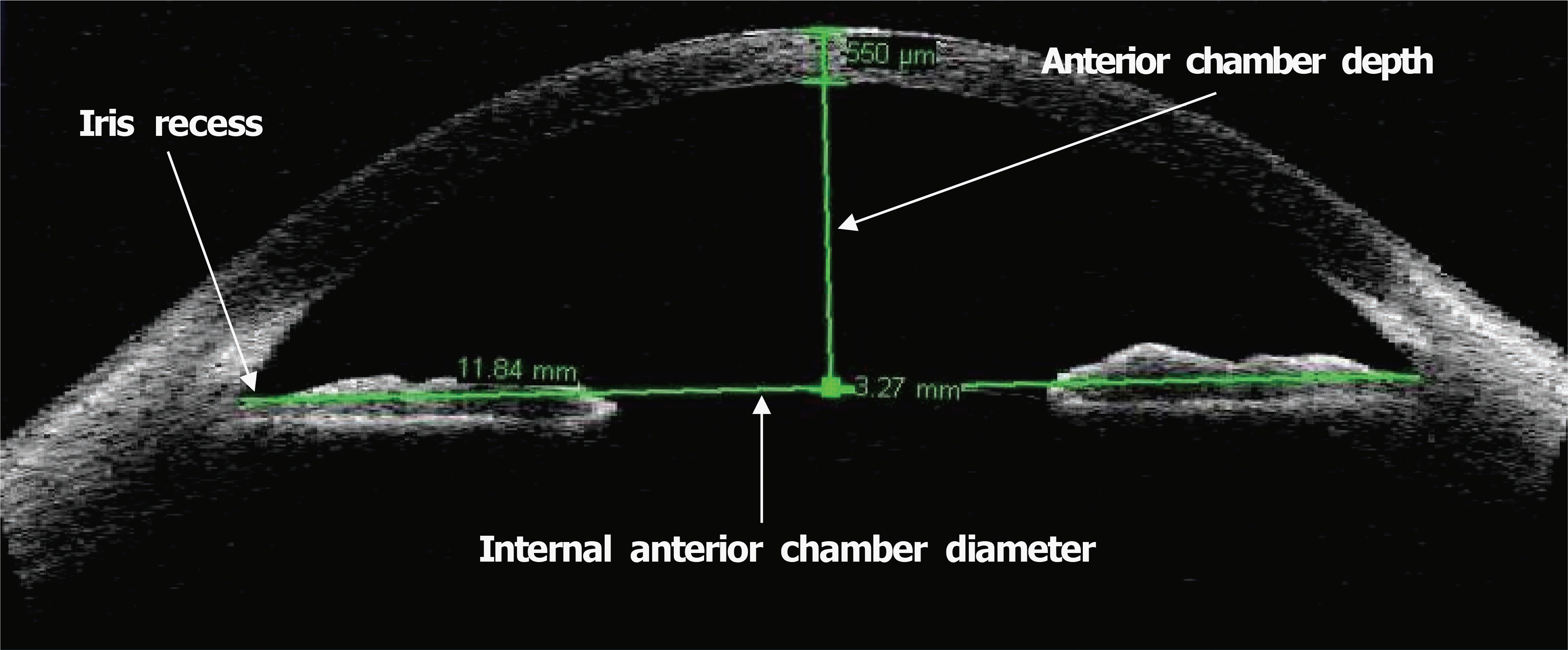 | Figure 1.Optical coherence tomography image (Visante OCT) with graphic tools for measurement of different anterior chamber dimensions. A caliper for anterior chamber depth and internal anterior chamber diameter measurements is used. Anterior chamber depth is measured between the corneal endothelium and a line joining the two opposite iris recesses. Internal anterior chamber diameter is measured between corresponding iris recesses. |
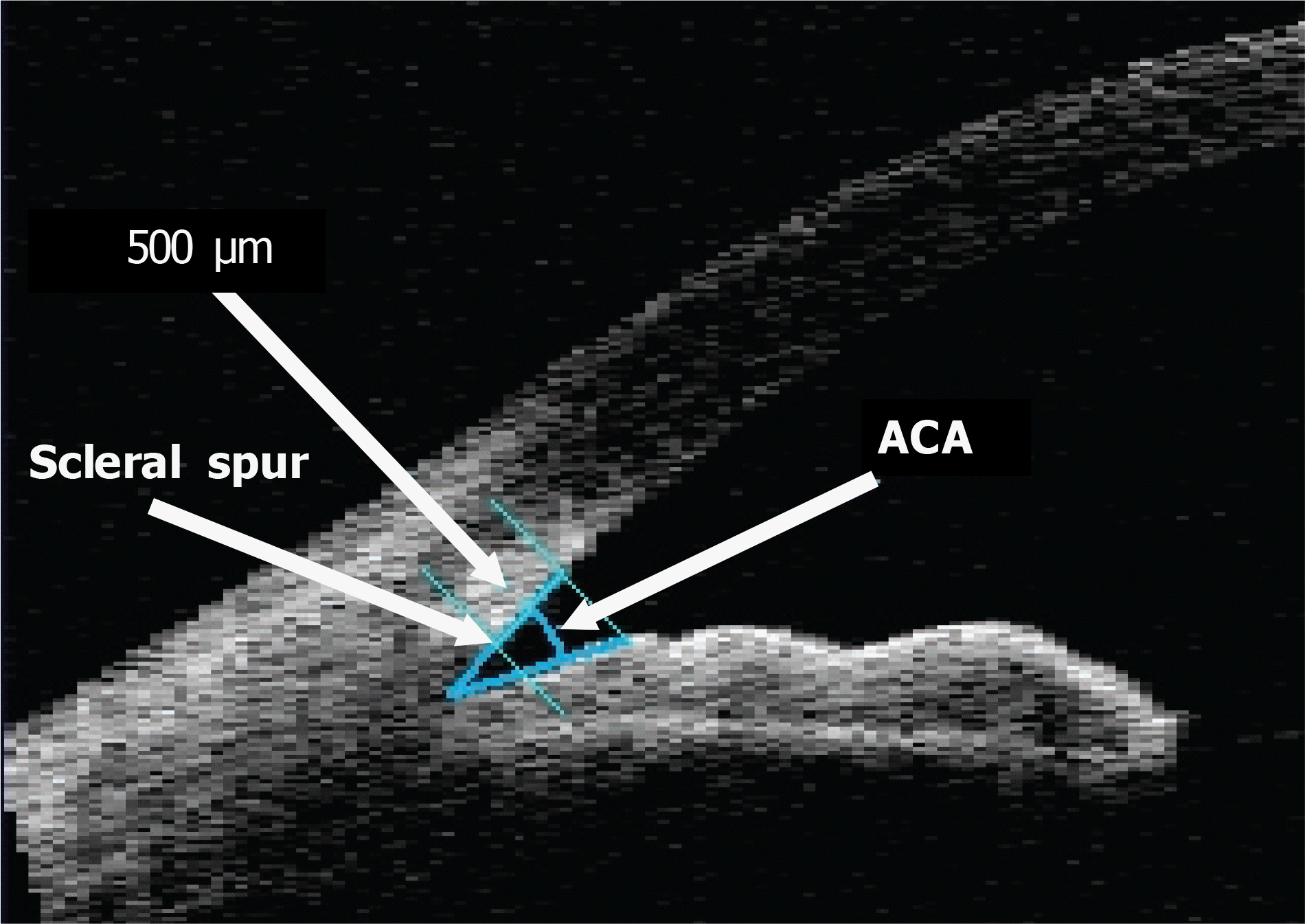 | Figure 2.Optical coherence tomography (Visante OCT) cross-sectional view through the anterior chamber angle region. Anterior chamber angle (ACA) is measured with the apex in the iris recess and the arms of the angle passing through a point on the trabecular meshwork at 500 µm from the scleral spur and the point on the iris perpendicularly opposite. |
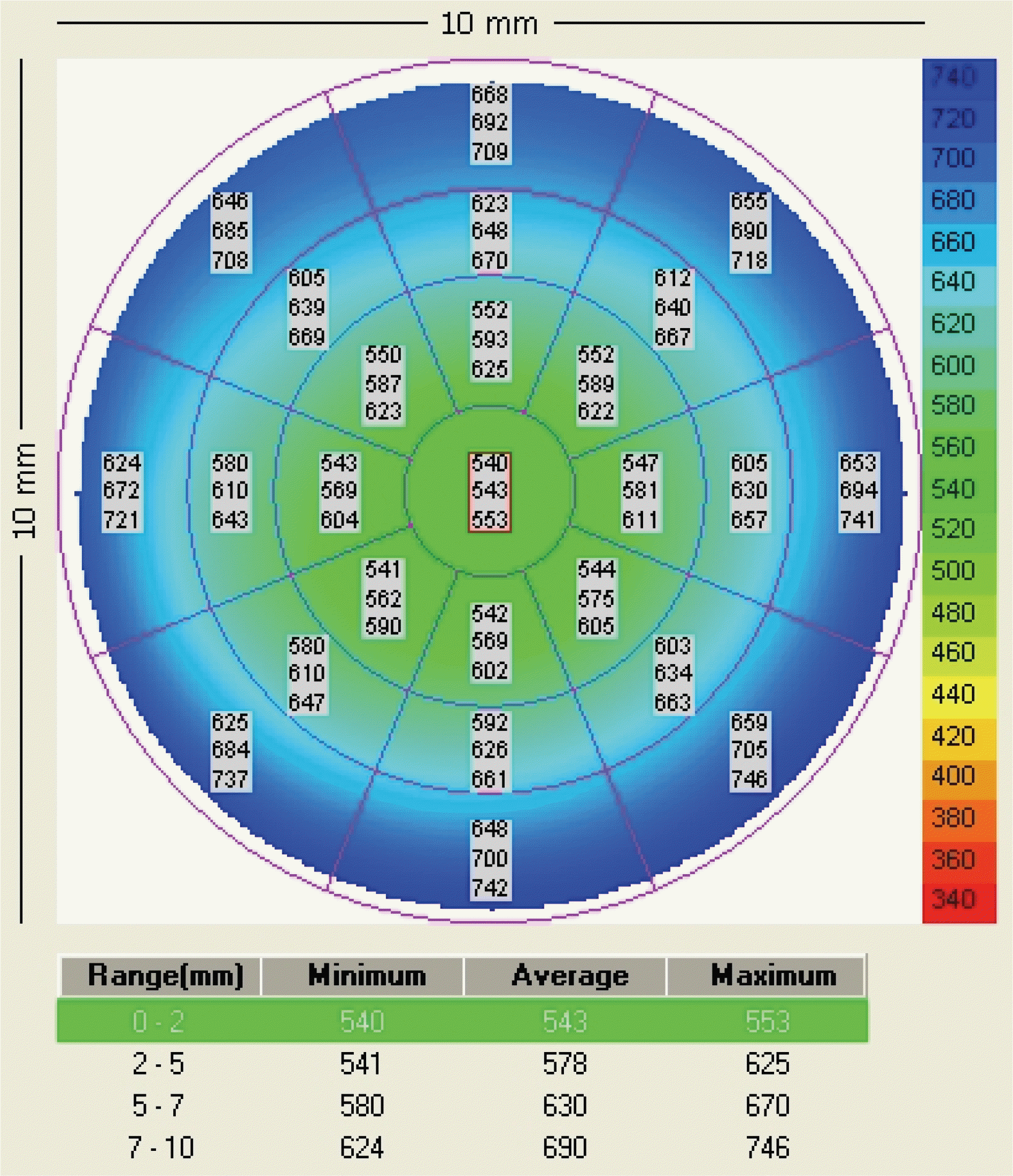 | Figure 3.An example of pachymetry mode of Visante OCT. Every sector has three values indicating minimum, average, and maximum corneal thickness in sequence from the above. Average value is the representative of the sector. |
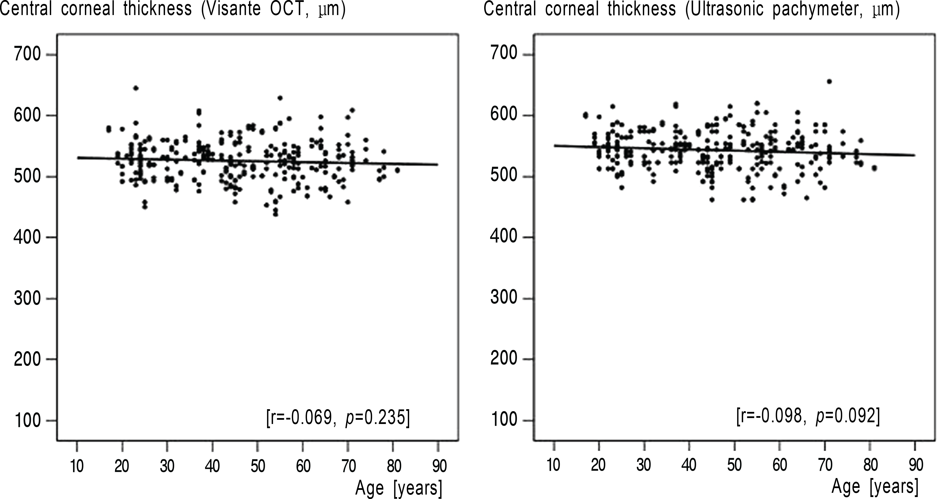 | Figure 4.Graphs show the results that there is no statistically significant relationship between central corneal thickness and age. |
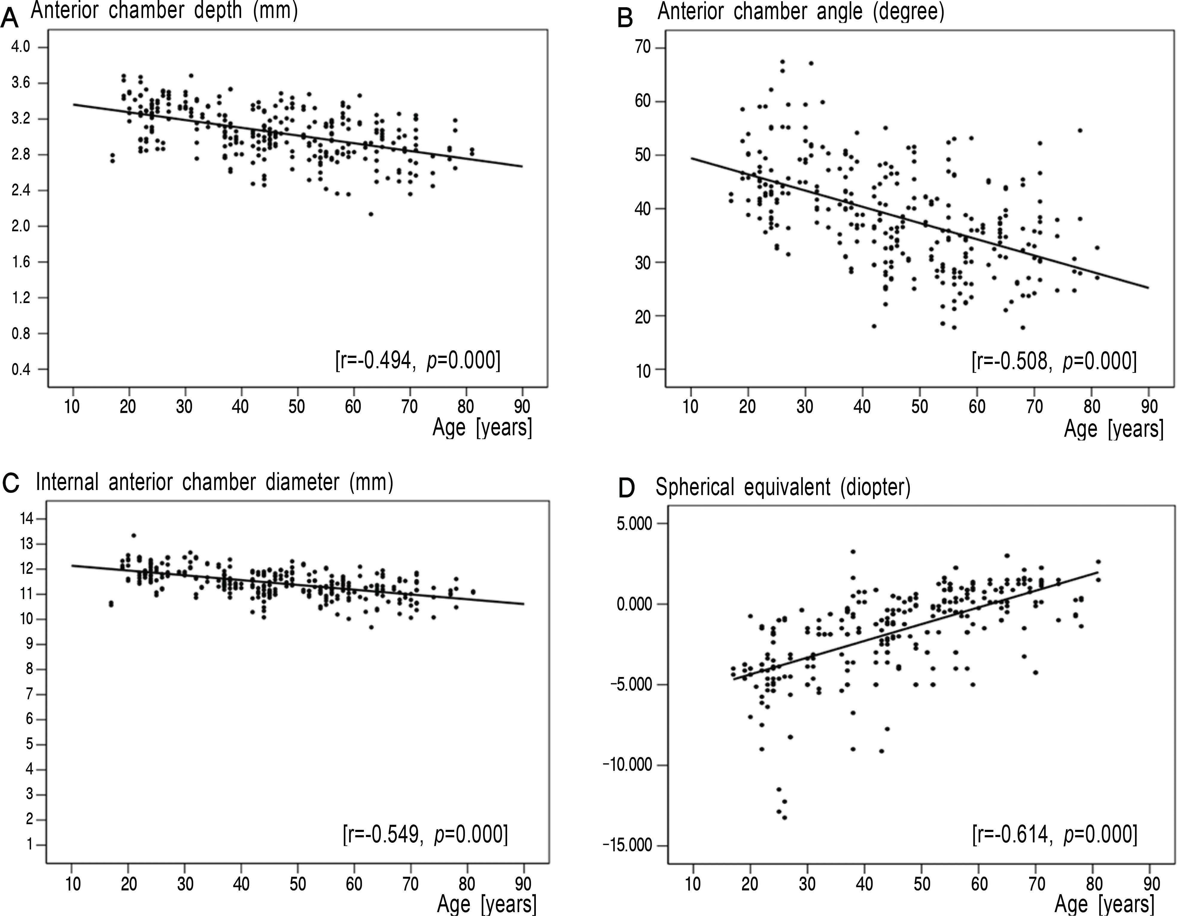 | Figure 5.Relationship between several parameters of the anterior segment and age. (A) Anterior chamber depth, (B) anterior chamber angle, and (C) internal anterior chamber diameter show a significant decrease in relation to age. (D) Spherical equivalent shows a significant shift from myopia to hyperopia in relation to age. |
Table 1.
Demographics & Refractive error of the subjects
Table 2.
Central corneal thickness measured by Visante OCT and ultrasonic pachymeter (mean± SD, µm)
| Age (years) |
Visante OCT |
Ultrasonic pachymeter |
||
|---|---|---|---|---|
| Male | Female | Male | Female | |
| 20∼29 | 535.2±30.4 | 522.4±32.4 | 551.8±24.9 | 543.6±31.3 |
| 30∼39 | 536.5±27.4 | 525.2±29.2 | 553.3±26.3 | 545.3±25.8 |
| 40∼49 | 529.1±30.3 | 517.2±32.3 | 547.6±30.6 | 534.4±34.4 |
| 50∼59 | 535.0±40.2 | 511.8±32.5 | 553.4±38.7 | 530.5±34.4 |
| 60∼ | 529.1±32.4 | 518.8±33.7 | 541.0±29.1 | 536.8±38.7 |
| Total | 533.0±32.2* | 518.8±32.0* | 549.5±30.2† | 537.8±33.5† |
Table 3.
Anterior chamber depth, anterior chamber angle, and internal anterior chamber diameter
| Age (years) |
Anterior chamber depth (mean±SD, mm) |
Anterior chamber angle (mean±SD, degree) |
Internal anterior chamber diameter (mean±SD, mm) |
|||
|---|---|---|---|---|---|---|
| Male | Female | Male | Female | Male | Female | |
| 20∼29 | 3.23±0.24 | 3.31±0.23 | 46.7±8.1 | 44.9±7.7 | 12.02±0.47 | 11.83±0.48 |
| 30∼39 | 3.13±0.27 | 3.16±0.22 | 46.9±8.8 | 41.6±6.6 | 11.69±0.48 | 11.67±0.44 |
| 40∼49 | 3.03±0.22 | 3.04±0.26 | 36.7±7.7 | 36.5±8.7 | 11.44±0.43 | 11.35±0.50 |
| 50∼59 | 3.01±0.21 | 2.90±0.29 | 34.9±9.5 | 31.5±7.8 | 11.35±0.42 | 11.13±0.44 |
| 60∼ | 2.89±0.23 | 2.86±0.28 | 34.8±5.4 | 33.4±9.1 | 11.13±0.48 | 11.02±0.53 |
| Total | 3.06±0.26* | 3.05±0.30* | 40.1±9.7† | 37.3±9.4† | 11.54±0.55‡ | 11.39±0.57‡ |
 XML Download