Abstract
Purpose
To analyze the effect of the augmented lateral tarsal strip for the correction of the paralytic ectropion in leprosy patients.
Methods
Ten leprosy patients (16 eyelids) with exposed keratitis and lagophthalmos from paralytic ectropion underwent surgery of the augmented lateral tarsal strip. Preoperative and postoperative vertical palpebral aperture, marginal reflex distance, lagophthalmos, and anterior segment findings were recorded at 3 and 6 months after surgery. Postoperative symptomatic and functional improvements were assessed at 6 months after surgery.
Results
There was a significant reduction between preoperative and postoperative measurements for vertical palpebral aperture (3.1±0.4 mm), lower marginal reflex distance (2.1±1.0 mm), and lagophthalmos (2.0±1.2 mm). Eye irritation symptoms and lid functions were improved in all patients. In a survey, the symptomatic, functional satisfaction was achieved in 90% of patients.
Go to : 
References
1. Rosenberg S, Goldfarb M. Management of paralytic ectropion. Ann Ophthalmol. 1981; 13:1063–5.
2. Ahn TG, Chung WS. Clinical experience of tarsal strip procedure. J Korean Ophthalmol Soc. 1990; 31:1489–94.
3. Foda HM. Surgical management of lagophthalmos in patients with facial palsy. Am J Otolaryngol. 1999; 20:391–5.

4. Chang L, Oliver J. A useful augmented lateral tarsal strip tarsorrhaphy for paralytic ectropion. Ophthalmology. 2006; 113:84–91.

5. Daniel E, Koshy S, Rao GS, Rao PS. Ocular complications in newly diagnosed borderline lepromatous and lepromatous leprosy patients: baseline profile of the Indian cohort. Br J Ophthalmol. 2002; 86:1336–40.

6. Toledano Fernández N, García Sáez S, Arteaga Sánchez A, Díaz Valle D. Bilateral lagophthalmos in lepromatous leprosy. Case report. Arch Soc Esp Oftalmol. 2002; 77:511–4.
7. Qian JG, Yan LB, Zhang GC. Long-term results after the correction of paralytic ectropion caused with leprosy in 115 eyes. Zhonghua Zheng Xing Wai Ke Za Zhi. 2004; 20:410–1.
8. Grauwin MY, Saboye J, Cartel JL. External canthopexy using the Edgerton-Montandon procedure in lagophthalmos of leprosy patients. Technique and indications. Apropos of 30 cases. Ann Chir Plast Esthet. 1996; 41:332–7.
9. Kumar P, Saxena RC, Srivastava SN. Leprotic paralytic lagophthalmos with ectropion and its surgical correction. Indian J Ophthalmol. 1986; 34:15–8.
10. Yoleri L, Songur E. Modified temporalis muscle transfer for paralytic eyelids. Ann Plast Surg. 1999; 43:598–605.

11. Snyder MC, Johnson PJ, Moore JF, Ogren FP. Early versus late gold weight implantation for rehabilitation of the paralytic eyelid. Laryngoscope. 2001; 111:2109–13.
12. Woo JM, Jeong SK, Park YG. Gold weight lid implantation for the management of facial nerve palsy. J Korean Ophthalmol Soc. 1995; 36:2067–73.
13. Anderson RL. Tarsal strip procedure for correction of eyelid laxity and canthal malposition in anophthalmic socket. Ophthalmology. 1981; 88:192–208.
Go to : 
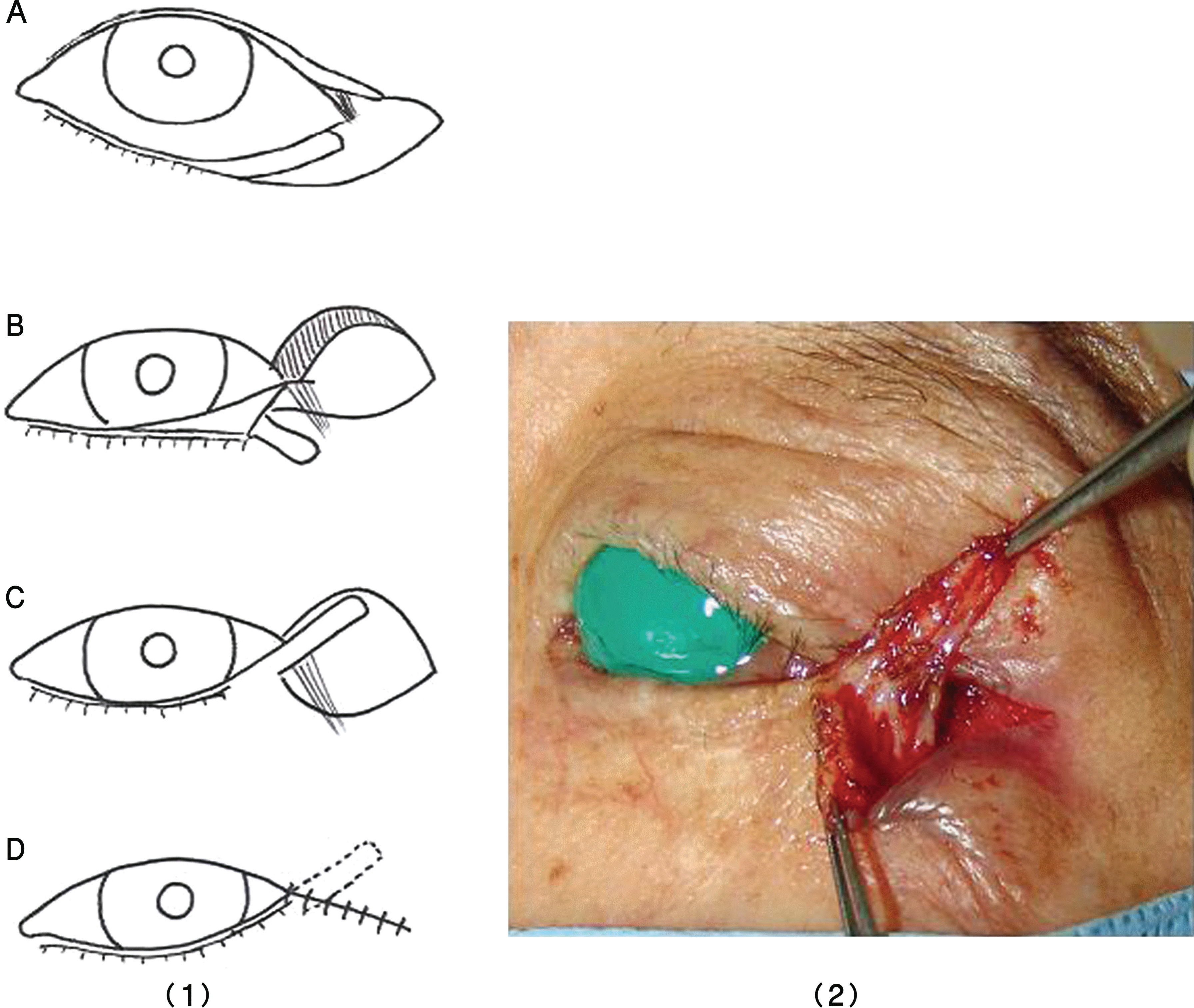
 | Figure 1.Diagram (1) & photograph (2) demonstrating the technique; (A) Lateral canthotomy and cantholysis. Formation of a long lateral tarsal strip; (B) The anterior and posterior lamella of the upper eyelid is split. Excision of a small triangular area of the upper anterior lamella is performed. Each posterior lamella of the upper and lower lids is reattached with 8‐0 Vicryl; C) The strip is attached to the superolateral orbital rim, overlapping the upper lateral eyelid; D) Orbicularis and skin are sutured with 6‐0 Prolene. |
 | Figure 2.Photographs of patients before and after surgery; (A-1) Preoperative view of the patient with paralytic ectropion of the left eye; (B-1) Preoperative view of the patient with paralytic ectropion of the right eye; (A-2, B-2) Six-month postoperative view. |
 | Figure 3.Analysis of the palpebral aperture (pal.), margin reflex distance 2 (MRD2), lagophthalmos (Lago.) before and after surgery at 3 and 6 months. |
Table 1.
Summary of the clinical data between before and 6 months after surgery
| Patient | Sex | Age | Preop visual acuity | Preop. anterior segment | Preop. Pal.‡(mm) | Preop. Lago.∏(mm) | Postop. visual acuity | Postop. anterior segment | Postop. Pal.(mm) | Postop. Lago. (mm) | Score# | |
|---|---|---|---|---|---|---|---|---|---|---|---|---|
| 1 | OD* | M | 67 | 0.2 | Exposure keratitis | 16 | 4 | 0.2 | Exposure keratitis | 2 | 70 | |
| OS* | 0.4 | PEE† | 13 | 2 | 0.4 | PEE | 2 | 60 | ||||
| 2 | OD | M | 67 | 0.02 | Corneal ulcer | 15 | 3 | 0.02 | Corneal opacity | 2 | 50 | |
| 3 | OD | F | 71 | 0.3 | Exposure keratitis | 16 | 4 | 0.5 | PEE | 2 | 80 | |
| 4 | OD | F | 70 | 0.3 | Exposure keratitis | 15 | 7 | 0.3 | Exposure keratitis | 4 | 70 | |
| OS | 0.2 | Corneal opacity | 16 | 5 | 0.2 | Corneal opacity | 2 | 60 | ||||
| 5 | OD | F | 53 | 0.2 | Corneal ulcer | 14 | 4 | 0.2 | Corneal opacity | 2 | 60 | |
| OS | 0.4 | Exposure keratitis | 13 | 2 | 0.4 | PEE | 1 | 60 | ||||
| 6 | OS | F | 82 | 0.2 | Exposure keratitis | 20 | 6 | 0.2 | Exposure keratitis | 3 | 90 | |
| 7 | OD | M | 78 | 0.3 | Exposure keratitis | 17 | 7 | 0.3 | Exposure keratitis | 4 | 80 | |
| OS | 0.3 | Exposure keratitis | 15 | 5 | 0.3 | Corneal opacity | 2 | 70 | ||||
| 8 | OD | M | 60 | 0.1 | Corneal opacity | 16 | 3 | 0.1 | Corneal opacity | 1 | 80 | |
| OS | 0.4 | Exposure keratitis | 16 | 4 | 0.4 | Exposure keratitis | 2 | 70 | ||||
| 9 | OD | M | 78 | 0.3 | Exposure keratitis | 13 | 4 | 0.3 | Exposure keratitis | 4 | 50 | |
| OS | 0.1 | Corneal opacity | 15 | 3 | 0.1 | Corneal opacity | 2 | 60 | ||||
| 10 | OD | F | 72 | 0.5 | Exposure keratitis | 16 | 5 | 0.7 | Exposure keratitis | 2 | 90 |
Table 2.
Measurements of the patients between preoperative and postoperative vertical palpebral aperture, margin reflex distance and lagophthalmos at 6 months (10 leprosy patients; 16 eyelids)
| Preoperative (A) | Postoperarive (B) | Difference between preop (A) and postop (B) | |
|---|---|---|---|
| Mean Pal.* (mm) | 15.0±1.7 mm | 12.4±1.0 mm | 3.1±0.4 mm |
| Mean MRD2† (mm) | 6.4±1.2 mm | 4.4±0.8 mm | 2.1±1.0 mm |
| Mean Largo.‡ (mm) | 3.8±1.7 mm | 2.2±0.9 mm | 2.0±1.2 mm |
Table 3.
Analysis between preoperative and postoperative palpebral aperture, margin reflex distance and lagophthalmo at 6 months according to severity
| Severity | Number of eye | Mean difference between preop and postop | |
|---|---|---|---|
| Pal.* (mm) | Large (≥16 mm) | 8 (50.0%) | 3.7±0.8 |
| Moderate (13<,<16 mm) | 5 (32.0%) | 2.2±0.5 | |
| Small (≤13 mm) | 3 (18.0%) | 0.8±0.4 | |
| MRD2† (mm) | Large (≥8 mm) | 5 (32.0%) | 3.1±0.8 |
| Moderate (5<,<8 mm) | 9 (55.5%) | 1.7±0.3 | |
| Small (≤5 mm) | 2 (12.5%) | 1.2±0.4 | |
| Largo.‡ (mm) | Severe (≥5 mm) | 6 (37.5%) | 3.0±0.0 |
| Moderate (2<,<5 mm) | 8 (50.0%) | 1.5±0.5 | |
| Mild (≤2 mm) | 2 (12.5%) | 0.2±0.3 |


 PDF
PDF ePub
ePub Citation
 Print
Print


 XML Download