

 PDF
PDF ePub
ePub Citation
 Print
Print


Abstract
Purpose
To assess the relationship between the internal diameter of the nasolacrimal duct and the success rate of silicone tube intubation in incomplete nasolacrimal duct obstruction patients with epiphora.
Methods
Dacryocystography was performed 80 patients with symptoms of epiphora and the anteroposterior (AP) diameters and the lateral diameters of the nasolacrimal ducts were measured. These measurements were compared between the group of patients who demonstrated improvement after the operation, and the group without symptom improvement.
Results
The mean length of the nasolacrimal duct in the group with nasolacrimal stenosis was 11.7±0.1 mm, the mean lateral diameter was 1.0±0.2 mm, and the mean AP diameter was 1.1±0.1 mm. The AP diameter of the symptomatic nasolacrimal duct in the group with successful postoperative results was 1.1±0.1 mm, and 0.9±0.1 mm in the group with unsuccessful results. The group with successful postoperative results had an average of 18.1±7.0% stenosis in the side of the eye with nasolacrimal duct stenosis compared with the asymptomatic side, and the group with unsuccessful postoperative results had an average of 34%±3.6% stenosis.
Conclusions
In cases with nasolacrimal duct stenosis, the success rate of silicone tube intubation was high when the AP diameter of the nasolacrimal duct was more than 1.1±0.1 mm and when the stenosis was less than 18.1±7.0% in the symptomatic side compared with the opposite side. These results are expected to help predict the success rates when preoperatively planning silicone tube intubation.
References
1. Kanski JJ. Clinical Ophthalmology: Disorders of the lacrimal drainage system. Oxford: Butterworth-Heinemann;1999. p. 43–52.
2. Nerad JA. Oculoplastic Surgery. St. Louis: Mosby;2001. p. 220–4.
3. Hurwitz JJ, Merkur S, DeAngelis D. Outcome of lacrimal surgery in older patients. Can J Ophthalmol. 2000; 35:18–22.

4. Nixon J, Birchall IW, Virjee J. The role of dacryocystography in the management of patinents with epiphora. Br J Radiol. 1990; 63:337–9.
5. Ewing AE. Roentgen ray demonstrations of the lacrimal abscess cavity. Am J Ophthalmol. 1909; 24:1–4.
6. Oum JS, Park JW, Choi YK, et al. Result of partial nasolacrimal duct obstruction after silicone tube intubation. J Korean Ophthalmol Soc. 2004; 45:1777–82.
7. Jones LT. An anatomical approach to problems of the eyelids and lacrimal apparatus. Arch Ophthalmol. 1961; 66:111–24.
8. Byon DS, Song CH, Shim JK, Shyn KH. Conjunctivochalasis as a cause of epiphora and its histopathological findings. J Korean Ophthalmol Soc. 1996; 37:400–4.
9. Lee SH, Kim SD, Kim JD. Silicone intubation for nasolacrimal duct obstruction in adult. J Korean Ophthalmol Soc. 1997; 38:185–9.
10. Jeong SK, Kim HD. Silicone tube intubation in acquired nasolacrimal duct obstruction. J Korean Ophthalmol Soc. 2000; 41:327–31.
11. Katowitz JA, Hollsten DA. Lacrimal surgery. New York: Churchill Livingstone;1998. p. 109–23.
12. Soll DB. Silicone intubation. An alternative to dacryocystorhi-nosyomy. Ophthalmology. 1978; 85:1259–66.
13. Anderson RL, Edwards JJ. Indications, complications and results with silicone stents. Ophthalmology. 1979; 86:1474–87.
14. Sohn HY, Hur J, Chung EH, Won IG. Clinical observation on silicone intubation in obstruction of lacrimal drainage system. J Korean Ophthalmol Soc. 1990; 31:135–40.
15. O'Donnell B, Shah R. Dacryocystorhinostomy for epiphora in the presence of a patient lacrimal system. Clin Experiment Ophthalmol. 2001; 29:27–9.
16. Mantynen J, Yoshitsugu M, Rautiainen M. Result of dacryocystorhinostomy in 96 patients. Acta Otolaryngal. 1997; 529:S187–9.
17. Lee HS, Lew HL, Yun YS. Classification of Nasolacrimal Duct Obstruction According to Dacryocystographic Finding and Its Clinical Significance. J Korean Ophthalmol Soc. 2003; 44:1475–82.
18. Weidenbecher M, Hosemann W, Buhr W. Endoscopic endonasal dacryocystorhinostomy: Results in 56 patients. Ann Otol Rhinol Laryngol. 1994; 103:363–7.
19. Boush GA, Lemke BN, Dorzbach RK. Results of endonasal laserassisted dacryocystorhinostomy. Ophthalmology. 1994; 101:955–9.
20. Crawford JS. Intubation of obstruction in the lacrimal system. Can J Ophthalmol. 1977; 12:289–92.
21. Dortzbach RK, France TD, Kushner BJ, Gonnering RS. Silicone intubation of the nasolacrimal duct in children. Am J Ophthalmol. 1982; 94:585–90.
22. Kushner BJ. Congenital nasolacrimal system obstruction. Arch Ophthalmol. 1982; 100:597–600.
23. Yu YS, Ham DI. Silicone intubation in children with nasolacrimal duct obstruction. J Korean Ophthalmol Soc. 1991; 32:409–14.
24. Cho KW. Lee SY, Kim SJ. Treatment of congenital Nasolacrimal duct obstruction using silicone intubation set. J Korean Ophthalmol Soc. 1995; 36:556–8.
25. Lee HS, Hwang WS, Byun YJ. Clinical results of silicone intubation for nasolacrimal duct obstruction in adult. J Korean Ophthalmol Soc. 1997; 38:1926–30.
26. Kim HD, Jeong SK. Silicone tube intubation in acquired nasolacrimal duct obstruction. J Korean Ophthalmol Soc. 2000; 41:327–31.
27. Angrist RC, Dortzbach RK. Silicone intubation for partial and total nasolacrimal duct obstruction in adults. Ophthal Plast Reconstr Surg. 1985; 1:51–4.
28. Ruby AJ, Lissner GS, O'Grady R. Surface reaction on silicone tubes used in the treatment of nasolacrimal drainage system obstruction. Ophthalmic Surg. 1991; 22:745–8.
29. Peter MS, Hugh DC. Head and Neck imaging. 3rd ed.Cicago: Mosby;1996. p. 1129–82.
30. Galloway JE, Kavic TA, Raflo GT. Digital subtraction dacryocystography: a new method of lacrimal system imaging. Ophthalmology. 1984; 91:956–62.
31. King SJ, Haigh SF. Technical report: Digital subtraction Dacryo-cystography. Clin Radiol. 1990; 42:351–3.
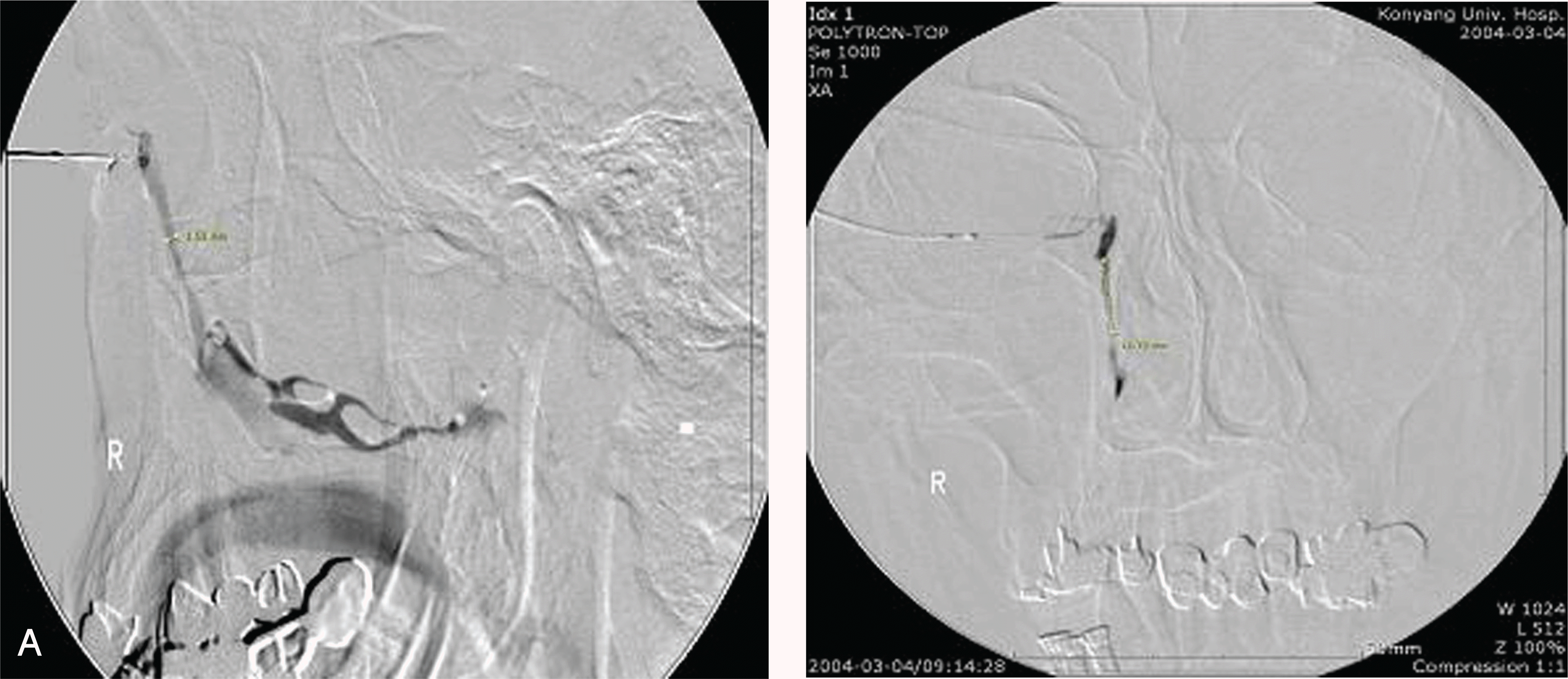
Table 1.
General characteristics of patients
| Subject | No. of patients(%) |
|---|---|
| Sex | |
| male | 23(28.7) |
| female | 57(71.3) |
| Age (years) | 29∼82(mean: 58.2) |
| Laterality of disease | |
| right | 34(42.5) |
| left | 46(57.5) |
| Period of disease (years) | 0.4∼12(mean=2.3) |
Table 2.
Nasolacrimal duct width and length in success group
| Normal (mm) | INLDO*(mm) | p-value | ||
|---|---|---|---|---|
| NLD† | anteroposterior width | 1.3±0.1 | 1.1±0.1 | <0.01 |
| right and left width | 1.3±0.2 | 1.0±0.2 | <0.01 | |
| vertical length | 11.9±0.5 | 11.7±0.8 | 0.09 |
Table 3.
Nasolacrimal duct width and length in failure group
| Normal (mm) | INLDO*(mm) | p-value | ||
|---|---|---|---|---|
| NLD† | anteroposterior width | 1.4±0.1 | 0.9±0.1 | <0.01 |
| right and left width | 1.4±0.1 | 1.1±0.2 | <0.01 | |
| vertical length | 11.8±0.9 | 11.7±1.5 | 0.83 |
Table 4.
Difference of nasolacrimal duct width and length between success and failure group
| Success (%) | Failure (%) | p-value | ||
|---|---|---|---|---|
| NLD* | anteroposterior width | 18.1±7.0 | 34.4±3.6 | <0.01 |
| right and left width | 23.1±3.4 | 28.5±4.3 | 0.75 | |
| vertical length | 1.6±0.2 | 0.8±0.1 | 0.85 |
 XML Download