

 PDF
PDF ePub
ePub Citation
 Print
Print


Abstract
Purpose
To report a case of a 9-year-old girl who complained of a floater symptom due to preretinal hemorrhage, subdural hemorrhage and arachnoid cyst.
Case summary
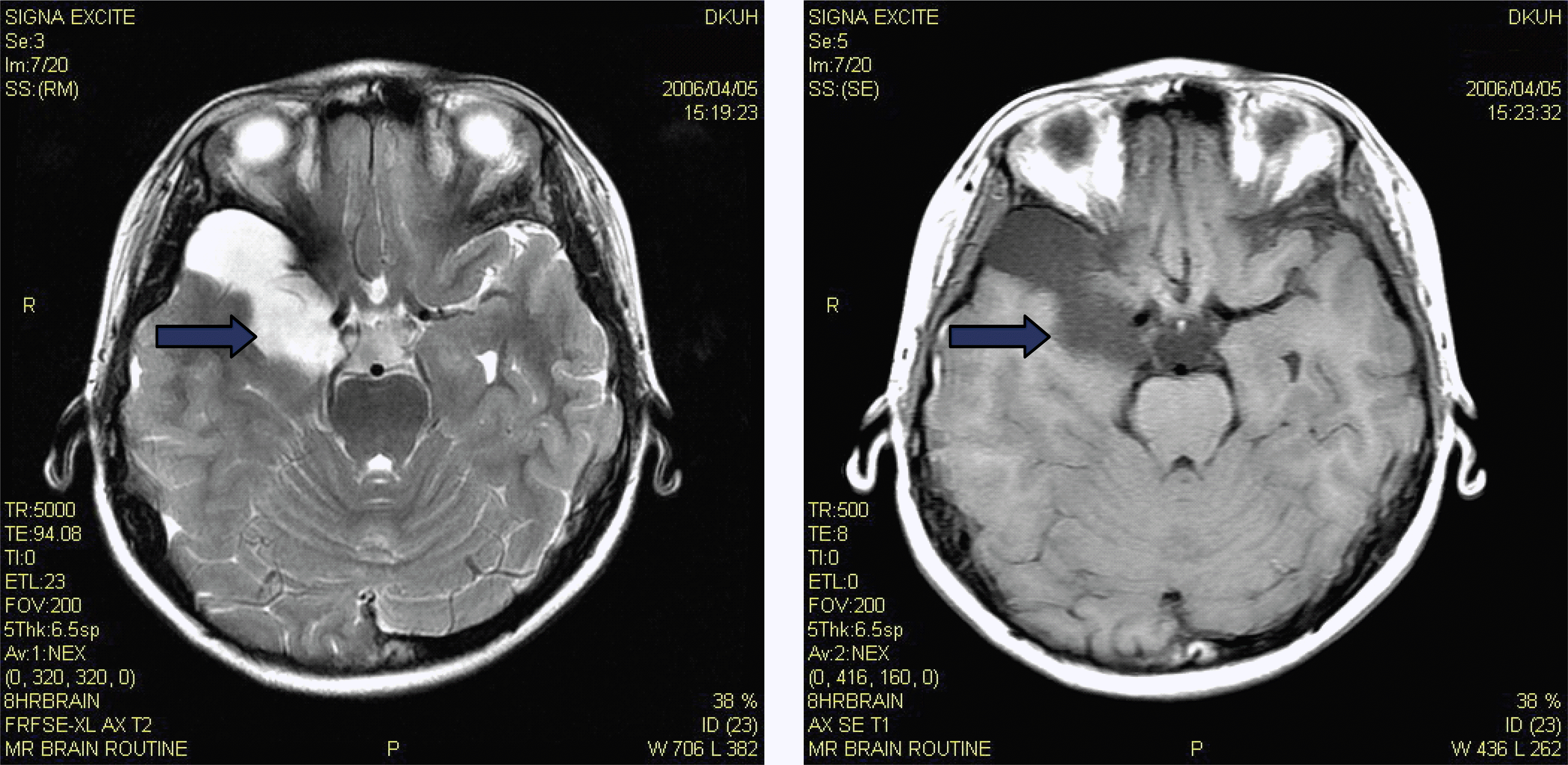
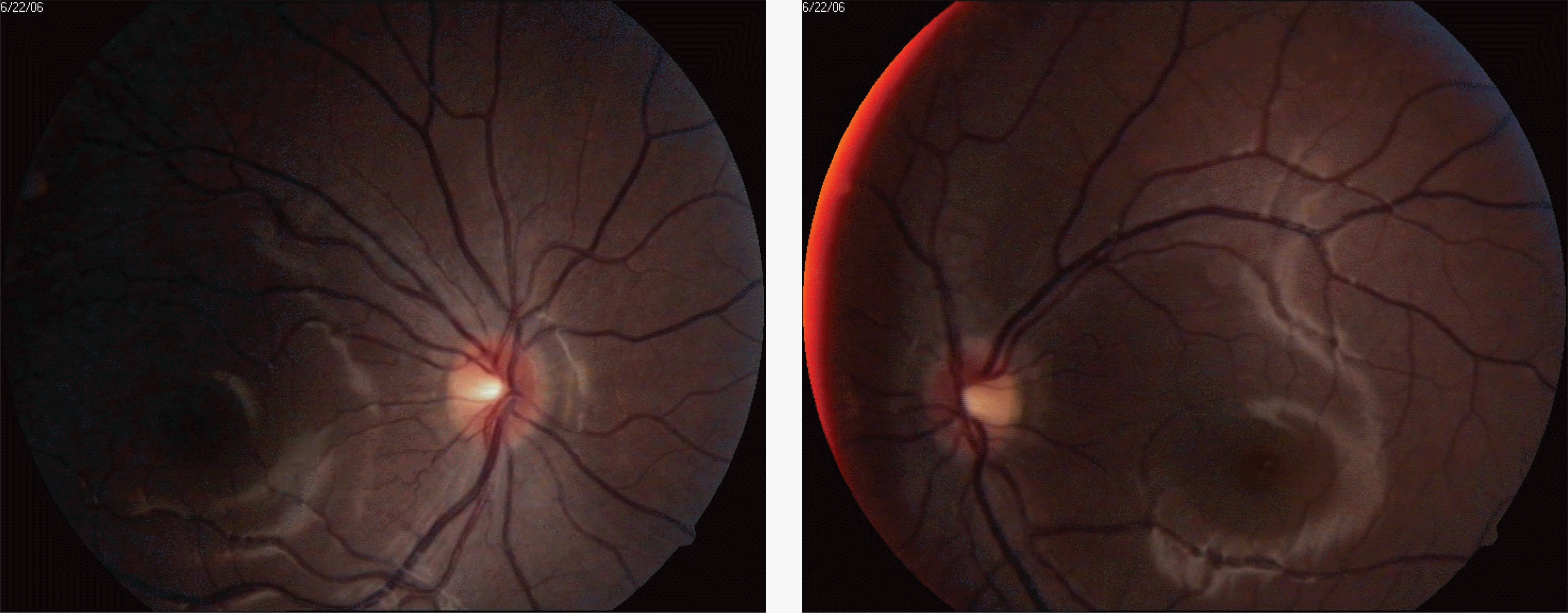
A 9-year-old girl presented to our clinic with floater symptom in her right eye and a headache. Her corrected visual acuities were 20/20 in both eyes. Preretinal hemorrhage around the optic disc in both eyes was observed. Brain MRI revealed subdural hemorrhage on the frontal, temporal, and parietal lobes, and the arachnoid cyst in the right fronto-temporal lobe. One year later, the preretinal hemorrhages were absorbed.
Conclusions
Arare case of concomitant preretinal and spontaneous subdural hemorrhage with arachnoid cyst was presented. The utilization of brain MRI is recommended to determine underlying causes when fundus examination reaveals retinal hemorrhage without trauma, systemic disease and neurologic symptom.
Go to : 
References
1. Poirrier AL, Ngosso-Tetanye I, Mouchamps M, Misson JP. Spontaneous arachnoid cyst rupture in a previously asymptomatic child. Eur J Paediatr Neurol. 2004; 8:247–51.
2. Clarkson JG, Flynn HW. Vitrectomy in Terson's syndrome. Am J Ophthalmol. 1980; 90:549–52.

3. Butterwirth-Heinemann. Clinical ophthalmology a systemic approach, 5th ed. Edinburg: Kanski J. 2003; 653.
4. Paton L. VII. Diseases of the nervous system: Ocular symptoms in subarachnoid haemorrhage. Trans Ophthalmol Soc U K. 1924; 110–124.
5. Cogan DG. Neurology of the visual system. Springfield: Charles C Thomas;1966. p. 184–93.
6. Manschot WA. Subarachnoid hemorrhage, intraocular symptoms and their pathogenesis. Am J Ophthalmol. 1954; 38:501–5.
7. Smith DC, Kearns TP, Sayer GP. Preretinal and optic nerve sheath hemorrhage: Preretinal and optic nerve sheath hemorrhage. Trans Am Acad Ophthalmol Otolaryngol. 1957; 61:201–11.
8. Weaver RG, Davis CH. Subhyaloid hemorrhage. Am J Ophthalmol. 1961; 52:257–9.
9. Morris DA, Henkind P. Relationship of intracranial optic nerve sheath and retinal hemorrhage. Am J Ophthalmol. 1967; 64:853–9.
10. Khan SG, Frenkel M. Intravitreal hemorrhage associated with rapid increased intracranial pressure (Terson's syndrome). Am J Ophthalmol. 1975; 80:37–43.
11. Shaw HE, Lander MB. Vitreous hemorrhage after intracranial hemorrhage. Am J Ophthalmol. 1975; 80:207–13.
12. Ogawa T, Kltaoka T, Dake Y, Amemiya T. Terson syndrome: A case report suggesting the mechanism of citreous hemorrhage. Ophthalmology. 2001; 108:1654–6.
13. Baker ML, Hand PJ, Tange D. Terson's syndrome in spontaneous spinal subarachnoid haemorrhage. J Clin Neurosci. 2008; 15:313–6.
14. Poirrier L, Ngosso-Tetanye I, Mouchamps M, Misson JP. Spontaneous arachnoid cyst rupture in a previously asymptomatic child: a case report. Eur J Paediatr Neurol. 2004; 8:247–51.
15. Killer HE, Flammer J. Unilateral papilledema caused by a fronto-temporo-parietal arachnoid cyst. Am J Ophthalmol. 2001; 132:589–91.
16. Yamauchi T, Saeki N, Yamamura A. Spontaneous disappearance of temporo-frontal arachnoid cyst in a child. Acta Neurochir (Wien). 1999; 141:537–40.
17. Dodd R, Barnes P, Huhn S. Spontneous resolution of a prepontine arachnoid cyst. Pediatr Neurosurgery. 2002; 37:152–7.
18. Cincu R, Agrawal A, Eiras J. Intracranial arachnoid cysts: Current concepts and treatment alternatives. Clin Neurol Neurosurg. 2007; 109:837–43.
19. Kang JK, Lee KS, Lee IW, et al. Shunt-independent surgical treatment of middle cranial fossa arachnoid cysts in children. Childs Nerv Syst. 2000; 16:111–6.
20. Albuquerque F, Ginnotta S. Arachnoid cyst rupture producing subdural hygroma and intracranialhypertension. Neurosurgery. 1997; 41:951–6.
Go to :
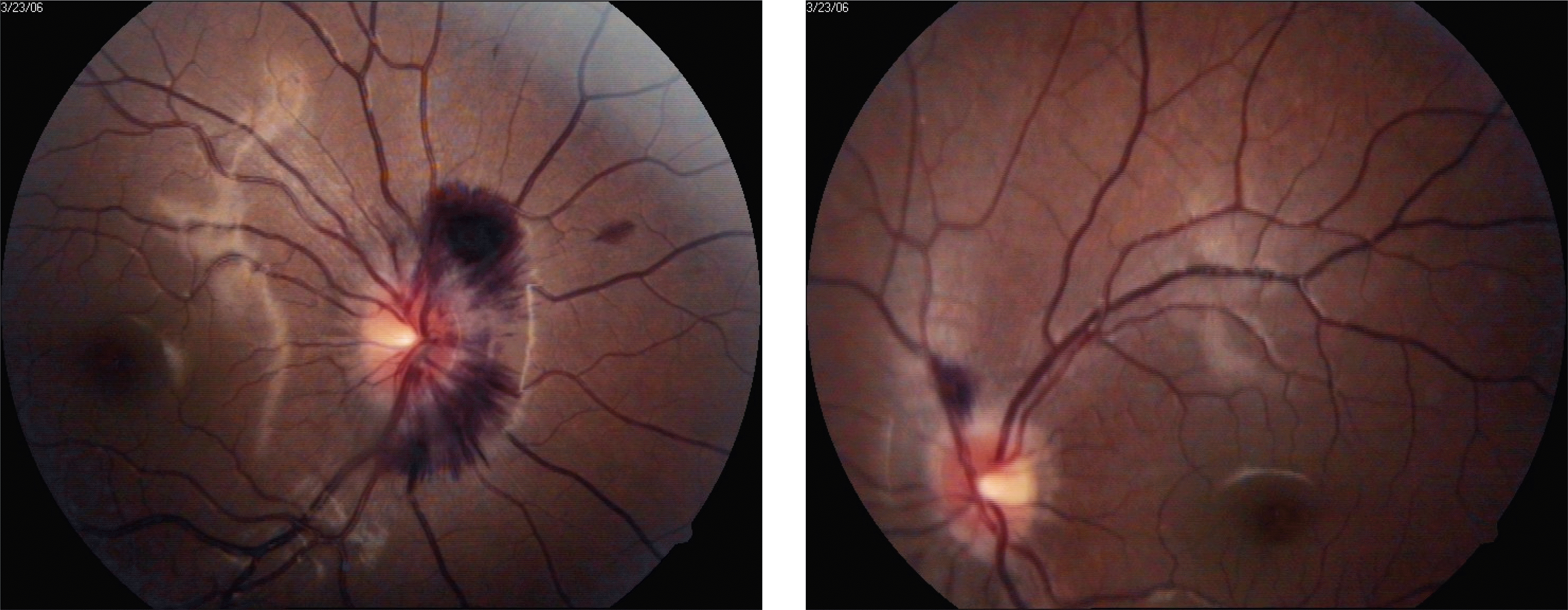 | Figure 1.The patient's fundus photograph at the first visit shows preretinal hemorrhage around the optic disc. |
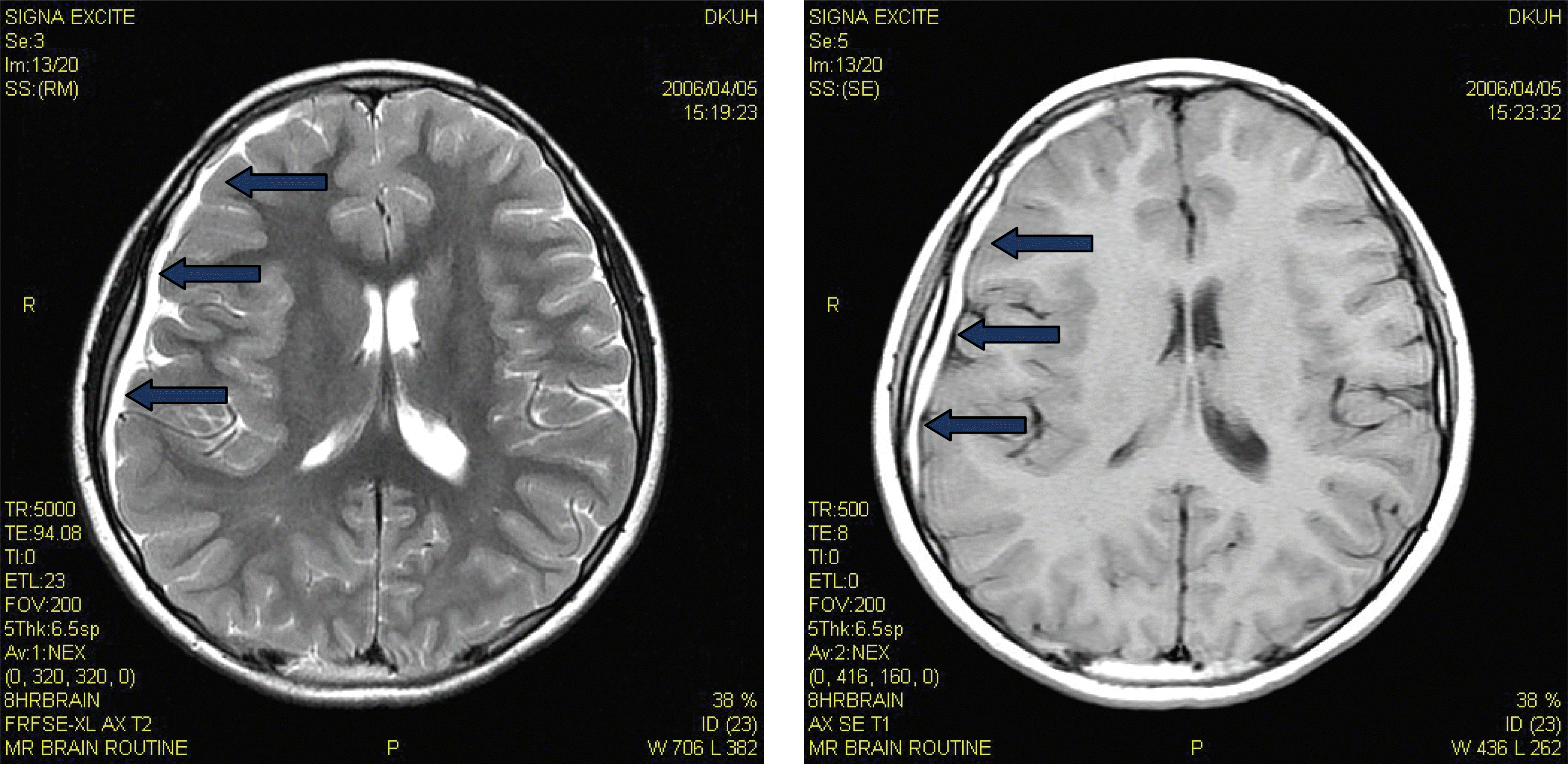 | Figure 2.The patient's transverse brain MRI section shows subacute subdural hemorrhage in the frontal, temporal and parietal lobes. |
 XML Download