

 PDF
PDF ePub
ePub Citation
 Print
Print


Abstract
Purpose
To evaluate the intraoperative and postoperative complications and outcomes of phacoemulsification of cataract in eyes with prior iriodotomy for acute angle closure glaucoma.
Methods
This retrospective case-control study included 30 eyes which underwent phacoemulsification. All 30 eyes had prior acute angle closure treated by laser iridotomy (ACG group). A second group that had phacoemusification for cataract only served as control (Control group).
Results
The ACG group showed significantly shallow anterior chamber (1.56±0.18 mm) and short axial length (22.32±0.60 mm). At 2 months after surgery, visual acuity was improved in both groups and IOP was significantly decreased in the ACG group. Posterior synechiae, small pupil and zonulysis were more commonly found in the ACG group than the control group. Intraoperative iris trauma and postoperative persistent mydriasis were also more common in the ACG group.
Conclusions
During cataract surgery in eyes with prior iriodotomy for acute angle closure glaucoma, there are many preoperative risk factors such as shallow anterior chamber, zonulysis, and small pupil. These increase the risk of posterior capsule rupture and bullous keratopathy. To reduce the complications and improve the visual acuity, a careful preoperative examination and proper intra- and post-operative management by skillful surgeons are needed.
Go to : 
References
1. Imaizumi M, Takaki Y, Yamashita H. Phacoemulsification and intraocular lens implantation for acute angle closure not treated or previously treated by laser iridotomy. J Cataract Refract Surg. 2006; 32:85–90.

2. Choong YF, Irfan S, Menage MJ. Acute angle closure glaucoma: an evaluation of a protocol for acute treatment. Eye. 1999; 13:613–6.
3. Tello C, Tran HV, Liebmann J, Ritch R. Angle closure: classification, concepts, and the role of ultrasound bio-microscopy in diagnosis and treatment. Semin Ophthalmol. 2002; 17:69–78.
4. Quigley HA. Long-term follow-up of laser iridotomy. Ophthalmology. 1981; 88:218–24.
5. Wilensky JT, Kaufman PL, Frohlichstein D. . Follow-up of angle-closure glaucoma suspects. Am J Ophthalmol. 1993; 115:338–46.
6. McMahan LB, Monica ML, Zimmerman TJ. Posterior chamber pseudophakes in glaucoma patients. Ophthalmic Surg. 1986; 17:146–50.
7. Tong JT, Miller KM. Intraocular pressure change after sutureless phacoemulsification and foldable posterior chamber lens implantation. J Cataract Refract Surg. 1998; 24:256–62.
8. Byun SW, Park CK, Hahn TW, Kim MS. Results of combined procedures versus two-stage operation using small incision in patients with cataract and glaucoma. J Korean Ophthalmol Soc. 1999; 40:2561–6.
9. Lai JS, Tham CC, Chan JC. The clinical outcomes of cataract extraction by phacoemulsification in eyes with primary angle-closure glaucoma (PACG) and co-existing cataract: a prospective case series. J Glaucoma. 2006; 15:47–52.
10. Jacobi PC, Dietlein TS, Lüke C. . Primary phacoe-mulsification and intraocular lens implantation for acute angle-closure glaucoma. Ophthalmology. 2002; 109:1597–603.
11. Khokhar S, Sindhu N, Pangtey MS. Phacoemulsification in filtered chronic angle closure glaucoma eyes. Clin Experiment Ophthalmol. 2002; 30:256–60.
12. Kim YH, Hyung S. Effect of cataract extraction in chronic angle-closure glaucoma patients. J Korean Ophthalmol Soc. 2007; 48:521–6.
13. Yun YM, Yim JH, Kim CS. Clinical factors that influence intraocular pressure change after cataract surgery in primary open-angle glaucoma and angle-closure glaucoma. J Korean Ophthalmol Soc. 2006; 47:85–96.
14. Erie JC, Hodge DO, Gray DT. The incidence of primary angle-closure glaucoma in Olmsted County, Minnesota. Arch Ophthalmol. 1997; 115:177–81.
15. Ritch R, Lowe RF. Angle-closure glaucoma: therapeutic overview. The Glaucomas. 2nd. St Louis: Mosby;1996. p. 1521.
16. Ritch R, Solomon LD. Argon Laser Peripheral Iridoplasty for Angle-Closure Glaucoma in Sibilings with Weill-Marchesani Syndrome. J Glaucoma. 1992; 1:243–7.
17. Lim KJ, Hyung SM, Youn DH. Ocular dimensions with aging in normal eyes. Korean J Ophthalmol. 1992; 6:19–31.
18. An JW, On KK, Kim JD. Biometric measurements in acute angle closure glaucoma. J Korean Ophthalmol Soc. 1993; 34:648–53.
19. Kim YW, Kim SD, Kim JD. Influence of lens factor and effect of selected cataract extraction on acute angle-closure glaucoma. J Korean Ophthalmol Soc. 2005; 46:1144–50.
20. Kubota T, Toguri I, Onizuka N, Matsuura T. Phacoe-mulsification and intraocular lens implantation for angle closure glaucoma after the relief of pupillary block. Ophthal-mologica. 2003; 217:325–8.
21. Markowitz SN, Morin JD. The endothelium in primary angle-closure glaucoma. Am J Ophthalmol. 1984; 98:103–4.
22. Bigar F, Witmer R. Corneal endothelial changes in primary acute angle-closure glaucoma. Ophthalmology. 1982; 89:596–9.
Go to :
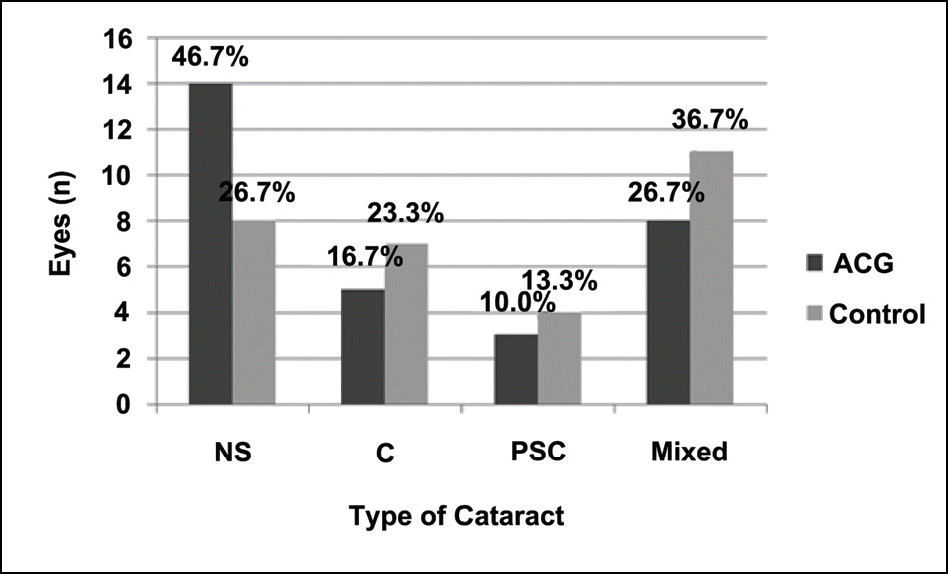 | Figure 1.Types of cataract in study and control patients. NS=nuclear sclerosis; C=cortical; PSC= posterior subcap sular; Mixed=NS/cortical; PSC/cortical or NS/PSC/Cortical. There are no statistical significant difference between the two groups (P>0.05 in all cases). |
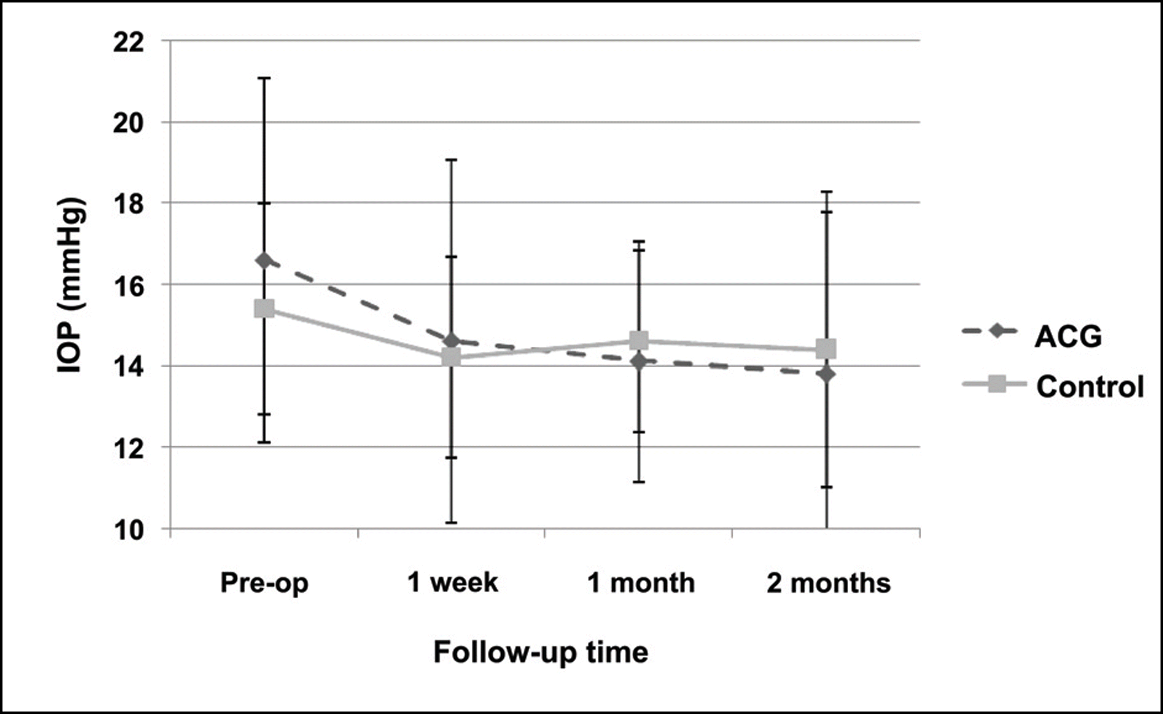 | Figure 2.Graphs demonstrating the course of the intraocular pressure (IOP) before and after phacoemulsification. At 1 and 2 months after surgery, intraocular pressure was significantly lowered in the angle-closure group (p=0.018, p=0.011). |
Table 1.
Preoperative and postoperative characteristics
| ∗ ACG | Control | p-value | |
|---|---|---|---|
| Age (years) | 69.07±8.95 (48 to 80) | 68.67±8.42 (50 to 81) | 0.859 |
| Gender Male | 6 | 9 | |
| Female | 24 | 21 | |
| IOP (mmHg) | 16.53±2.97 (12 to21) | 15.53±2.52 (9 to 20) | 0.296 |
| † UCVA (logMAR) | 0.80±0.11 (0.39 to 2.00) | 0.74±0.42 (0.30 to 1.4 | 40)0.586 |
| ‡ BCVA (logMAR) | 0.63±0.42 (0.09 to 2.00) | 0.60±0.40 (0.49 to 1.4 | 40)0.763 |
| Medications (number) | 1.33±1.09 (0 to 3) | 0 | 0.000 |
| Nucleus opacity score (§ LOCS III) | 3.27±0.98 (2 to 6) | 3.27±0.98 (2 to 6) | 1.000 |
Table 2.
Preoperative biometric characteristics
| ∗ ACG | Control | P-value |
|---|---|---|
| Spherical equivalent (D) -2.09±2.96 (-4.25 to +3.75) | -1.46±3.35 (-4.50 to +3.00) | 0.564 |
| Cylindrical refraction (D)1.24±1.28 (plano to 4.25) | 1.46±1.46 (plano to 3.25) | 0.613 |
| A-scan biomicroscopy | ||
| ACD (mm)1.56±0.18 (1.17 to 1.85) | 2.56±0.35 (1.89 to 3.32) | 0.002 |
| Lens thickness (mm)5.72±0.29 (5.32 to 6.27) | 5.26±0.99 (3.52 to 6.26) | 0.022 |
| Axial length (mm) 22.32±0.60 (20.9 to 23.9) | 23.48±1.03 (21.86 to 25.49) | 0.054 |
| † LT to ‡ AL ratio factor 0.26±0.01 (0.24 to 0.27) | 0.23±0.04 (0.15 to 0.26) | 0.001 |
| § IOL power (D) 22.90±1.53 (20.5 to 26) | 20.39±2.25 (16 to 23.5) | 0.248 |
| Endothelial cell count (mm2)2268.87±531.39 (1256 to 2932) | 2592.40±372.69 (1814 to 3267) | 0.004 |
Table 3.
Preoperative ocular characteristics
Table 4.
Intraoperative observation and complications
| Complications | ∗ ACG | Control | P-value |
|---|---|---|---|
| Iris trauma | 7 (23.3%) | 1 (3.3%) | 0.023 |
| Hyphema | 4 (13.3%) | 0 (0%) | 0.038 |
| Unsuccessful † CCC | 2 (6.7%) | 1 (3.3%) | 0.554 |
| Posterior capsule rupture | 3 (10.0%) | 1 (3.3%) | 0.301 |
| Without anterior vitrectomy | 1 (3.3%) | 1 (3.3%) | |
| With anterior vitrectomy | 2 (6.7%) | 0 (0%) | |
| Radial tear during capsulorhexis or phacoemulsification | 3 (10.0%) | 0 (0%) | 0.076 |
| Dropped nuclear fragment | 1 (3.3%) | 0 (0%) | 0.313 |
Table 5.
Intraoperative additional procedures
| Additional procedures | ∗ ACG | Control | P-value |
|---|---|---|---|
| Synechiolysis | 9 (30.0%) | 0 (0%) | 0.001 |
| Tension ring implantation | 2 (6.7%) | 0 (0%) | 0.150 |
| † IOL implantation into the sulcus | 3 (10.0%) | 1 (3.3%) | 0.301 |
| IOL scleral fixation | 2 (6.7%) | 0 (0%) | 0.150 |
Table 6.
Postoperative complications.
| Complications | ∗ ACG | Control | P-value |
|---|---|---|---|
| Persistent mydriasis | 7 (23.3%) | 0 (0%) | 0.005 |
| Corneal edema | 8 (26.7%) | 3 (10.0%) | 0.095 |
| Hyphema | 4 (13.3%) | 0 (0%) | 0.038 |
| Vitreous tractional membrane requiring Nd:YAG | 2 (6.7%) | 0 (0%) | 0.150 |
| Increased in intraocular pressure (≥25 mmHg) | 2 (6.7%) | 0 (0%) | 0.150 |
| Bullous keratopathy | 1 (3.3%) | 0 (0%) | 0.313 |
 XML Download