Abstract
Purpose
To evaluate the efficacy of photodynamic therapy (PDT) with verteporfin for polypoidal choroidal vasculopathy (PCV) in Korean patients.
Methods
Clinical data of patients who were treated with PDT for PCV and followed up for more than 6 months were collected from 14 hospitals around the country. The changes in the best corrected visual acuity, angiographic outcome, retinal thickness measured by optical coherence tomography (OCT), and adverse effects of treatment were evaluated.
Results
Eighty six patients (86 eyes) were recruited (male: 75.6%, age: 65.9±8.3 years, mean follow-up: 14.8±10.2 months). The mean logMAR visual acuity at baseline was 0.55±0.32 and did not show any statistically significant difference from the final mean logMAR visual acuity (0.53±0.54) (p =0.639). The mean treatment session number of PDT was 2±1.2. Visual acuity stabilized or improved in 70.9% of patients. Visual acuity improved by more than 2 lines in 33 eyes (38.4%) and worsened by more than 2 lines in 21 eyes (24.4%) of patients. Vascular leakage decreased in 62.5% of patients in fluorescein angiography and polypoidal lesions disappeared or were reduced in 57.3% of patients in indocyanine green angiography. There was no systemic adverse effect of PDT, but increased subretinal hemorrhage after PDT occurred in 10 eyes (11.6%).
Go to : 
References
1. Stern RM, Zakov ZN, Zegarra H, Gutman FA. Multiple recurrent serosanguineous retinal pigment epithelial detachments in black women. Am J Ophthalmol. 1985; 100:560–9.

2. Yannuzzi LA, Sorenson J, Spaide RF, Lipson B. Idiopathic polypoidal choroidal vasculopathy (IPCV). Retina. 1990; 10:1–8.

3. Uyama M, Matsubara T, Fukushima I, et al. Idiopathic polypoidal choroidal vasculopathy in Japanese patients. Arch Ophthalmol. 1999; 117:1035–42.

4. Moorthy RS, Lyon AT, Rabb MF, et al. Idiopathic polypoidal choroidal vasculopathy of the macula. Ophthalmology. 1998; 105:1380–5.

5. Uyama M, Wada M, Nagai Y, et al. Polypoidal choroidal vasculopathy: natural history. Am J Ophthalmol. 2002; 133:639–48.
6. Kwok AK, Lai TY, Chan CW, et al. Polypoidal choroidal vasculopathy in Chinese patients. Br J Ophthalmol. 2002; 86:892–7.

7. Sho K, Takahashi K, Yamada H, et al. Polypoidal choroidal vasculopathy: incidence, demographic features, and clinical characteristics. Arch Ophthalmol. 2003; 121:1392–6.
8. Lee WK, Kwon SI. Polypoidal choroidal vasculopathy. J Korean Ophthalmol Soc. 2000; 42:2573–84.
9. Yannuzzi LA, Wong DW, Sforzolini BS, et al. Polypoidal choroidal vasculopathy and neovascularized age-related macular degeneration. Arch Ophthalmol. 1999; 117:1503–10.

10. Spaide RF, Yannuzzi LA, Slakter JS, et al. Indocyanine green videoangiography of idiopathic polypoidal choroidal vasculopathy. Retina. 1995; 15:100–10.

11. Tateiwa H, Kuroiwa S, Gaun S, et al. Polypoidal choroidal vasculopathy with large vascular network. Graefes Arch Clin Exp Ophthalmol. 2002; 240:354–61.

12. Spaide RF. ICG videography of idiopathic polypoidal choroidal vasculopathy. Yannuzzi LA, Flower RW, Slakter JS, editors. Indocyanine green angiography. 1st ed.St. Louis: Mosby;1997. 1:chap. 25.
13. Yannuzzi LA, Ciardella A, Spaide RF, et al. The expanding clinical spectrum of idiopathic polypoidal choroidal vasculopathy. Arch Ophthalmol. 1997; 115:478–85.

14. Lafaut BA, Leys AM, Snyers B, et al. Polypoidal choroidal vasculopathy in Caucasians. Graefes Arch Clin Exp Ophthalmol. 2000; 238:752–9.

15. Ahuja RM, Stanga PE, Vingerling JR, et al. Polypoidal choroidal vasculopathy in exudative and haemorrhagic pigment epithelial detachments. Br J Ophthalmol. 2000; 84:479–84.

16. Wen F, Chen C, Wu D, Li H. Polypoidal choroidal vasculopathy in elderly Chinese patients. Graefes Arch Clin Exp Ophthalmol. 2004; 242:625–9.

17. Yannuzzi LA, Nogueira FB, Spaide RF, et al. Idiopathic polypoidal choroidal vasculopathy: a peripheral lesion. Arch Ophthalmol. 1998; 116:382–3.
18. Gomez-Ulla F, Gonzalez F, Torreiro MG. Diode laser photo-coagulation in idiopathic polypoidal choroidal vasculopathy. Retina. 1998; 18:481–3.

19. Yuzawa M, Mori R, Haruyama M. A study of laser photocoagulation for polypoidal choroidal vasculopathy. Jpn J Ophthalmol. 2003; 47:379–84.

20. Nishijima K, Takahashi M, Akita J, et al. Laser photocoagulation of indocyanine green angiographically identified feeder vessels to idiopathic polypoidal choroidal vasculopathy. Am J Ophthalmol. 2004; 137:770–3.

21. Quaranta M, Mauget-Faÿsse M, Coscas G. Exudative idiopathic polypoidal choroidal vasculopathy and photodynamic therapy with verteporfin. Am J Ophthalmol. 2002; 134:277–80.

22. Hussain N, Hussain A, Natarajan S. Role of photodynamic therapy in polypoidal choroidal vasculopathy. Indian J Ophthalmol. 2005; 53:101–4.

23. Spaide RF, Donsoff I, Lam DL, et al. Treatment of polypoidal choroidal vasculopathy with photodynamic therapy. Retina. 2002; 22:529–35.

24. Silva RM, Figueira J, Cachulo ML, et al. Polypoidal choroidal vasculopathy and photodynamic therapy with verteporfin. Graefes Arch Clin Exp Ophthalmol. 2005; 243:973–9.

25. Mauget-Faÿsse M, Quaranta-El Maftouhi M, De La Marniė rre E, Leys A. Photodynamic therapy with verteporfin in the treatment of exudative idiopathic polypoidal choroidal vasculopathy. Eur J Ophthalmol. 2006; 16:695–704.

26. Eandi CM, Ober MD, Freund KB, et al. Selective photodynamic therapy for neovascular age-related macular degeneration with polypoidal choroidal neovascularization. Retina. 2007; 27:825–31.

27. Chan WM, Lam DS, Lai TY, et al. Photodynamic therapy with verteporfin for symptomatic polypoidal choroidal vasculopathy: one-year results of a prospective case series. Ophthalmology. 2004; 111:1576–84.
28. Akaza E, Yuzawa M, Matsumoto Y, et al. Role of photodynamic therapy in polypoidal choroidal vasculopathy. Jpn J Ophthalmol. 2007; 51:270–7.

29. Otani A, Sasahara M, Yodoi Y, et al. Indocyanine green angiography: guided photodynamic therapy for polypoidal choroidal vasculopathy. Am J Ophthalmol. 2007; 144:7–14.

30. Gomi F, Ohji M, Sayanagi K, et al. One-year outcomes of photodynamic therapy in age-related macular degeneration and polypoidal choroidal vasculopathy in Japanese patients. Ophthalmology. 2008; 115:141–6.

31. Akaza E, Mori R, Yuzawa M. Long-term results of photodynamic therapy of polypoidal choroidal vasculopathy. Retina. 2008; 28:717–22.

32. Lee SC, Seong YS, Kim SS, et al. Photodynamic therapy with verteporfin for polypoidal choroidal vasculopathy of the macula. Ophthalmologica. 2004; 218:193–201.

33. Lee PY, Kim KS, Lee WK. Photodynamic therapy with verteporfin in polypoidal choroidal vasculopathy. J Korean Ophthalmol Soc. 2004; 45:216–27.
34. Blumenkranz MS, Bressler NM, Bressler SB, et al. Verteporfin therapy for subfoveal choroidal neovascularization in agerelated macular degeneration: three-year results of an openlabel extension of 2 randomized clinical trials-TAP report no.5. Arch Ophthalmol. 2002; 120:1307–14.
35. Yu HG, Kang SW, Nam WH, et al. Photodynamic therapy for choroidal neovascularization secondary to age-related macular degeneration. J Korean Ophthalmol Soc. 2007; 48:789–98.
36. Blinder KJ, Blumenkranz MS, Bressler NM, et al. Verteporfin therapy of subfoveal choroidal neovascularization in pathologic myopia: 2-year results of a randomized clinical trial-VIP report no. 3. Ophthalmology. 2003; 110:667–73.
37. Chung SE, Kang JH, Kang SW. Chronic central serous chorioretinopathy: photodynamic therapy. J Korean Ophthalmol Soc. 2007; 48:279–84.
38. Song MH, Lee PY, Kim KS, Lee WK. The effect of photodynamic therapy in chronic central serous chorioretinopathy. J Korean Ophthalmol Soc. 2007; 48:1048–56.

39. Lee JW, Kim IT. Epidemiologic and clinical characteristics of polypoidal choroidal vasculopathy in Korean patients. J Korean Ophthalmol Soc. 2007; 48:63–74.
40. Byeon SH, Lee SC, Oh HS, et al. Incidence and clinical patterns of polypoidal choroidal vasculopathy in Korean patients. Jpn J Ophthalmol. 2008; 52:57–62.

41. Ross RD, Gitter KA, Cohen G, Shomaker KS. Idiopathic polypoidal choroidal vasculopathy associated with retinal arterial macroaneurysm and hypertensive retinopathy. Retina. 1996; 16:105–11.

42. Okubo A, Sameshima M, Uemura A, et al. Clinicopathological correlation of polypoidal choroidal vasculopathy revealed by ultrastructural study. Br J Ophthalmol. 2002; 86:1093–8.

43. Lip PL, Hope-Ross MW, Gibson JM. Idiopathic polypoidal choroidal vasculopathy: a disease with diverse clinical spectrum and systemic associations. Eye. 2000; 14:695–700.

44. Ladas ID, Rouvas AA, Moschos MM, et al. Polypoidal choroidal vasculopathy and exudative age-related macular degeneration in Greek population. Eye. 2004; 18:455–9.

45. Schlő tzer-Schrehardt U, Viestenz A, Naumann GO, et al. Dose- related structural effects of photodynamic therapy on choroidal and retinal structures of human eyes. Graefes Arch Clin Exp Ophthalmol. 2002; 240:748–57.
46. Chan WM, Lam DSC, Lai TY, et al. Choroidal vascular remodelling in central serous chorioretinopathy after indocyanine green guided photodynamic therapy with verteporfin: a novel treatment at the primary disease level. Br J Ophthalmol. 2003; 87:1453–8.

47. Lee PY, Lee WK. Changes of network vessels after photo-dynamic therapy in polypoidal choroidal vasculopathy. J Korean Ophthalmol Soc. 2006; 47:1751–8.
48. Lee WK, Lee PY, Lee SK. Photodynamic therapy for polypoidal choroidal vasculopathy: vaso-occlusive effect on the branching vascular network and origin of recurrence. Jpn J Ophthalmol. 2008; 52:108–15.

Go to : 
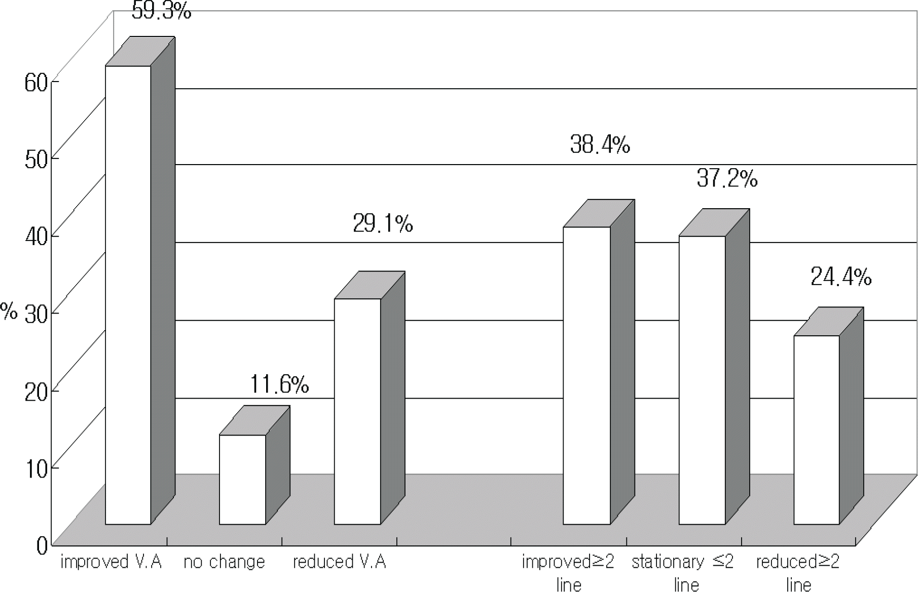 | Figure 1.Changes of visual acuity after photodynamic therapy in polypoidal choroidal vasculopathy. V.A=visual acuity. |
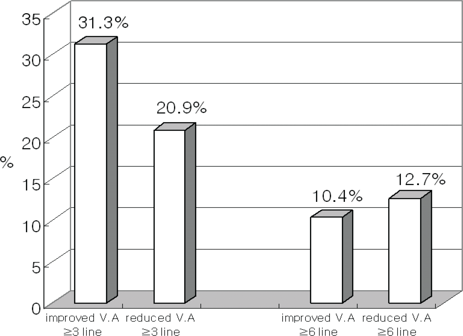 | Figure 2.Proportion of moderate (more than 3 lines) and severe (more than 6 lines) visual loss after photodynamic therapy in polypoidal choroidal vasculopathy. V.A=visual acuity. |
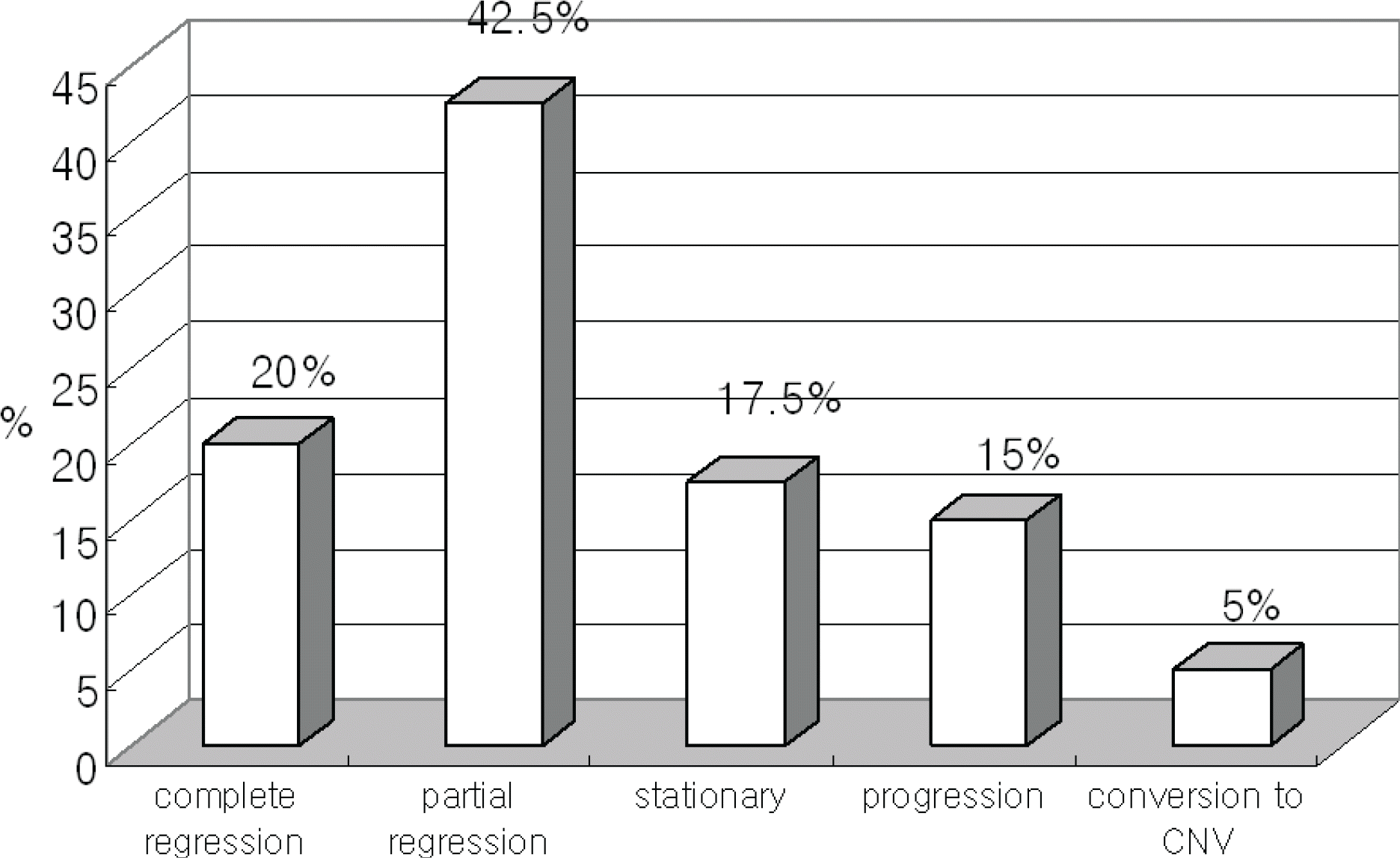 | Figure 3.Fluorescein angiographic outcomes of polypoidal choroidal vasculopathy after photodynamic therapy. |
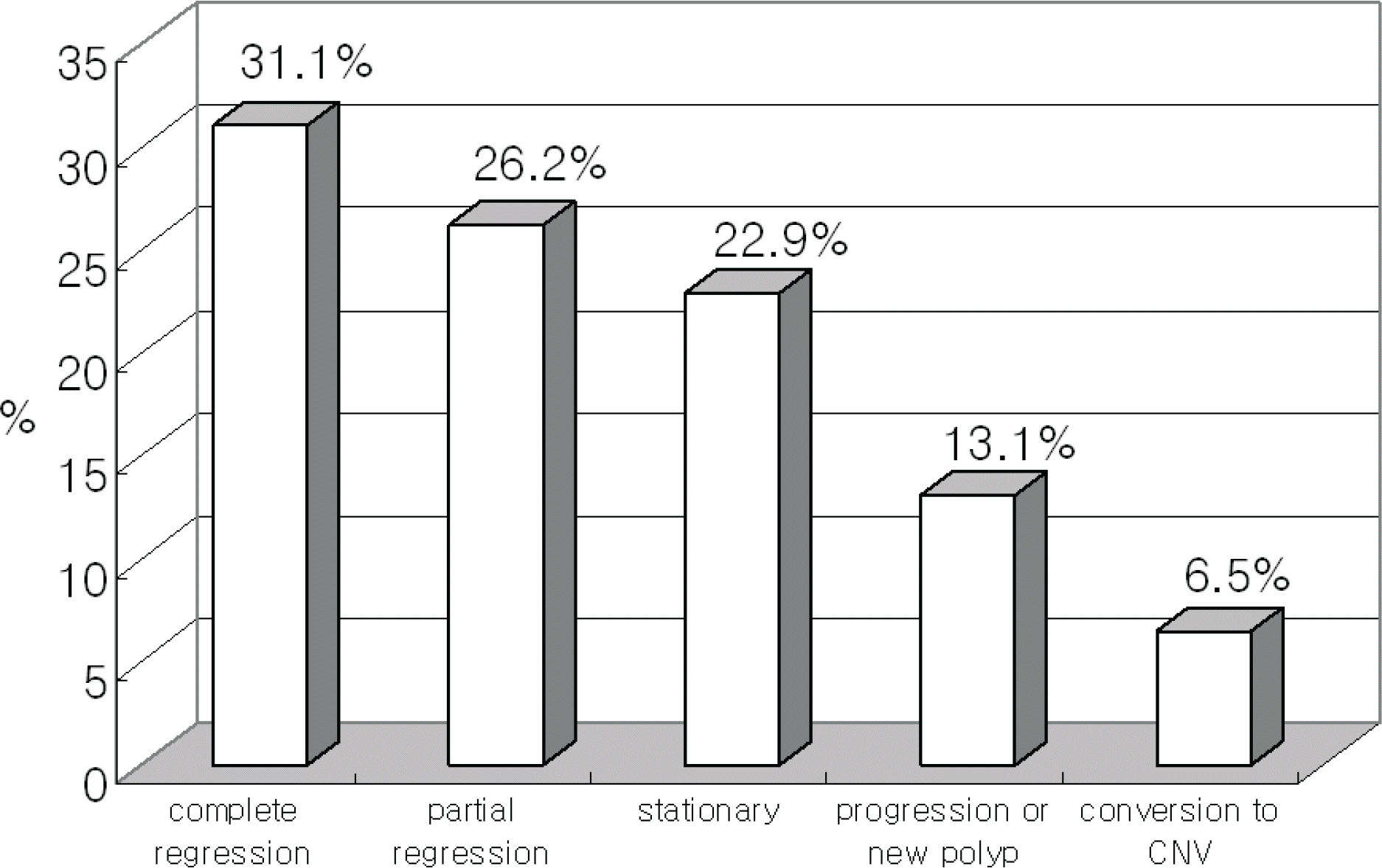 | Figure 4.Final activities of polyp in indocyanine green angiography after photodynamic therapy in polypoidal choroidal vasculopathy. |
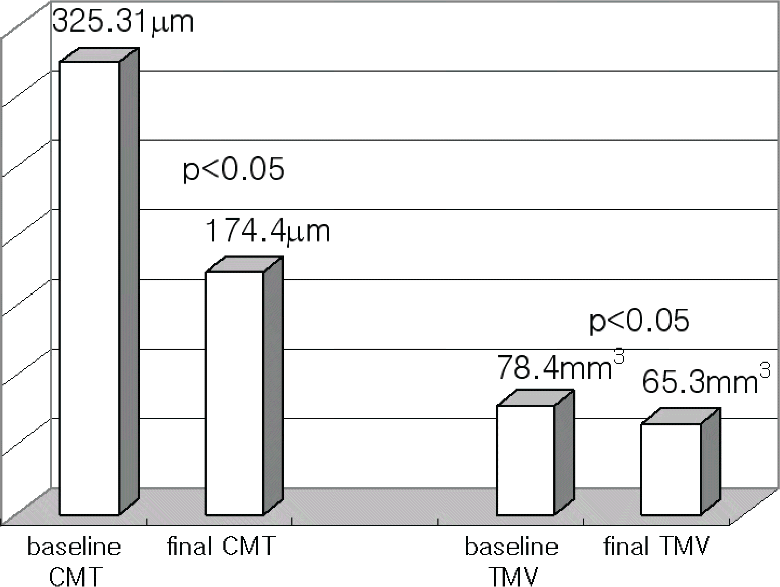 | Figure 5.Changes of central macular thickness (CMT) and total macular volume (TMV) in optical coherent tomogram after photodynamic therapy in polypoidal choroidal vasculopathy. |
Table 1.
Demographics and baseline characteristics of patients
| No. of patients (n) | 86 |
|---|---|
| Age (years) | 65.9±8.3 |
| Male/Female (%) | 65/21 (75.6%/24.4%) |
| Right/Left eye (%) | 45/41 (52.3%/47.7%) |
| Follow up (months) | 14.8±10.2 |
| Location of polyp (%) | Subfovea-33 (38.4%), Juxtafovea-53 (61.6%) |
| Number of polyps | 3.7±2.4 (range 1-15) |
| Status of fellow eye | ARMD: 22, PCV: 4, Disciform scar: 1, |
| Retinal detachment: 1, ERM: 1, POAG: 1 | |
| Systemic disease | HTN: 18, DM: 9, Angina:1, Tbc: 1, |
| Allergy:1, Stomach ca.: 1 | |
| Number of PDT treatment | 2.0±1.2 (range 1-8) |
| Baseline GLD (μm) | 2814.5±1194.8 |
| Laser spot size at 1st PDT (μm) | 3706.3±1099 |
| Bseline BCVA (mean± SD/median) | 0.55±0.32 LogMAR/0.5 LogMAR |
| Final BCVA (mean± SD/median) | 0.53±0.54 LogMAR/0.395 LogMAR (p=0.693*) |
Table 2.
Comparison between subfoveal and juxtafoveal polypoidal choroidal vasculopthy
| subfovea | juxtafovea | p* | |
|---|---|---|---|
| No. of Patients (n) | 33 | 53 | |
| Age (years) | 68.2±8.2 | 64.6±8.2 | 0.053 |
| Right/Left (eyes) | 17/16 | 28/25 | 0.905 |
| Male/Female (eyes) | 28/5 | 37/16 | 0.13 |
| Follow up (months) | 15.8±9.1 | 14.2±10.8 | 0.479 |
| No. of Polyp | 4±2.96 | 3.5±2.15 | 0.396 |
| No. of Retreatment | 2.24±1.28 | 1.87±1.19 | 0.172 |
| Baseline BCVA (LogMAR) | 0.55±0.26 | 0.56±0.36 | 0.812 |
| Final BCVA (LogMAR) | 0.61±0.61 | 0.48±0.49 | 0.282 |
| Changes of BCVA (LogMAR) | −0.06±0.62 | 0.08±40.47 | 0.226 |
| Baseline GLD (μm) | 2879.2±1090.8 | 2774.2±1263.8 | 0.694 |
| Laser spot size at 1st PDT (μm) | 3792.9±1123.8 | 3652.4±1090.6 | 0.567 |
| Final FAG activity (n) | n=32 | n=48 | 0.245 |
| Complete regression | 3 (9.4%) | 13 (27.1%) | |
| Partial regression | 13 (40.6%) | 21 (43.8%) | |
| Stationary | 8 (25%) | 6 (12.5%) | |
| Progression | 6 (18.8%) | 6 (12.5%) | |
| Conversion to CNV | 2 (6.3%) | 2 (4.2%) | |
| Final ICG activity of polyp (n) | n=22 | n=39 | 0.438 |
| Complete regression | 6 (27.3%) | 13 (33.3%) | |
| Partial regression | 5 (22.7%) | 11 (28.2%) | |
| Stationary | 6 (27.3%) | 8 (20.5%) | |
| Progression or new polyp | 3 (13.6%) | 5 (12.8%) | |
| Conversion to CNV | 2 (9.1%) | 2 (5.1%) |


 PDF
PDF ePub
ePub Citation
 Print
Print


 XML Download