

 PDF
PDF ePub
ePub Citation
 Print
Print


Abstract
Purpose
To evaluate the efficacy and safety of partial fluid air exchange at the end of 23-gauge transconjunctival sutureless vitrectomy to prevent postoperative hypotony.
Methods
Fifty-five eyes in 49 consecutive patients who underwent partial fluid air exchange at the end of 23-gauge sutureless vitrectomy by a single surgeon at Gil Hospital between August 2007 and February 2008 were recruited for this study. Intraocular pressure (IOP), visual acuity and post-operative complications were evaluated.
Results
Surgical indications included proliferative diabetic retinopathy (n=31), epiretinal membrane (n=9), nondiabetic vitreous hemorrhage (n=5), vitreous opacities (n=3), and others (n=7). Two eyes showed hypotony (<6 mmHg) on postoperative day 1 and resolved within a week without any supplemental procedures. Other complications included choroidal detachment in 1 eye, hyphema in 1 eye, and transient IIOP in 2 eyes. In 38 eyes in which combined cataract surgery was performed, air bubble-related complications including iris capture by the IOL in 3 eyes (7.9%) and opacification of the posterior capsule in 11 eyes (28.9%) occurred. No case of retinal detachment or endophthalmitis was observed. The final best corrected visual acuity was 20/40 or better in 14 eyes (25.5%).
Go to : 
References
1. Lakhanpal RR, Humayun MS, de Juan E Jr, et al. Outcomes of 140 consecutive cases of 25-gauge transconjunctival surgery for posterior segment disease. Ophthalmology. 2005; 112:817–24.

2. Theelen T, Verbeek AM, Tilanus MA, van den Biesen PR. A novel technique for self-sealing, wedge-shaped pars plana sclerotomies and its features in ultrasound biomicroscopy and clinical outcome. Am J Ophthalmol. 2003; 136:1085–92.
3. Yanyali A, Celik E, Horozoglu F, Nohutcu AF. Corneal topographic changes after transconjunctival (25-gauge) sutureless vitrectomy. Am J Ophthalmol. 2005; 140:939–41.
4. Fine HF, Iranmanesh R, Iturralde D, Spaide RF. Outcomes of 77 consecutive cases of 23-gauge transconjunctival vitrectomy surgery for posterior segment disease. Ophthalmology. 2007; 114:1197–200.
5. Chen E. 25-Gauge transconjunctival sutureless vitrectomy. Curr Opin Ophthalmol. 2007; 18:188–93.
6. Fujii GY, De Juan E Jr, Humayun MS, et al. Initial experience using the transconjunctival sutureless vitrectomy system for vitreoretinal surgery. Ophthalmology. 2002; 109:1814–20.
7. Fujii GY, De Juan E Jr, Humayun MS, et al. A new 25-gauge instrument system for transconjunctival sutureless vitrectomy surgery. Ophthalmology. 2002; 109:1807–12.
8. Byeon SH, Chu YK, Lee SC, et al. Problems associated with the 25-gauge transconjunctival sutureless vitrectomy system during and after surgery. Ophthalmologica. 2006; 220:259–65.
9. Shimada H, Nakashizuka H, Mori R, et al. 25-gauge scleral tunnel transconjunctival vitrectomy. Am J Ophthalmol. 2006; 142:871–3.
10. Kunimoto DY, Kaiser RS. Incidence of endophthalmitis after 20- and 25-gauge vitrectomy. Ophthalmology. 2007; 114:2133–7.
11. Scott IU, Flynn HW Jr, Dev S, et al. Endophthalmitis after 25-gauge and 20-gauge pars plana vitrectomy: incidence and outcomes. Retina. 2008; 28:138–42.
12. Kim MJ, Park KH, Hwang JM, et al. The safety and efficacy of transconjunctival sutureless 23-gauge vitrectomy. Korean J Ophthalmol. 2007; 21:201–7.
13. Eckardt C. Transconjunctival sutureless 23-gauge vitrectomy. Retina. 2005; 25:208–11.
14. Lopez-Guajardo L, Pareja-Esteban J, Teus-Guezala MA. Oblique sclerotomy technique for prevention of incompetent wound closure in transconjunctival 25-gauge vitrectomy. Am J Ophthalmol. 2006; 141:1154–6.
15. Amato JE, Akduman L. Incidence of complications in 25-gauge transconjunctival sutureless vitrectomy based on the surgical indications. Ophthalmic Surg Lasers Imaging. 2007; 38:100–2.
16. Gupta OP, Weichel ED, Regillo CD, et al. Postoperative complications associated with 25-gauge pars plana vitrectomy. Ophthalmic Surg Lasers Imaging. 2007; 38:270–5.
17. Inoue M, Shinoda K, Shinoda H, et al. Two-step oblique incision during 25-gauge vitrectomy reduces incidence of postoperative hypotony. Clin Experiment Ophthalmol. 2007; 35:693–6.
18. Sim DA, Wong R, Griffiths MF. Injecting an air bubble at the end of sutureless cataract surgery to prevent inflow of ocular surface fluid. Eye. 2007; 21:1444–5.
19. Tewari A, Shah GK, Fang A. Visual outcomes with 23-gauge transconjunctival sutureless vitrectomy. Retina. 2008; 28:258–62.
20. Demetriades AM, Gottsch JD, Thomsen R, et al. Combined phacoemulsification, intraocular lens implantation, and vitrectomy for eyes with coexisting cataract and vitreoretinal pathology. Am J Ophthalmol. 2003; 135:291–6.
21. Scharwey K, Pavlovic S, Jacobi KW. Combined clear corneal phacoemulsification, vitreoretinal surgery, and intraocular lens implantation. J Cataract Refract Surg. 1999; 25:693–8.
22. Suzuki Y, Sakuraba T, Mizutani H, et al. Postoperative refractive error after simultaneous vitrectomy and cataract surgery. Ophthalmic Surg Lasers. 2000; 31:271–5.
Go to :
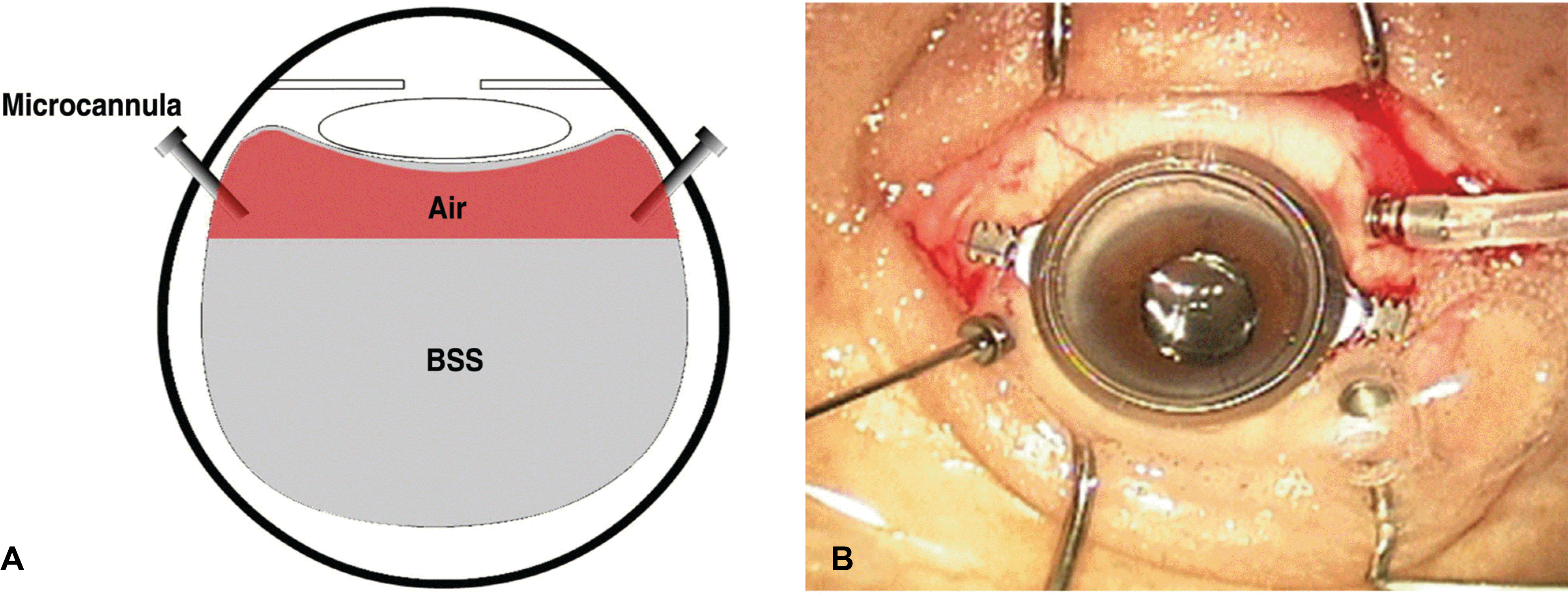 | Figure 1.Partial fluid air exchange at the end of 23-gauge sutureless vitrectomy. (A) Schematic picture of the procedure. (B) Intraoperative photograph showing vitreous cavity being replaced by air. Air-bubbles are popping out through the cannula. |
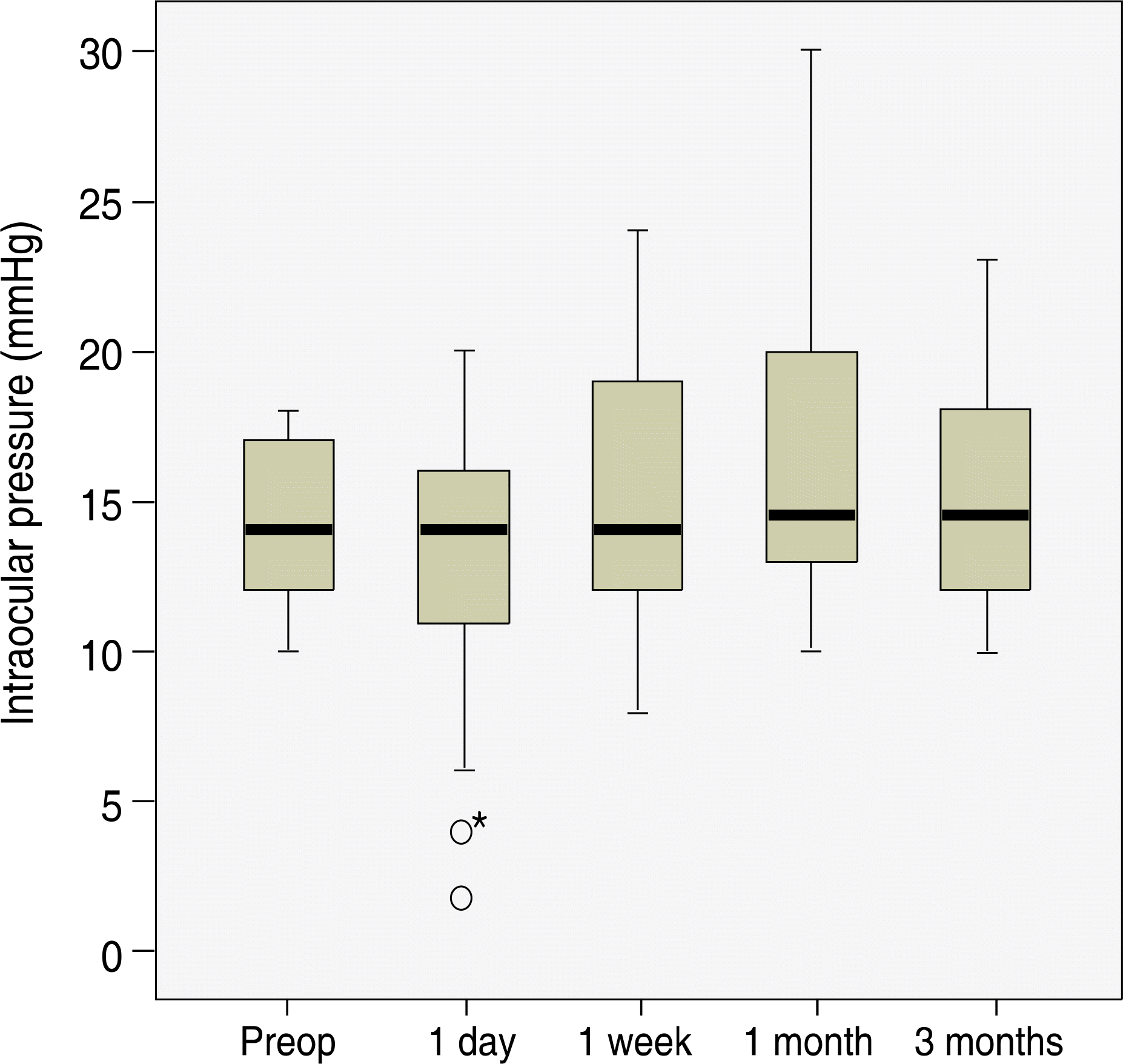 | Figure 2.Preoperative and postoperative intraocular pressure (IOP) changes. There were no significant differences in IOP among the measurements. * Two cases of hypotony were observed at postoperative 1 day. |
Table 1.
Indications for surgery
Table 2.
Summary of preoperative and postoperative visual acuity by surgical indication
| Indication (n) | Preop† VA§ (logMARП± SD#) | Final Postop‡ VA (logMAR± SD) | p value* |
|---|---|---|---|
| PDR** (n=31)†† | 1.06±0.2 | 0.82±0.3 | 0.02 |
| ERM†† (n=9) | 0.7±0.3 | 0.3±0.2 | <0.01 |
| Non-clearing vitreous hemorrhage (n=5) | 1.4±0.5 | 0.3±0.2 | <0.01 |
| Other§§ (n=10) | 0.54±0.4 | 0.31±0.3 | 0.03 |
| Total (n=55) | 1.1±0.5 | 0.5±0.3 | <0.01 |
Table 3.
Postoperative complications
| Complication | No. of Eyes | % |
|---|---|---|
| Leakage-related complication | ||
| Hypotony* | 2/55 | 3.6 |
| Choroidal detachment | 1/55 | 1.8 |
| Combined surgery-related complication† | ||
| Opacification of posterior capsule | 11/38 | 28.9 |
| Iris capture by intraocular lens optic | 3/38 | 7.9 |
| Fibrinous anterior chamber inflammation | 6/38 | 15.8 |
| Hyphema | 1/38 | 2.6 |
 XML Download