

 PDF
PDF ePub
ePub Citation
 Print
Print


Abstract
Purpose
To investigate the clinical usefulness of conjunctival brush cytology (CBC) in the diagnosis of dry eye syndrome.
Methods
Conjunctival impression cytology (CIC) was performed on the right eye and CBC was performed on the left eye in 24 patients with dry eye syndrome (9 patients with Sjӧ gren's syndrome (SS) and 15 patients with non-Sjӧ gren syndrome (Non-SS)) and 7 control subjects. The grade of squamous metaplasia was analyzed and the correlation between the grade and tear surface parameters was evaluated.
Results
The squamous metaplasia grade score in CIC and CBC were 2.44±0.73 and 1.56±1.01 in SS patients (p=0.047), 1.53±0.74 and 0.80±0.78 in Non-SS patients (p=0.017), and 0.43±0.54 and 0.14±0.38 (p=0.250) in control subjects, re-spectively. In SS patients, the score correlated significantly with the symptom score, basal tear secretion, break-up time (BUT) and keratoepitheliopathy score in CBC (p<0.05), but did not correlate with the tear surface parameters in CIC. In Non-SS patients, the score correlated significantly with the symptom score, basal tear secretion, BUT and keratoepitheliopathy score in CBC, and with the keratoepitheliopathy score in CIC (p<0.05).
Go to : 
References
1. Mckelvie P. Ocular surface impression cytology. Adv Ana Pathol. 2003; 10:328–37.

2. Anagnostopoulou-Fotinopoulou I, Rammou-Kinia R. Cytobrush sampling in conjunctival cytology. Diagn Cytopathol. 1993; 9:113–5.
3. Tsubota K, Kajiwara K, Ugajin S, Hasegawa T. Conjunctival brush cytology. Acta Cytol. 1990; 34:233–5.
4. Singh R, Joseph A, Umapathy T, et al. Impression cytology of the ocular surface. Br J Ophthalmol. 2005; 89:1655–9.
5. Albietz JM, Bruce AS. The conjunctival epithelium in dry eye subtypes: effect of preserved and non-preserved topical treatments. Curr Eye Res. 2001; 22:8–18.
6. Nelson JD, Wright JC. Conjunctival goblet cell densities in ocular surface disease. Arch Ophthalmol. 1984; 102:1049–51.
7. Takano Y, Fukagawa K, Dogru M, et al. Inflammatory cells in brush cytology samples correlate with the severity of corneal lesions in atopic keratoconjunctivitis. Br J Ophthalmol. 2004; 88:1504–5.
8. Pflugfelder SC, Solomon A, Stern ME. The diagnosis and management of dry eye: a twenty-five-year review. Cornea. 2000; 19:644–9.
9. Calonge M, Diebold Y, Sáez V, et al. Impression cytology of the ocular surface: a review. Exp Eye Res. 2004; 78:457–72.
10. Yağ mur M, Ersöz C, Ersöz TR, Varinli S. Brush technique in ocular surface cytology. Diagn Cytopathol. 1997; 17:88–91.
11. Adams GG, Dilly PN. Differential staining of ocular goblet cells. Eye. 1989; 3:840–4.
12. Vitali C, Bombardieri S, Jonsson R, et al. Classification criteria for Sjögren's syndrome: a revised version of the European criteria proposed by the American-European Consensus Group. Ann Rheum Dis. 2002; 61:554–8.
13. Nelson JD. Impression cytology. Cornea. 1988; 7:71–81.
14. Tseng SC. Staging of conjunctival squamous metaplasia by impression cytology. Ophthalmology. 1985; 92:728–33.
15. McKelvie PA, Daniell M. Impression cytology following mitomycin C therapy for ocular surface squamous neoplasia. Br J Ophthalmol. 2001; 85:1115–9.
16. Danjo Y, Lee M, Horimoto K, Hamano T. Ocular surface damage and tear lactoferrin in dry eye syndrome. Acta Ophthalmol (Copenh). 1994; 72:433–7.
17. Kunert KS, Tisdale AS, Gipson IK. Goblet cell numbers and epithelial proliferation in the conjunctiva of patients with dry eye syndrome treated with cyclosporine. Arch Ophthalmol. 2002; 120:330–7.
18. Egbert PR, Lauber S, Maurice DM. A simple conjunctival biopsy. Am J Ophthalmol. 1977; 84:798–801.
19. Nelson JD, Havener VR, Cameron JD. Cellulose acetate impressions of the ocular surface; dry eye states. Arch Ophthalmol. 1983; 101:1869–72.
20. Nelson JD. Ocular surface impressions using cellulose acetate filter material. Ocular pemphigoid. Surv Ophthalmol. 1982; 27:67–9.
21. Tole DM, McKelvie PA, Daniell M. Reliability of impression cytology for the diagnosis of ocular surface squamous neoplasia employing the Biopore membrane. Br J Ophthalmol. 2001; 85:154–8.
22. Caccamise WC. Early detection of xerophthalmia by impression cytology. Arch Ophthalmol. 1986; 104:970–2.
23. Puangsricharern V, Tseng SC. Cytologic evidence of corneal diseases with limbal stem cell deficiency. Ophthalmology. 1995; 102:1476–85.
24. Pepose JS. Improved impression cytology techniques for the immunopathological diagnosis of superficial viral infections. Br J Ophthalmol. 1998; 82:1097.
25. Keijser S, Missotten GS, De Wolff-Rouendaal D, et al. Impression cytology of melanocytic conjunctival tumours using the Biopore membrane. Eur J Ophthalmol. 2007; 17:501–6.
26. Kristensen GB, Hølund B, Grinsted P. Efficacy of the cyto-brush versus the cotton swab in the collection of endocervical cells. Acta Cytol. 1989; 33:849–51.
Go to :
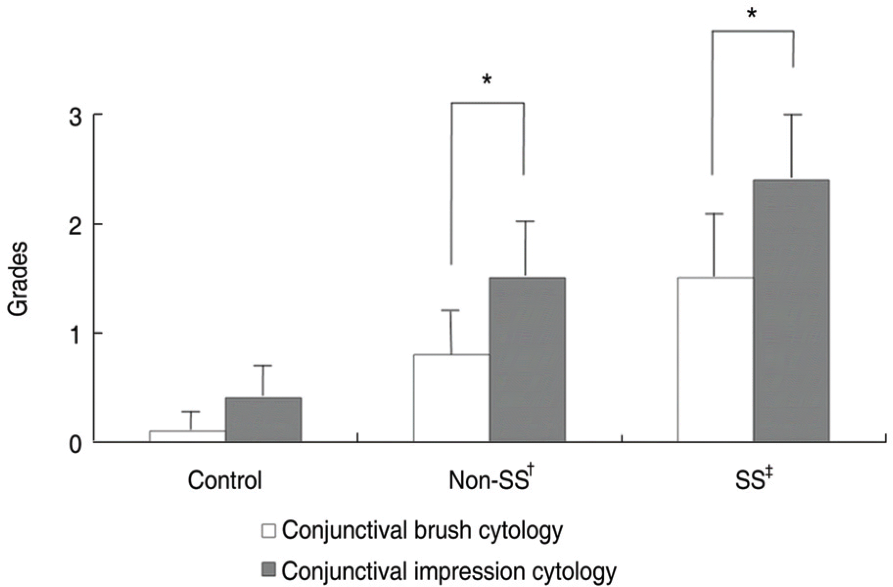 | Figure 1.Comparison of the grade of conjuctival squamous metaplasia between conjunctival brush and impression cytology. * p value<0.05; † Non-SS: Non-Sjӧgren's syndrome; ‡ SS: Sjӧgren's syndrome. |
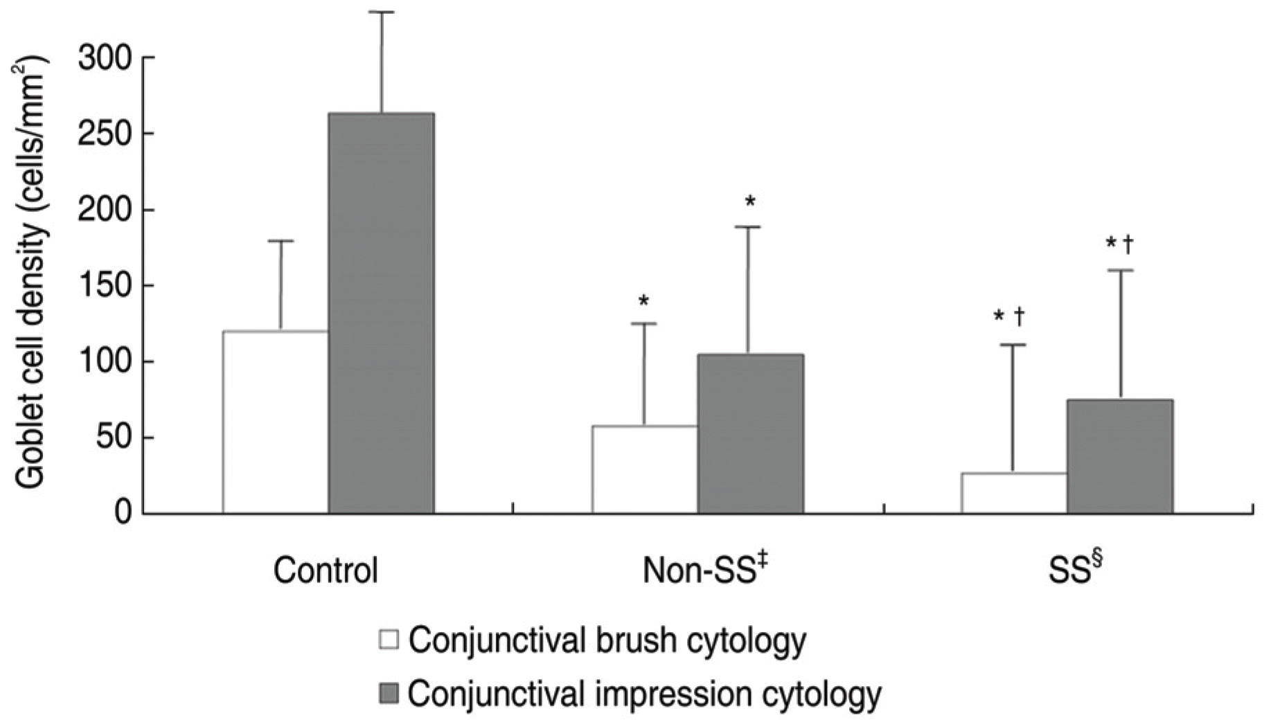 | Figure 2.Goblet cell density of controls, Non-SS, SS patients in conjunctival brush and impression cytology.* p<0.05 compared with control; † p<0.05 compared with Non-SS; ‡ Non-SS=Non-Sjӧgren's syndrome; § SS= Sjӧgren's syndrome. |
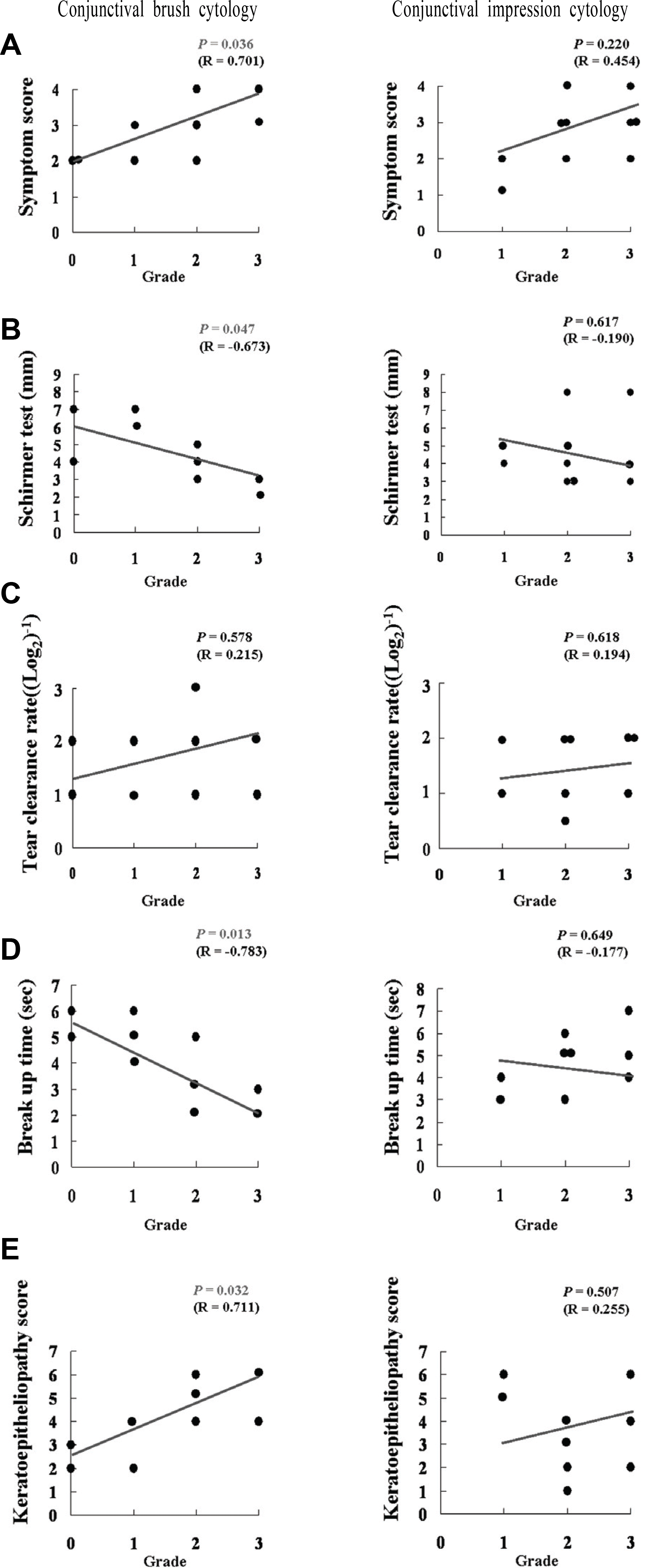 | Figure 3.Correlation between squamous metaplasia grades by conjuctival brush and impression cytology and tear surface parameters, including symptom score (A), basal tear secretion (B), tear clearance rate (C), tear break up time (D), and keratoepitheliopathy score (E) in Sjӧgren syndrome patients. |
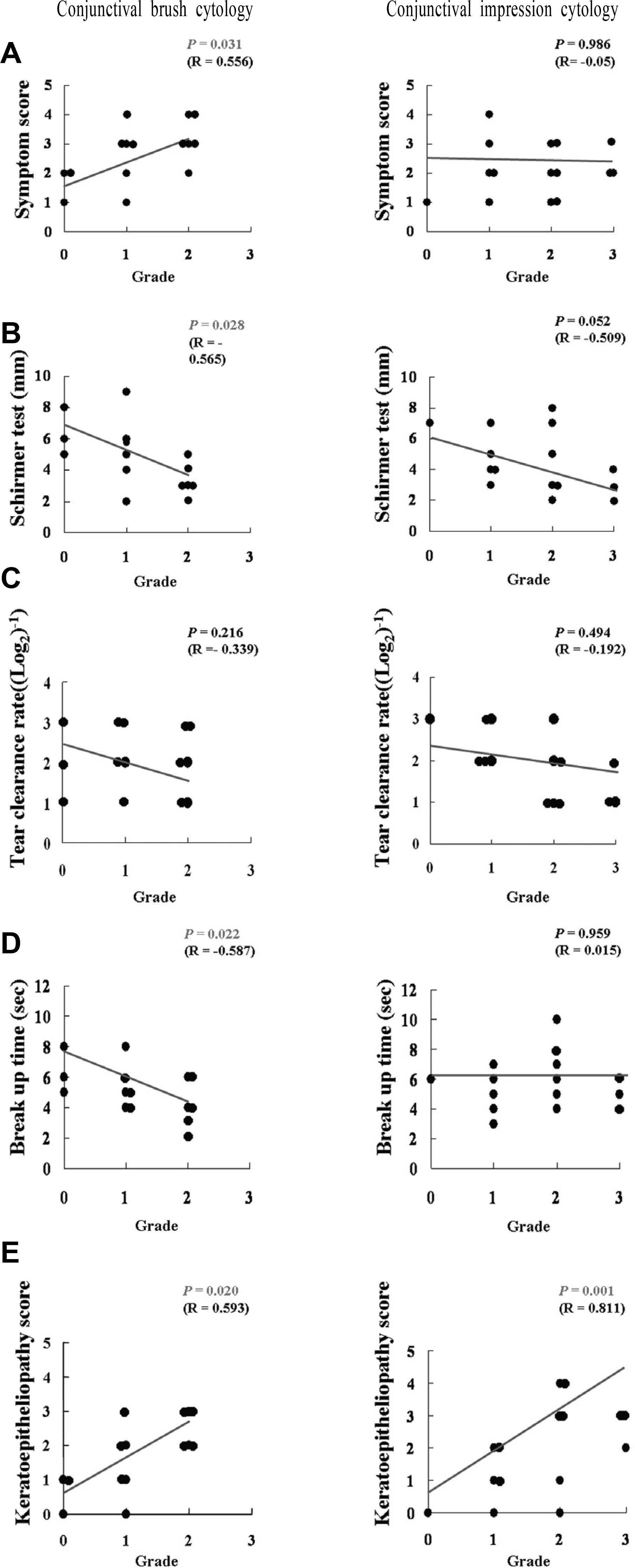 | Figure 4.Correlation between squamous metaplasia gradesconjuctival brush and impression cytology and tear surface parameters including symptom score (A), basal tear secretion (B), tear clearance rate (C), tear break up time (D), and keratoepitheliopathy score (E) in Non-Sjӧgren syndrome patients. |
Table 1.
Dermographic data in subjects for conjunctival impression cytology and brush cytology
| SS§ | Non-SS∥ | Control | |
|---|---|---|---|
| Patients number | 9 | 15 | 7 |
| Male/Female | 0/9 | 6/9 | 3/4 |
| Age (years) | 50.4±14.6 | 41.8±13.9 | 41.4±16.4 |
| Symptom score | 2.67±0.87* | 2.00±0.93† | 0.14±0.38 |
| Basal secretion test (mm) | 4.44±1.59* | 5.33±1.95† | 10.57±1.62 |
| Tear clearance rate ((Log2)-1) | 2.39±0.61*‡ | 3.57±0.90 | 4.00±0.39 |
| Break up time (sec) | 4.89±1.27*‡ | 5.67±1.40† | 10.71±1.25 |
| Keratoepitheliopathy score | 3.89±1.45*‡ | 1.07±0.80† | 0.14±0.38 |
 XML Download