Abstract
Purpose
To report the results of femtosecond laser-assisted‘mushroom-shaped wound-configurized keratoplasty’in 6 patients based on the results of experimental models in rabbits’and enucleated porcine eyes.
Methods
Mushroom-shaped donor corneal grafts were designed and transplanted in 3 rabbit eyes and 10 enucleated porcine eyes. The histologic findings were observed and wound burst pressure compared between penetrating keratoplasty (PKP) and mushroom-shaped keratoplasty. The‘mushroom-shaped wound-configurized keratoplasty’was subsequently performed in 6 eyes of 6 patients.
Results
In all eyes of the rabbits, histologic findings showed stable wound structure. In the porcine models, mushroom-shaped keratoplasty showed a better resistance to IOP than PKP after complete suture. Patients who underwent the surgery showed an improvement of over 2 lines in Snellen visual acuity after the operation. The mean follow-up period was approximately 11 months. Complete suture removal was performed within 6 months and accurate and stable wound structure was identified.
Go to : 
References
1. McNeill JI, Kaufman HE. A double running suture technique for keratoplasty: earlier visual rehabilitation. Ophthalmic Surg. 1977; 8:58–61.
2. Davison JA, Bourne WM. Results of penetrating keratoplasty using a double running suture technique. Arch Ophthalmol. 1981; 99:1591–5.

3. McNeill JI, Wessels IF. Adjustment of single continuous suture to control astigmatism after penetrating keratoplasty. Refract Corneal Surg. 1989; 5:216–23.

4. Musch DC, Meyer RF, Sugar A, Soong HK. Corneal astigmatism after penetrating keratoplasty. Ophthalmology. 1989; 96:698–703.

5. Van Meter WS, Gussler JR, Soloman KD, Wood TO. Postkeratoplasty astigmatism control. Ophthalmology. 1991; 98:177–83.

6. Assil KK, Zarnegar SR, Schanzlin DJ. Visual outcome after penetrating keratoplasty with double continuous or combined interrupted and continuous suture wound closure. Am J Ophthalmol. 1992; 114:63–71.

7. De Molfetta V, Brambilla M, De Casa N. . Residual corneal astigmatism after perforating keratoplasty. Ophthal-mologica. 1980; 179:316–21.

8. Perlman EM. An analysis and interpretation of refractive errors after penetrating keratoplasty. Ophthalmology. 1981; 88:39–45.

9. Samples JR, Binder PS. Visual acuity, refractive error, and astigmatism following corneal transplantation for pseudophakic bullous keratopathy. Ophthalmology. 1985; 92:1554–60.
10. Oh JW, Hahn TW, Park CK, Kim JH. Comparison of Corneal Astigmatism after Keratoplasty with 3 Kinds of Suture Techniques. J Korean Ophthalmol Soc. 1998; 39:2569–74.
11. Kim KE, Joo CK. Changes in Astigmatism after Suture Removal in Penetrating Keratoplasty. J Korean Ophthalmol Soc. 2003; 44:284–8.
12. Musch DC, Meyer RF, Sugar A. The effect of removing running sutures on astigmatism after penetrating keratoplasty. Arch Ophthalmol. 1988; 106:488–92.

13. Binder PS. The effect of suture removal on postkeratoplasty astigmatism. Am J Ophthalmol. 1988; 105:637–45.

14. Lin DT, Wilson SE, Reidy JJ. . Topographic changes that occur with 10-0 nylon suture removal following keratoplasty. Refract Corneal Surg. 1990; 6:21–5.
15. Mader TH, Yuan R, Lynn MJ. . Changes in keratometric astigmatism after suture removal more than one year after penetrating keratoplasty. Ophthalmology. 1993; 100:119–27.

16. Davis EA, Azar DT, Jakobs FM, Stark WJ. Refractive and keratometric results after triple procedure: experience with early and late suture removal. Ophthalmology. 1998; 105:624–30.
17. Kim KS, Kim MS. The Effect of PRK and LASIK for the Correction of Postkeratoplasty Astigmatism. J Korean Ophthalmol Soc. 2004; 45:376–82.
18. Binder PS, Abel R Jr, Polack FM, Kaufman HE. Keratoplasty wound separations. Am J Ophthalmol. 1975; 80:109–15.

19. Farley MK, Pettit TH. Traumatic wound dehiscence after penetrating keratoplasty. Am J Ophthalmol. 1987; 104:44–9.

20. Rehany U, Rumelt S. Ocular trauma following penetrating keratoplasty. Arch Ophthalmol. 1998; 116:1282–6.

21. Tseng SH, Lin SC, Chen FK. Traumatic wound dehiscence after penetrating keratoplasty. Cornea. 1999; 18:553–8.

22. Melles GR, Lander F, van Dooren BT. . Preliminary clinical results of posterior lamellar keratoplasty through a sclerocorneal pocket incision. Ophthalmology. 2000; 107:1850–6.
23. Alio JL, Shah S, Barraquer C. . New techniques in lamellar keratoplasty. Curr Opin Ophthalmol. 2002; 13:224–9.

24. Busin M. A new lamellar wound configuration for penetrating keratoplasty surgery. Arch Ophthalmol. 2003; 121:260–5.

25. Busin M, Arffa RC. Microkeratome-assisted Mushroom keratoplasty with minimal endothelial replacement Am J Ophthalmol. 2005; 140:138–40.
26. Hoffart L, Proust H, Matonti F. . Short-term results of penetrating keratoplasty performed with the Femtec femtosecond laser. Am J Ophthalmol. 2008; 146:50–5.

27. Por YM, Cheng JY, Parthasarathy A. . Outcomes of femtosecond laser-assisted penetrating keratoplasty. Am J Ophthalmol. 2008; 145:772–4.

28. Cheng YY, Tahzib NG, van Rij G, van Cleynenbreugel H, Pels E, Hendrikse F, Nuijts R. Femtosecond laser-assisted inverted mushroom keratoplasty. Cornea. 2008; 27:679–85.

29. Steinert RF, Ignacio TS, Sarayba MA. “Top Hat”-shaped penetrating keratoplasty using the femtosecond laser. Am J Ophthalmol. 2007; 143:689–91.

30. Seitz B, Behrens A, Langenbucher A. . Experimental 193-nm excimer laser trephination with divergent angles in penetrating keratoplasty. Cornea. 1998; 17:410–6.
31. Farid M, Kim M, Steinert RF. Results of penetrating keratoplasty performed with a femtosecond laser zigzag incision initial report. Ophthalmology. 2007; 114:2208–12.

32. Slade SG. Applications for the femtosecond laser in corneal surgery. Curr Opin Ophthalmol. 2007; 18:338–41.

33. Bahar I, Kaiserman I, McAllum P, Rootman D. Femtosecond laser-assisted penetrating keratoplasty: stability evaluation of different wound configurations. Cornea. 2008; 27:209–11.
34. Price MO, Price FW Jr. Efficacy of topical cyclosporine 0.05% for prevention of cornea transplant rejection episodes. Ophthalmology. 2006; 113:1785–90.

35. Filatov V, Steinert RF, Talamo JH. Postkeratoplasty astigmatism with single running suture or interrupted sutures. Am J Ophthalmol. 1993; 115:715–21.

36. Claesson M, Armitage WJ. Astigmatism and the impact of relaxing incisions after penetrating keratoplasty. J Refract Surg. 2007; 23:284–9.

37. Imaizumi T. Movement of corneal endothelium after penetrating keratoplasty. Observation of sex chromatin as a cell marker. Nippon Ganka Gakkai Zasshi. 1990; 94:928–36.
38. Groh MJ, Seitz B, Kuchle M, Naumann GO. Clearing of the host cornea after penetrating keratoplasty for pseudophakic bullous keratopathy. Klin Monatsbl Augenheilkd. 1999; 215:275–80.
39. Langenbucher A, Seitz B, Nguyen NX, Naumann GO. Corneal endothelial cell loss after nonmechanical penetrating keratoplasty depends on diagnosis: a regression analysis. Graefes Arch Clin Exp Ophthalmol. 2002; 240:387–92.

40. Patel SV, Hodge DO, Bourne WM. Corneal endothelium and postoperative outcomes 15 years after penetrating keratoplasty. Am J Ophthalmol. 2005; 139:311–9.

41. Gil SY, Park CK, Hahn TW. Evaluation of Donor Corneal Endothelium after Keratoplasty. J Korean Ophthalmol Soc. 2006; 47:519–24.
42. Price FW Jr, Price MO. Femtosecond laser shaped penetrating deratoplasty: one-year results utilizing a top-hat configuration. Am J Ophthalmol. 2008; 145:210–4.
Go to : 
 | Figure 1.In porcine eyes on artificial chamber, wound burst pressures were recorded by digital manometer after 4, 8, 16 sutures. (A: Conventional PKP, B: Mushroom-shaped keratoplasty) |
 | Figure 2.Donor cornea mounted in an artificial chamber under the femtosecond laser and applanation was done. |
 | Figure 3.Illustration of the femtosecond laser-shaped penetrating keratoplasty “Mushroom-shaped” graft configuration. |
 | Figure 4.Histologic examination of rabbit’s cornea show accurate and stable wound structure. |
 | Figure 5.Postoperative slit-lamp photograph of patient 2 after ‘mushroom-shaped’ keratoplasty. Note the clear central cornea with well attached peripheral flange. |
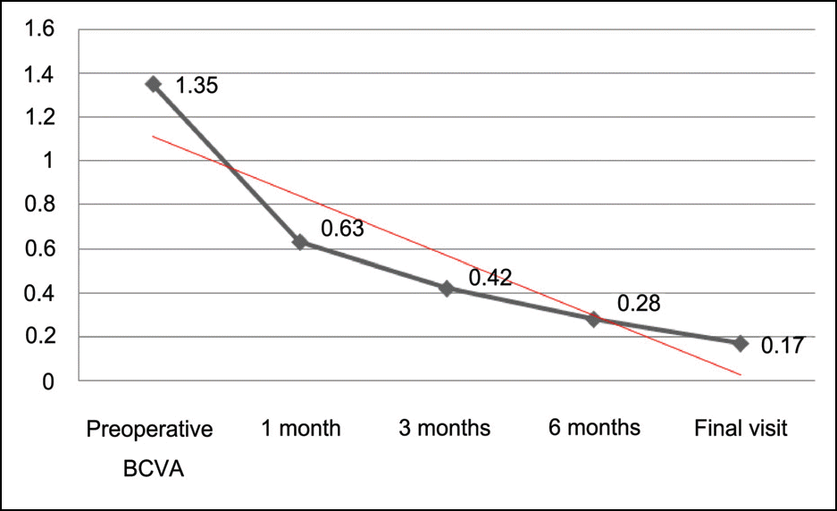
 | Figure 6.Changes of mean best corrected visual acuity (BCVA) after Femtosecond laser-assisted ‘mushroom- shaped’ wound-configurized keratoplasty. |
 | Figure 7.Ultrasound biomicroscopic finding (above) and anterior OCT (below) of patient’s cornea show well attached stable wound structure as in rabbits’ model |
Table 1.
Summary of age, gender, treated eye, diagnosis, preoperative BCVA, data of donor corneal cut of patients
| Patient No. | Sex /Age (yr) | Diagnosis | Treated eye | Preoperative BCVA∗(LogMAR) | design of donor graft (Anterior/Posterior/depth)(mm) |
|---|---|---|---|---|---|
| 1 | M/36 | Corneal opacity d/t corneal injury | Right | 0.4 | 8.2 / 6 / 0.32 |
| 2 | M/27 | Keratoconus Hydrops | Left | 2 | 8.6 / 6 / 0.3 |
| 3 | M/40 | Corneal opacity d/t corneal injury | Left | 1.6 | 8.2 / 6 / 0.35 |
| 4 | M/28 | Keratoconus Hydrops | Left | 2 | 8.6 / 6 / 0.32 |
| 5 | M/18 | Keratoconus | Right | 1.1 | 8.2 / 6 0.3 |
| 6 | M/27 | Keratoconus | Left | 1 | 8.2 / 7 / 0.35 |
Table 2.
Mean burst pressure for traditional PKP and mushroom keratoplasty in porcine corneas
| No. stitches | Traditional PKP∗ (n=5) | Mushroom keratoplasty (n=5) | P-value |
|---|---|---|---|
| 4 | 1.2±1.1 | 2.4±0.55 | 0.10 |
| 8 | 16.6±6.23 | 27.4±7.37 | 0.10 |
| 16 | 78.4±19.71 | 133±24.6 | 0.008 |
Table 3.
Summary of preoperative and postoperative endothelial cell count, time of suture removal and follow-up period
Table 4.
Summary of refractive results, spherical equivalent, postoperative BCVA, corneal thickness, intraocular pressure after keratoplasty
| Patient | 6 months Sim K’s astigmatism (Diopter) | Spherical equivalent at final visit (Diopter) | Postoperative BCVA∗ at final visit (LogMAR) | Corneal thickness at final visit (µm) | Postoperative IOP (mmHg) |
|---|---|---|---|---|---|
| 1 | 3.1 | 1.25 | 0.2 | 554 | 11~22 |
| 2 | 1.5 | -4 | 0.1 | 610 | 12~20 |
| 3 | 4 | 1.75 | 0 | 543 | 10~15 |
| 4 | 3.8 | -10 | 0.2 | 511 | 12~18 |
| 5 | 2.8 | -5.75 | 0.2 | 509 | 11~19 |
| 6 | 2.9 | -12 | 0.3 | 630 | 15~20 |
| Mean± SD | 3.0±0.9 | -4.79±5.7 | 0.17±0.1 | 559±50 | 15.4±4.2 |


 PDF
PDF ePub
ePub Citation
 Print
Print


 XML Download