Abstract
Purpose
To report a case of epithelial ingrowth treatment by surgical excision of epithelial tissues and intracameral 5-fluorouracil injection.
Case summary
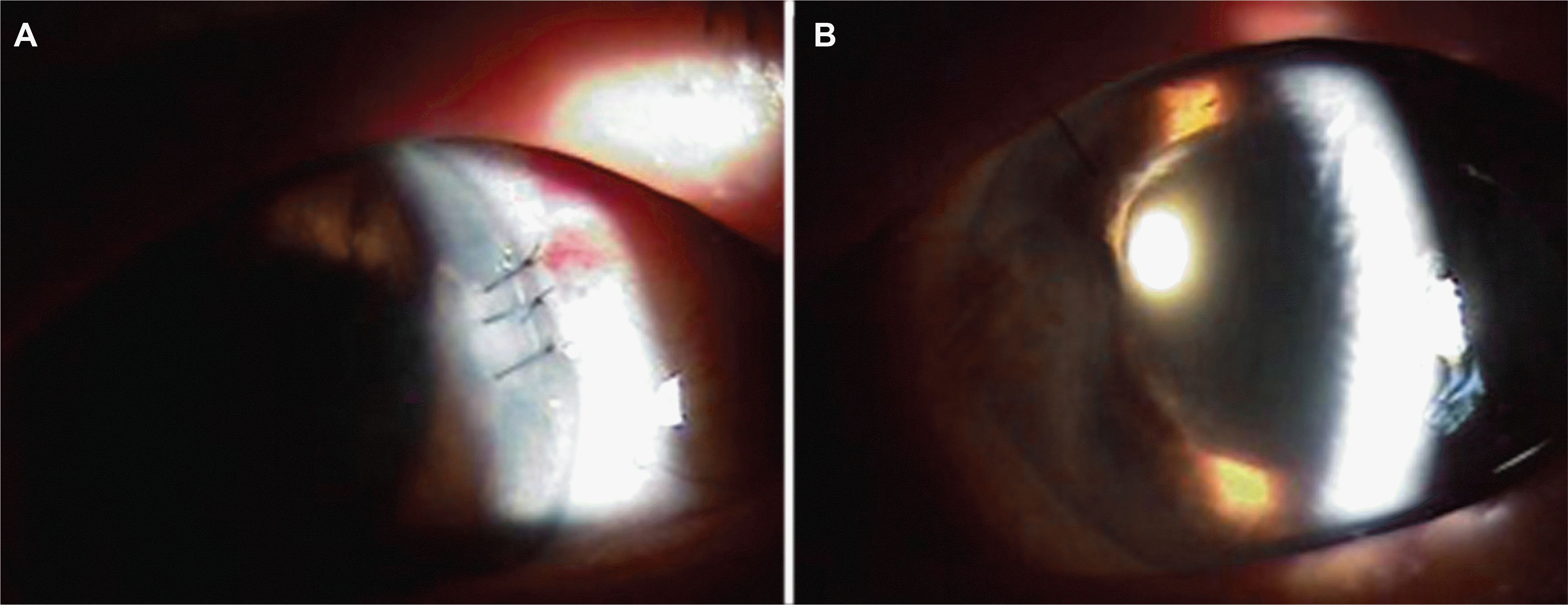
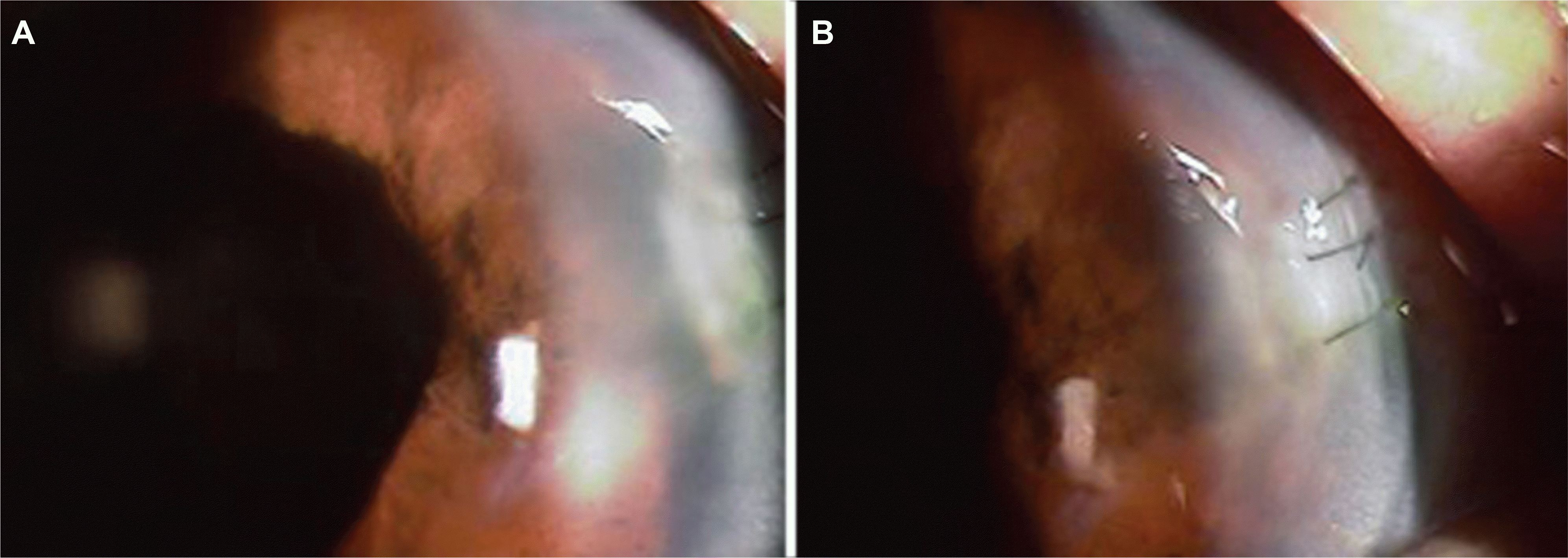
A 70-year-old female patient who underwent phacoemulsification through clear cornea incision in both eyes 2 years before, was referred for her left ocular pain and corneal edema of 3 months’ duration. Diffuse sheet-like epithelium grew from the clear cornea incision site to the pupil margin lesion of the iris. The epithelial tissues were excised and 5-fluorouracil was injected intracamerally. There were no recurrences for 2 months.
References
1. Vargas LG, Vroman DT, Solomon KD, et al. Epithelial Down-growth after Clear Corena Phacoemulsification: report of two cases and review of the literature. Ophthalmology. 2002; 109:2331–5.
2. Sullivan GL. Epithelization of the anterior chamber following cataract extraction: a new approach to treatment. Trans Am Acd Ophthalmol. 1958; 56:606–54.
3. Long JC, Tyner GS. Three cases of epithelial invasion of the anterior chamber treated surgically. Arch Ophthalmol. 1957; 58:396–400.

4. Brown SI. Treatment of advanced epithelial downgrowth. Trans Am Acd Ophthalmol. 1973; 77:618–22.
5. Friedman AH. Radical anterior segment surgery for epithelial invasion of the anterior chamber: report of three cases. Trans Am Acd Ophthalmol. 1977; 83:216–23.
6. Stark WJ, Michels RG, Maumenee AE, Cupples H. Surgical management of epithelial ingrowth. Am J Ophthalmol. 1978; 85:772–80.

7. Maumenee AE, Paton D, Morse PH, Butner R. Review of 40 histologically proven cases of epithelial downgrowth following cataract extraction and suggested surgical management. Am J Ophthalmol. 1970; 69:598–603.

8. Knauf HP, Rowsey JJ, Margo CE. Cystic epithelial down-growth clear-corneal cataract extraction. Arch Ophthalmol. 1997; 22:330–5.
9. Chen SH, Pineda R 2nd. Epithelial and fibrous downgrowth: mechanism of disease. Ophthalmol Clin North Am. 2008; 15:41–8.
10. Jadav DS, Rhlander NR, Vold SD, et al. Endoscopic photo-coagulation in the management of epithelial downgrowth. Cornea. 2008; 27:601–4.

11. Weiner MJ, Trentacoste J, Pon DM, Albert DM. Epithelial downgrowth: a 30-year clinicopathological review. Br J Ophthalmol. 1989; 73:6–11.

12. Kim SK, Ibarra MS, Syed NA, et al. Development of epithelilal downgrowth several decades after intraocular surgery. Cornea. 2005; 24:108–9.
13. Cho JS, Ko MK, Shin JC. Epithelial ingrowth of anterior chamber and anterior surface of vitreous. Korean J Ophthalmol. 1998; 12:118–21.

14. Lai MM, Haller JA. Resolution of epithelial ingrowth in a patient treated with 5-fluorouracil. Am J Ophthalmol. 2002; 133:562–4.

15. Tomlins PJ, Savant V, Quinlan M. Failure of intracameral fluorouracil to resolve an epithelial ingrowth following clear corneal cataract surgery. J Cataract Refract Surg. 2007; 33:923–4.

16. Lee BL, Gaton DD, Weinreb RN. Epithelial downgrowth following phacoemulsification through a clear cornea. Arch Ophthalmol. 1999; 117:283.

17. Shikh AA, Damji KF, Mintsioulis G, et al. Bilateral epithelial downgrowth managed in one eye with intraocular 5-fluor-ouracil. Arch Ophthalmol. 2008; 120:1396–8.
18. Srinivasan S, Jones DH, Jay JL, Roberts F. Epithelial down-growth following clear cornea phacoemulsification in a buphthalmic eye. Br J Ophthalmol. 2004; 88:152–3.

19. Kim SW, Byun YJ, Kim EK, Kim TI. Treatment of epithelial ingrowth after laser in situ keratomilusis using amniotic membrane patch. J Korean Ophthalmol Soc. 2007; 48:230–7.
20. Lim TH, Kim MJ, Kim TI, Tchah HW. Lamellar keratoplasty and restoration of traumatic dislocation of LASIK flap using human fibrin adhesive. J Korean Ophthalmol Soc. 2005; 46:1741–6.
21. Hunyor AP, Davis Belcher III. BSC. Epithelial and Fibrous Invasion of the Eye. Steinert RF, Fine IH, Gimbel HV, editors. Cataract Surgery: Technique, Complications, Management. 2nd ed.Philadelphia: Saunders;2004. chap. 46.
Figure 1.
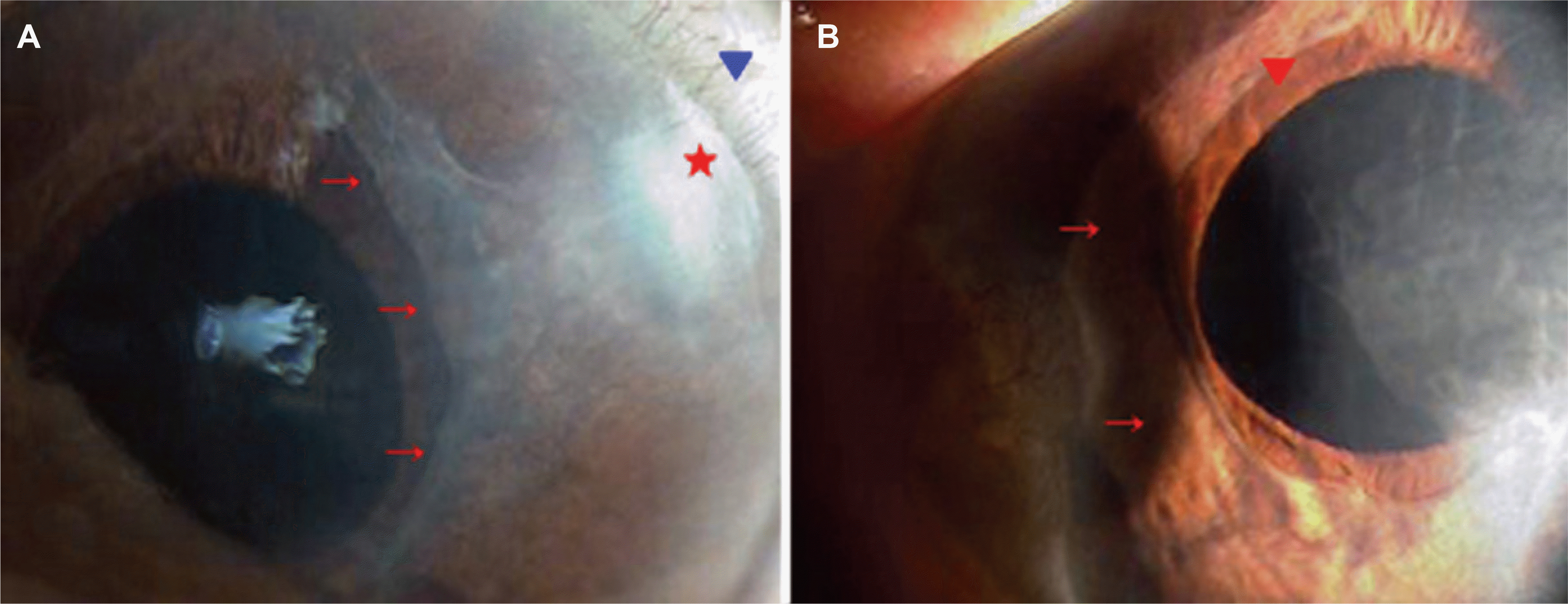
Anterior segment photograph shows diffuse sheet-like epithelial membrane covers from clear cornea incision site to around the pupil margin. Red star is the old clear corneal incision site and vascularization of the incision site (blue arrow head) (A). Everted iris is shown (red arrow head) and diffuse corneal edema. All red arrows indicate the diffuse sheet-like membrane (B).

Figure 2.
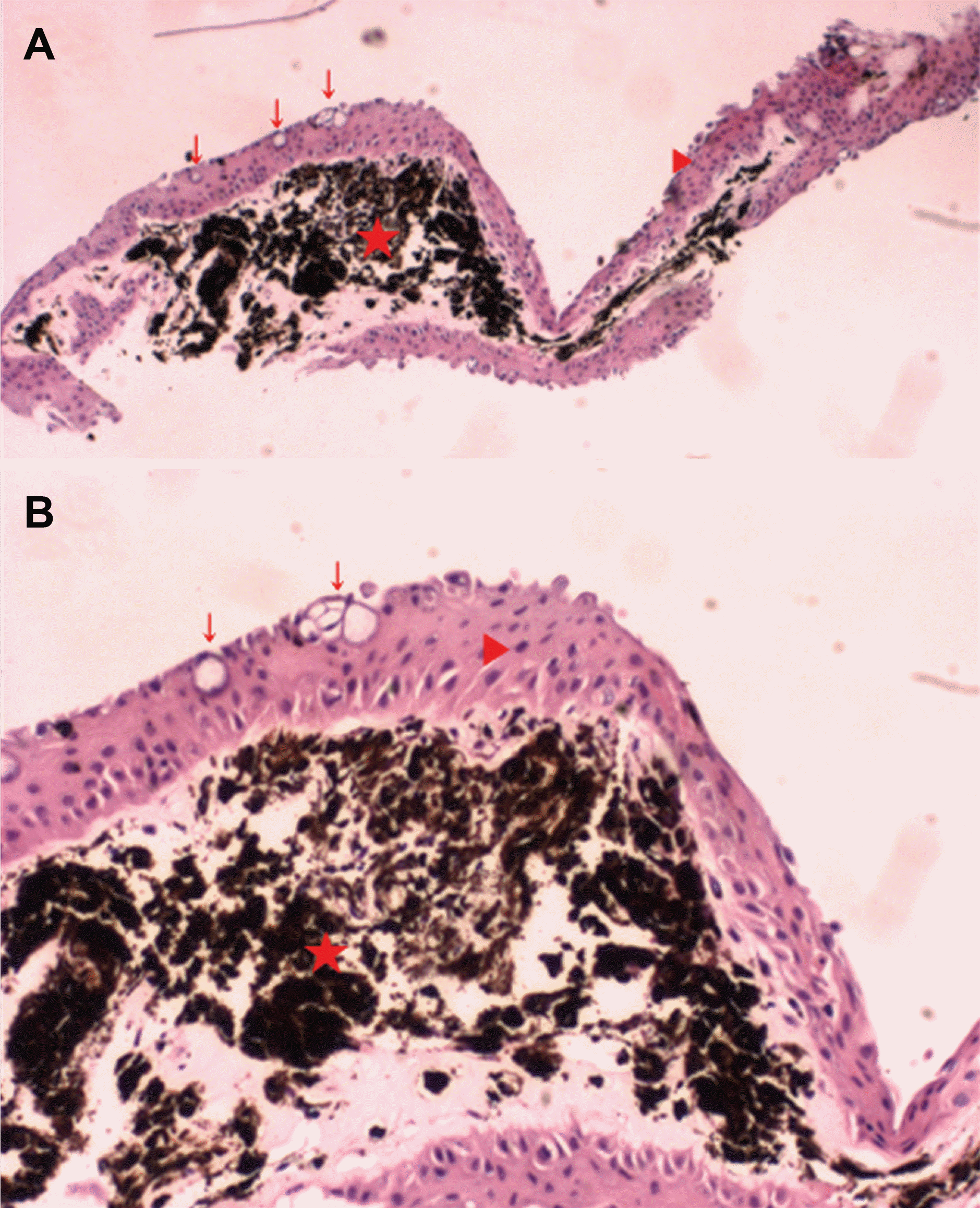
Microscopic examination of the excised tissue shows that black pigmented lesion is a stroma of the iris (red star) is surrounded by stratified epithelium (red arrow head) contains conjunctival origin goblet cells (red arrows). (H&E staining ×40 A, ×100 B)


 PDF
PDF ePub
ePub Citation
 Print
Print


 XML Download