Abstract
Case summary
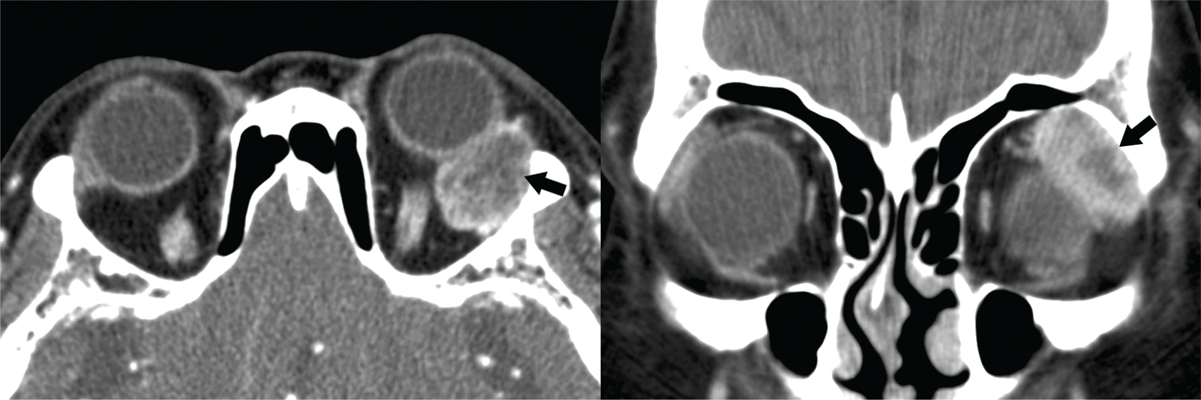
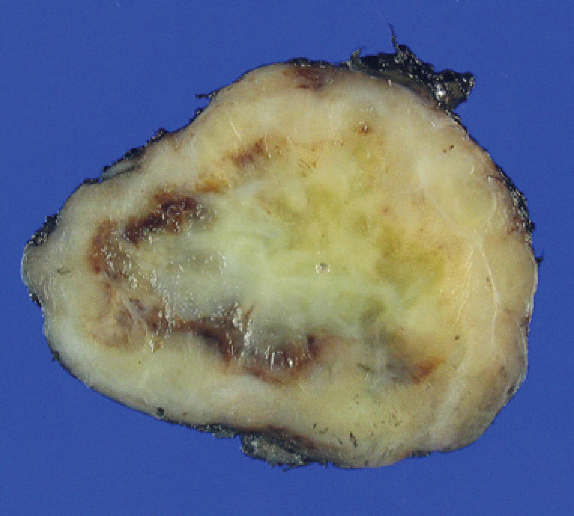
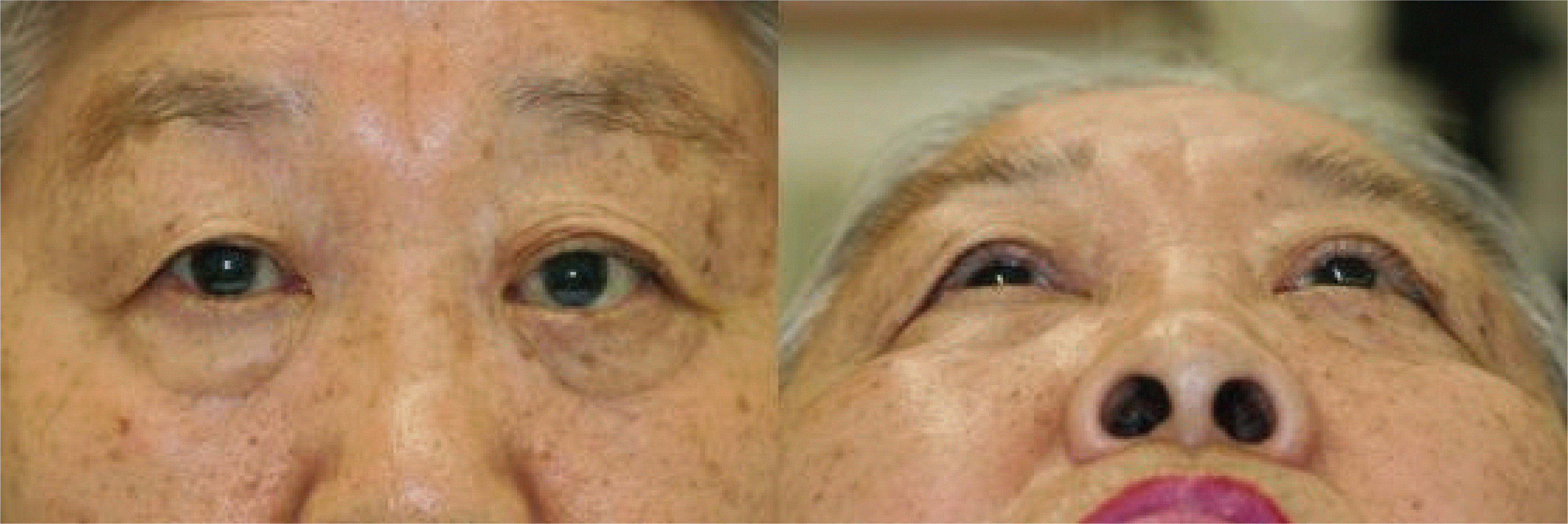
A 69‐ year‐ old woman presented with a 3‐ month history of proptosis in her left eye. Intraocular pressure was 17 mmHg in her right eye and 23 mmHg in her left eye. There was a left hypotropia on upgaze. A fundus examination showed retinal folds in the superotemporal area in her left eye. Computed tomography revealed a 2.6 cm‐ sized well‐ defined enhancing solid mass in the superotemporal extraconal space of the left orbit, pushing her left eye forward. Lateral orbitotomy, tumor removal, and biopsy were performed. Pathological findings showed a fascicular pattern of benign spindle cells with mild cellular pleomorphism and hyaline degeneration, without mitotic figures. Immunohistochemical stain was positive with smooth muscle actin (SMA), which was compatible with orbital leiomyoma.
References
1. Lee JG, Kim J, Chung H. Leiomyoma in the ciliary body resected Ab externo. J Korean Ophthalmol Soc. 2000; 41:2285–90.
2. Jolly SS, Brownstein S, Jordan DR. Leiomyoma of the anterior orbit and eyelid. Can J Ophthalmol. 1995; 30:366–70.
3. Neetens A, Smet H. Orbital leiomyoma. Bull Soc Belge Ophthal. 1984; 210:73–7.
5. Arat YO, Font R, Chaudhry I, Boniuk M. Leiomyoma of the orbit and periocular region: a clinicopathologic study of four cases. Ophthal Plast Reconstr Surg. 2005; 21:16.
6. Badoza D, Weil D, Zarate J. Orbital leiomyoma: a case report. Ophthal Plast Reconstr Surg. 1999; 15:460.
7. Betharia SM, Arora R, Kishore K, et al. Leiomyoma of the orbit. Indian J Ophthalmol. 1991; 39:35–7.
8. Gündüz K, Günalp I, Erden E, Erekul S. Orbital leiomyoma: report of a case and review of the literature. Surv Ophthalmol. 2004; 49:237–42.

9. Henderson JW, HarrisonJr EG. Vascular leiomyoma of the orbit: report of a case. Trans Am Acad Ophthalmol Otolaryngol. 1970; 74:970–4.
10. Jakobiec FA, Howard GM, Rosen M, Wolff M. Leiomyoma and leiomyosarcoma of the orbit. Am J Ophthalmol. 1975; 80:1028–42.

11. Jakobiec FA, Jones IS, Tannenbaum M. Leiomyoma: an unusual tumour of the orbit. Br J Ophthalmol. 1973; 57:825.

12. Kulkarni V, Rajshekhar V, Chandi SM, Maroon JC. Orbital apex leiomyoma with intracranial extension. Commentary. Surg Neurol. 2000; 54:327–30.
13. Merani R, Khannah G, Mann S, Ghabrial R. Orbital leiomyoma: a case report with clinical, radiological and pathological correlation. Clin Experiment Ophthalmol. 2005; 33:408–11.

14. Saga T, Takeuchi T, Tagawa Y. Orbital leiomyoma accompanied by orbital pseudotumor. Jpn J Ophthalmol. 1982; 26:175–82.
16. van den Broek PP, de Faber J, Kliffen M, Paridaens D. Anterior orbital leiomyoma possible pulley smooth muscle tissue tumor. Arch Ophthalmol. 2005; 123.
18. Wiechens B, Werner JA, Luttges J, et al. Primary orbital leiomyoma and leiomyosarcoma. Ophthalmologica. 1999; 213.

19. Wolter JR. Hemangio-leiomyoma of the orbit. Eye Ear Nose Throat Monthly. 1965; 44:42–6.
Figure 2.
Orbital CT findings of the patient: A well-defined solid mass (arrow) was found in the superotemporal extraconal space of the left orbital cavity. The tumor was enhanced in centripetal pattern from the periphery to the central area. Invasion to the adjacent structures was not noted.

Figure 3.
Gross pathologic finding of the excised mass: a 2.6 cm-sized encapsulated white irregular solid mass was dissected from superotemporal extraconal space of the left orbit.

Figure 4.
Light micrographs of the excised mass: (A) Microscopic features of leiomyoma. The tumor is composed of benign spindle cells showing mild cellular pleomorphism, hyaline degeneration, without mitotic figures (H&E, ×400) (B) Immunohistochemical staining of SMA shows irregular packets and fascicles of spindle cells. (SMA, ×400) (C-F) Immunohistochemical staining was negative for desmin, CD34, S-100 and ALK. (desmin, CD34, S-100, ALK, ×400)


 PDF
PDF ePub
ePub Citation
 Print
Print


 XML Download