Abstract
Purpose
This study was performed to evaluate the macular and peripapillary retinal nerve fiber layer (RNFL) thickness and to evaluate their association with glaucomatous visual field change.
Methods
Forty normal eyes of 24 subjects, 30 ocular hypertension eyes of 15 patients, 30 pre-perimetric glaucoma eyes of 18 patients and 90 open angle glaucoma eyes of 59 patients. The macularand peripapillary RNFL thickness were measured by the optical coherence tomography (Stratus OCT TM model 3000, Carl Zeiss Meditec) and visual field tests were performed by the Humphrey’s automated perimetry.
Results
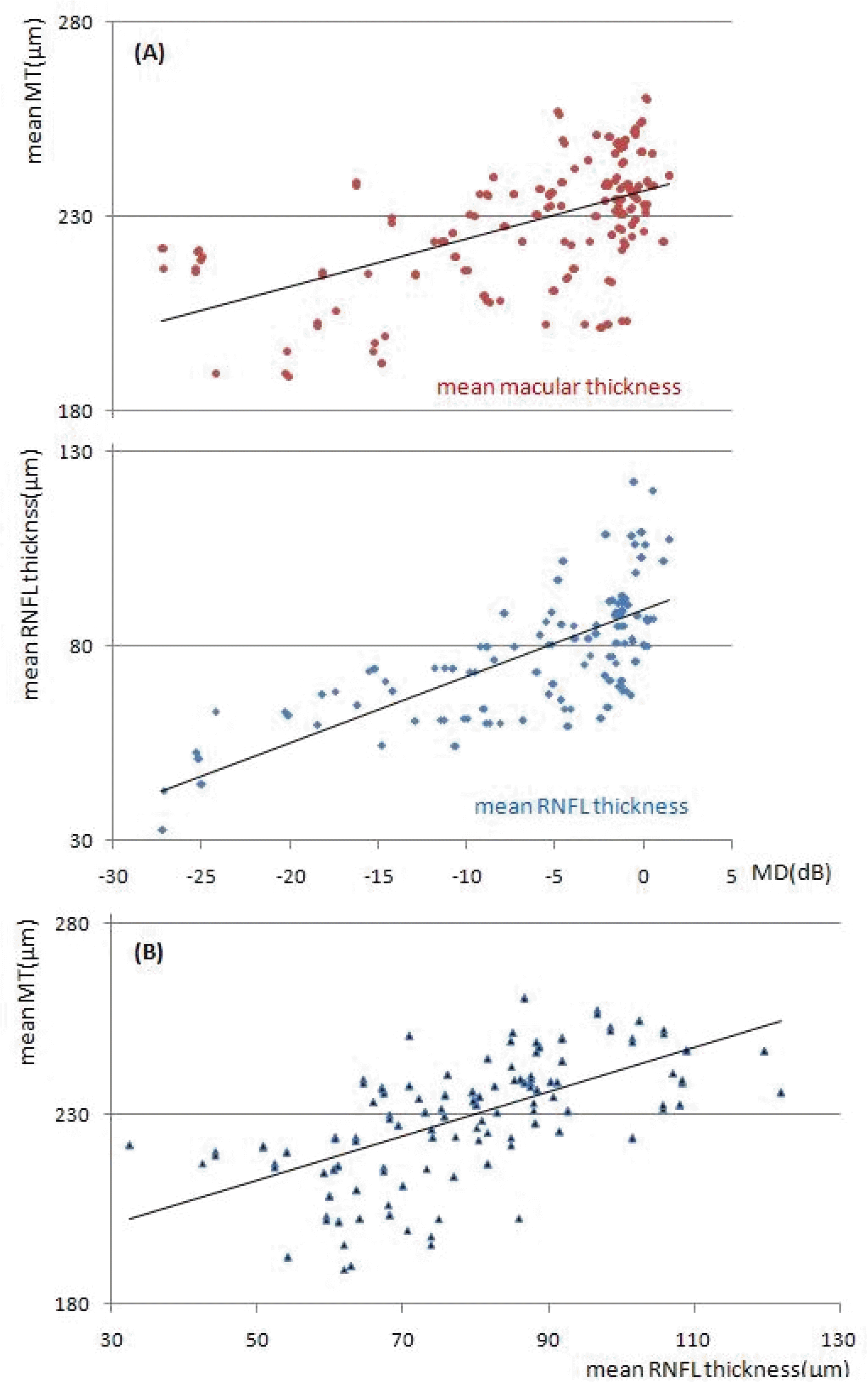
There was a significant decrease of both the macular (p<0.05) and peripapillary RNFL thickness (p<0.001) in the open angle glaucoma group compared with the normal group. In 190 eyes, statistically significant positive relationship was demonstrated between mean deviation (MD) and all areas of peripapillary RNFL thickness (p<0.001) as well as between MD and all areas of macular thickness except the fovea, central ring (p<0.01).
Go to : 
References
1. Quigley HA, Addicks EM, Green WR. Optic nerve damage in human glaucoma. III. Q uantitative correlation of nerve fiber loss and visual field defect in glaucoma, ischemic neuropathy, papilledema, and toxic neuropathy. Arch Ophthalmol. 1982; 100:135–46.
2. Quigley HA, Miller NR, George T. Clinical evaluation of nerve fiber layer atrophy as an indicator of glaucomatous optic nerve damage. Arch Ophthalmol. 1980; 98:1564–71.

3. Sommer A, Miller NR, Pollack I, et al. The nerve fiber layer in the diagnosis of glaucoma. Arch Ophthalmol. 1977; 95:2149–56.

4. Quigley HA, Dunkelberger GR, Green WR. Retinal ganglion cell atrophy correlated with automated perimetry in human eyes with glaucoma. Am J Ophthalmol. 1989; 107:453–64.

5. Harwerth RS, Carter Dawson L, Shen F, et al. Ganglion cell losses underlying visual field defects from experimental glaucoma. Invest Ophthalmol Vis Sci. 1999; 40:2242–50.
6. Zeimer R, Asrani S, Zou S, et al. Quantitative detection of glaucomatous damage at the posterior pole by retinal thickness mapping. A pilot study. Ophthalmology. 1998; 105:224–31.
7. Greenfield DS, Bagga H, Knighton RW. Macular thickness changes in glaucomatous optic neuropathy detected using optical coherence tomography. Arch Ophthalmol. 2003; 121:41–6.

8. Medeiros FA, Zangwill LM, Bowd C, et al. Evaluation of retinal nerve fiber layer, optic nerve head, and macular thickness measurements for glaucoma detection using optical coherence tomography. Am J Ophthalmol. 2005; 139:44–55.

9. Izatt JA, Hee MR, Swanson EA, et al. Micrometer-scale resolution imaging of the anterior eye in vivo with optical coherence tomography. Arch Ophthalmol. 1994; 112:1584–9.

11. Baumann M, Gentile RC, Liebmann JM, et al. Reproducibility of retinal thickness measurements in normal eyes using optical coherence tomography. Ophthalmic Surg Lasers. 1998; 29:280–5.

12. Schuman JS, Pedut-Kloizman T, Hertzmark E, et al. Reproducibility of nerve fiber layer thickness measurements using optical coherence tomography. Ophthalmology. 1996; 103:1889–98.

13. Hee MR, Izatt FA, Swanson EA, et al. Optical coherence tomography of the human retina. Arch Ophthalmol. 1995; 113:325–32.

14. Schuman JS, Hee MR, Puliafito CA, et al. Quantification of nerve fiber layer thickness in normal and glaucomatous eyes using optical coherence tomography. Arch Ophthalmol. 1995; 113:586–96.

15. Blumenthal EZ, Williams JM, Weinreb RN, et al. Reproducibility of nerve fiber layer thickness measurements by use of optical coherence tomography. Ophthalmology. 2000; 107:2278–82.
16. Zangwill LM, Wiliams J, Berry CC, et al. A comparison of optical coherence tomography and retinal nerve fiber layer-photography for detection of nerve fiber layer damage in glaucoma. Ophthalmology. 2000; 107:1309–15.
17. Hoh ST, Greenfield DS, Mistlberger A, et al. Optical coherence tomography and scanning laser polarimetry in normal, ocular hypertensive, and glaucomatous eyes. Am J Ophthalmol. 2000; 129:129–35.

18. Bowd CA, Zangwill LM, Berry CC, et al. Detecting early glaucoma by assessment of retinal nerve fiber layer thickness and visual function. Invest Ophthalmol Vis Sci. 2001; 42:1993–2003.
19. El Beltagi TA, Bowd C, Boden C, et al. Retinal nerve fiber layer thickness measured with optical coherence tomography is related to visual function in glaucomatous eyes. Ophthalmology. 2003; 110:2185–91.

20. Kim YN, Kang JH, Kim JS, et al. Correlation between retinal nerve fiber layer thickness and visual field in normal tension glaucoma. J Korean Ophthalmol Soc. 2005; 46:1532–9.
21. Ma KT, Lee SH, Hong S, et al. Relationship between the retinal thickness analyzer and the GDx VCC scanning laser polarimeter, Stratus OCT optical coherence tomograph, and Heidelberg retina tomograph II confocal scanning laser ophthalmoscopy. Korean J Ophthalmol. 2008; 22:10–7.

22. Paunescu LA, Schman JS, Price LL, et al. Reproducibility of nerve fiber thickness, macular thickness, and optic nerve head measurements using stratus OCT. Invest Ophthalmol Vis Sci. 2004; 45:1716–24.
23. Hess DB, Asrani SG, Bhide MG, et al. Macular and retinal nerve fiber layer analysis of normal and glaucomatous eyes in children using optical coherence tomography. Am J Ophthalmol. 2005; 139:509–17.

24. Wollstein G, Schman JS, Price LL, et al. Optical coherence tomography macular and peripapillary retinal nerve fiber layer measurements and automated visual fields. Am J Ophthalmol. 2004; 138:218–25.
25. Leung CK, Chan WM, Yung WH, et al. Comparison of macular and peripapillary measurements for the detection of glaucoma. an optical coherence tomography study. Ophthalmology. 2005; 112:391–400.
26. Bagga H, Greenfield DS, Knighton RW. Macular symmetry testing for glaucoma detection. J Glaucoma. 2005; 14:358–63.

27. Lederer DE, Schuman JS, Hertzmark E, et al. Analysis of macular volume in normal and glaucomatous eyes using optical coherence tomography. Am J Ophthalmol. 2003; 135:838–43.

28. Mrugacz M, Bakunowicz-Lazarczyk A, Sredzinska-Kita D. Use of optical coherence tomography in myopia. J Pediatr Ophthalmol Strabismus. 2004; 41:159–61.

29. Asrani S, Zou S, d'Anna S, et al. Noninvasive mapping of the normal retinal thickness at the posterior pole. Ophthalmology. 1999; 106:269–73.
Go to : 
 | Figure 1.(A) Relationship between the mean deviation (MD) (dB) and the mean peripapillary retinal nerve fiber layer (RNFL) thickness (μm), n=190; Pearson correlation coefficient=0.554 (p <0.001), and relationship between the mean deviation (MD) (dB) and the mean macular thickness (μm), n=190; Pearson correlation coefficient=0.513 ( p <0.001). (B) Relationship between the mean macular thickness (μm) and the mean peripapillary retinal nerve fiber layer (RNFL) thickness (μm), n=190; Pearson correlation coefficient=0.513 (p <0.001); *MD=mean deviation; † dB=decibel; ‡ RNFL=retinal nerve fiber layer. |
Table 1.
Clinical characteristics of the subjects
| Normal (n=40 eyes) | OHTП(n=30) | Pre-perimetric (n=30) | Early defect (n=30) | Moderate defect (n=30) | Severe defect (n=30) | p* | |
|---|---|---|---|---|---|---|---|
| Age (yrs) (mean± SD) | 58.7±7.2 | 58.9±7.8 | 59.4±8.5 | 61.4±6.6 | 61.1±9.0 | 60.5±8.0 | 0.282 |
| Sex (M:F) | 23:17 | 14:16 | 17:13 | 16:14 | 18:12 | 18:12 | 0.571 |
| MD‡(mean± SD) | -1.21±1.49 | -0.90±0.91 p=0.458† | -0.99±1.07 p=0.607† | -3.61±1.75 p<0.001† | -9.25±1.67 p<0.001† | -19.32±4.89 p<0.001† | <0.001 |
| PSD§(mean± SD) | 1.87±1.13 | 2.28±1.34 p=0.301† | 2.52±1.36 p=0.106† | 4.51±1.69 p<0.001† | 9.25±3.28 p<0.001† | 12.1±1.95 p<0.001† | <0.001 |
Table 2.
The peripapillary retinal nerve fiber layer thickness of the study populations
| Peri- papillary RNFL‡ thickness | Normal (n=40 eyes) | OHT§(n=30) | Pre-perimetric (n=30) | Early defect (n=30) | Moderate defect (n=30) | Severe defect (n=30) | p* |
|---|---|---|---|---|---|---|---|
| Superior (µm) | 114.6±20.9 | 110.5±15.7 p=0.245† | 105.6±15.5 p=0.153† | 86.9±15.6 p<0.001† | 82.3±12.6 p<0.001† | 76.4±19.3 p<0.001† | <0.001 |
| Inferior (µm) | 124.2±18.8 | 119.8±14.8 p=0.352† | 99.6±12.2 p<0.001† | 91.8±20.4 p<0.001† | 71.5±19.9 p<0.001† | 61.5±20.8 p<0.001† | <0.001 |
| Nasal (µm) | 69.1±14.1 | 67.9±10.5 p=0.456† | 57.9±13.6 p<0.001† | 60.6±10.7 p=0.028† | 70.8±12.5 p<0.001† | 50.1±9.1 p<0.001† | <0.001 |
| Temporal (µm) | 70.1±7.4 | 67.4±9.3 p=0.273† | 61.9±12.4 p=0.011† | 57.9±13.6 p<0.001† | 52.2±11.9 p=0.035† | 48.1±14.1 p<0.001† | <0.001 |
| Mean (µm) | 94.5±12.4 | 90.4±11.2 p=0.237† | 77.1±19.1 p=0.001† | 74.6±9.2 p<0.001† | 70.1±9.5 p<0.001† | 59.3±11.6 p<0.001† | <0.001 |
Table 3.
The macular thickness in normal, OHT, pre-perimetric glaucoma, open angle glaucoma (early, moderate, severe defect) groups
| Macular thickness (MT)(µm) | Normal (n=40 eyes) | OHT§(n=30) | Pre-perimetric (n=30) | Early defect (n=30) | Moderate defect (n=30) | Severe defect (n=30) | p* |
|---|---|---|---|---|---|---|---|
| fovea (µm) | 157.5±19.7 | 152.1±14.2 p=0.436‡ | 156.5±23.1 p=0.881‡ | 152.7±18.9 p=0.221‡ | 154.9±16.1 p=0.472‡ | 151.0±17.9 p=0.278‡ | 0.597 |
| 1 mm (µm) | 192.1±19.5 | 182.7±15.2 p=0.181‡ | 187.9±16.7 p=0.485‡ | 176.9±14.2 p=0.005† | 178.4±13.2 p=0.014† | 177.6±13.7 p=0.011† | 0.006 |
| Superior (3 mm)(µm) | 268.2±11.9 | 263.6±12.2 p=0.250‡ | 259.8±10.3 p=0.027† | 245.9±15.6 p<0.001† | 256.1±16.1 p=0.006† | 235.9±34.2 p<0.001† | <0.001 |
| Inferior (3 mm)(µm) | 268.2±12.1 | 263.8±18.1 p=0.292‡ | 250.9±10.3 p<0.001† | 247.5±19.3 p<0.001† | 245.4±12.5 p<0.001† | 224.0±17.3 p<0.001† | <0.001 |
| Nasal (3 mm)(µm) | 272.4±12.5 | 265.9±11.4 p=0.143‡ | 258.1±12.3 p=0.001† | 250.0±17.9 p<0.001† | 259.8±14.1 p<0.001† | 243.6±26.9 p<0.001† | <0.001 |
| Temporal (3 mm)(µm) | 258.4±12.4 | 254.4±16.7 p=0.349‡ | 252.3±11.1 p=0.126† | 241.5±15.1 p<0.001† | 236.5±14.5 p<0.001† | 224.6±14.5 p<0.001† | <0.001 |
| Superior (6 mm)(µm) | 235.9±14.0 | 237.8±12.0 p=0.736‡ | 225.1±14.8 p=0.023† | 218.5±15.5 p<0.001† | 216.4±14.0 p<0.001† | 207.8±8.8 p<0.001† | <0.001 |
| Inferior (6 mm)(µm) | 220.9±11.8 | 222.0±18.1 p=0.785‡ | 202.4±16.1 p=0.023† | 200.3±14.3 p<0.001† | 197.5±11.5 p<0.001† | 185.9±14.2 p<0.001† | <0.001 |
| Nasal (6 mm)(µm) | 250.0±13.8 | 248.5±11.4 p=0.763‡ | 233.5±15.7 p=0.001† | 231.1±19.4 p<0.001† | 230.1±13.5 p<0.001† | 205.7±26.2 p<0.001† | <0.001 |
| Temporal (6 mm)(µm) | 219.5±12.6 | 217.7±14.1 p=0.785‡ | 207.7±12.2 p=0.005† | 204.0±10.1 p<0.001† | 201.3±10.1 p<0.001† | 186.8±11.5 p<0.001† | <0.001 |
| Mean MT (µm) | 242.8±10.1 | 238.6±11.3 p=0.349‡ | 230.9±10.9 p=0.001† | 224.0±14.1 p<0.001† | 224.6±9.8 p<0.001† | 210.2±15.1 p<0.001† | <0.001 |
Table 4.
Correlation between MD, PSD and macular thickness in 190 eyes
| Macular thickness | Pearson correlation coefficient (MD†) | Pearson correlation coefficient (PSD‡) |
|---|---|---|
| fovea | 0.048 (p=0.635*) | -0.010 (p=0.917*) |
| 1 mm | 0.009 (p=0.933*) | -0.050 (p=0.610*) |
| Superior (3 mm) | 0.279 (p=0.005*) | -0.422 (p<0.001*) |
| Inferior (3 mm) | 0.353 (p<0.001*) | -0.483 (p<0.001*) |
| Nasal (3 mm) | 0.270 (p=0.005*) | -0.322 (p=0.001*) |
| Temporal (3 mm) | 0.449 (p<0.001*) | -0.599 (p<0.001*) |
| Superior (6 mm) | 0.483 (p<0.001*) | -0.602 (p<0.001*) |
| Inferior (6 mm) | 0.420 (p<0.001*) | -0.507 (p<0.001*) |
| Nasal (6 mm) | 0.363 (p<0.001*) | -0.492 (p<0.001*) |
| Temporal (6 mm) | 0.490 (p<0.001*) | -0.600 (p<0.001*) |
| Mean macular thickness | 0.513 (p<0.001*) | -0.596 (p<0.001*) |
Table 5.
Correlation between MD, PSD and RNFL thickness in 190 eyes
| RNFL thickness | Pearson correlation coefficient (MD†) | Pearson correlation coefficient (PSD‡) |
|---|---|---|
| Superior | 0.579 (p<0.001*) | -0.524 (p<0.001*) |
| Inferior | 0.374 (p<0.001*) | -0.392 (p<0.001*) |
| Nasal | 0.399 (p<0.001*) | -0.291 (p<0.001*) |
| Lateral | 0.485 (p<0.001*) | -0.485 (p<0.001*) |
| Mean RNFL§ | 0.554 (p<0.001*) | -0.492 (p<0.001*) |
Table 6.
Comparison of superior and inferior inner, outer macular thickness, retinal nerve fiber layer thickness in open angle glaucoma 20 eyes with unilateral (superior only) visual field defect
| MT† superior (3 mm)(µm) | MT inferior (3 mm)(µm) | p* | |
|---|---|---|---|
| Superior VF‡ defect(n=20) | 245.70±25.26 | 223.40±14.52 | 0.026 |
| MT superior (6 mm)(µm) | MT inferior (6 mm)(µm) | p* | |
| Superior VF defect(n=20) | 208.30±15.56 | 179.50±10.45 | <0.001 |
| RNFL superior (µm) | RNFL inferior (µm) | p* | |
| Superior VF defect(n=20) | 88.90±9.54 | 65.90±15.95 | 0.001 |


 PDF
PDF ePub
ePub Citation
 Print
Print


 XML Download