

 PDF
PDF ePub
ePub Citation
 Print
Print


Abstract
Purpose
To report the results of strabismus surgery in five patients diagnosed with myasthenia gravis with strabismus.
Case summary
The authors retrospectively analyzed the effect and timing of surgery for patients who developed strabismus after being diagnosed for myasthenia gravis. Cases 1 and 2 were female myasthenia gravis patients, eight and 45 years of age, who underwent surgery after symptoms developed for correction of exotropia at 33 months and ten years after diagnosis, respectively. Case 3 was a 33-year-old male hyperthyroidism patient who had an exotropia operation six years after his strabismus diagnosis. Cases 4 and 5 were a 22– year-old female and a 50-year-old male patient, who underwent surgery for correction of left hypotropia at 14 months and ten months after diagnosis, respectively. Eventually, a total of three cases of horizontal deviation and two cases of vertical deviation had successful outcomes resulting within ten prism diopters. The patients in cases 4 and 5, both of whom had vertical deviations, experienced a short wait time from the date of symptom presentation until they were able to receive surgery. In particular, case 5 developed left hypertropia two months prior to surgery and the strabismus angle increased until six months prior to surgery. However, the hypertropia stabilized afterwards and the patient finally obtained orthophoria after a left inferior rectus advancement operation.
Go to : 
References
1. Vincent A, Willcox N, Hill M, et al. Determinant spreading and immune responses to acetylcholine receptors in myasthenia gravis. Immunol Rev. 1998; 164:157–68.

2. Heckmann JM, Owen EP, Little F. Myasthenia gravis in South Africans: racial differences in clinical manifestations. Neuromuscul Disord. 2007; 17:929–34.
3. Oöpik M, Puksa L, Luus S-M, et al. Clinical and laboratory- reconfirmed myasthenia gravis: a population-based study. Eur J Neurol. 2008; 15:246–52.
4. Mantegazza R, Baggi F, Antozzi C, et al. Myasthenia gravis (MG): epidemiological data and prognostic factors. Ann N Y Acad Sci. 2003; 998:413–23.
5. Zhang X, Yang M, Xu J, et al. Clinical and serological study of myasthenia gravis in HuBei Province, China. J Neurol Neurosurg Psychiatry. 2007; 78:386–90.
6. Emilia-Romagna Study Group on Clinical and Epidemiological Problems in Neurology. Incidence of myasthenia gravis in the Emilia- Romagna region: a prospective multicenter study. Neurology. 1998; 51:255–8.
7. Morris OC, O'Day J. Strabismus surgery in the management of diplopia caused by myasthenia gravis. Br J Ophthalmol. 2004; 88:832.
8. Davidson JL, Rosenbaum AL, McCall LC. Strabismus surgery in patients with myasthenia. J Pediatr Ophthalmol Strabismus. 1993; 30:292–5.
9. Bentley CR, Dawson E, Lee JP. Active management of patients with ocular manifestations of myasthenia gravis. Eye. 2001; 15:18–22.
10. Mein J, Harcourt B. Diagnosis and management of ocular motility disorders. Oxford: Blackwell;1986. p. 324–6.
11. Glaser JS. Clinical ophthalmology. Philadelphia: Lipincott;1988. p. 31–7.
12. Miller NR. Myopathies and disorders of neuromuscular transmission. Walsh and Hoyt's clinical neuro-ophthalmology. 4th ed.St Louis: Mosby;1987. p. 859–60.
13. Acheson JF, Elston JS, Lee JP, Fells P. Extraocular muscle surgery in myasthenia gravis. Br J Ophthalmol. 1991; 75:232–5.
14. Bartley GB, Fatourechi V, Kadrmas EF, et al. Clinical features of Graves' ophthalmopathy in an incidence cohort. Am J Ophthalmol. 1996; 121:284–90.
15. Brodsky MC, Pollock SC, Buckley EG. Neural misdirection in congenital ocular fibrosis syndrome: implications and pathogenesis. J Pediatr Ophthalmol Strabismus. 1989; 26:159–61.
16. Christensen PB, Jensen TS, Tsiropoulos I, et al. Associated autoimmune diseases in myasthenia gravis. A population-based study. Acta Neurol Scand. 1995; 91:192–5.
Go to :
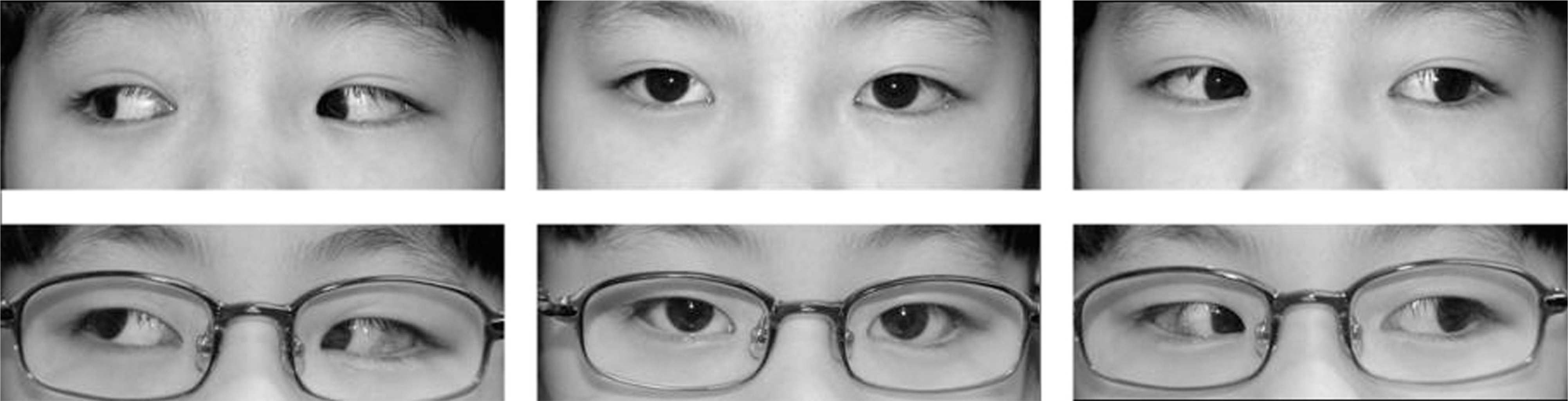 | Figure 1.Preoperative (top) and postoperative (bottom) photographs of patient 1. The patient showed 10ΔX 4ΔRH at 7 months after the surgery. |
 | Figure 2.Preoperative (top) and postoperative (bottom) photographs of patient 2. Note large angle exotropia and adduction deficit in both eyes. The patient showed 2ΔX at 1 month after the surgery. |
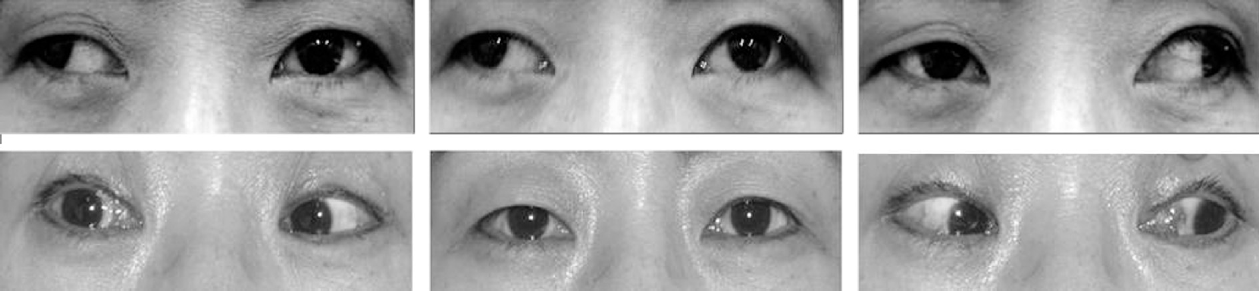
 | Figure 3.Preoperative (top) and postoperative (bottom) photographs of case 3. associated with thyroid-related orbitopathy. Note the pattern like divergence fixus. |
 | Figure 4.Preoperative (top) and postoperative (bottom) photographs of patient 4. The patient showed orthotropia 3 months after the surgery (bottom). |
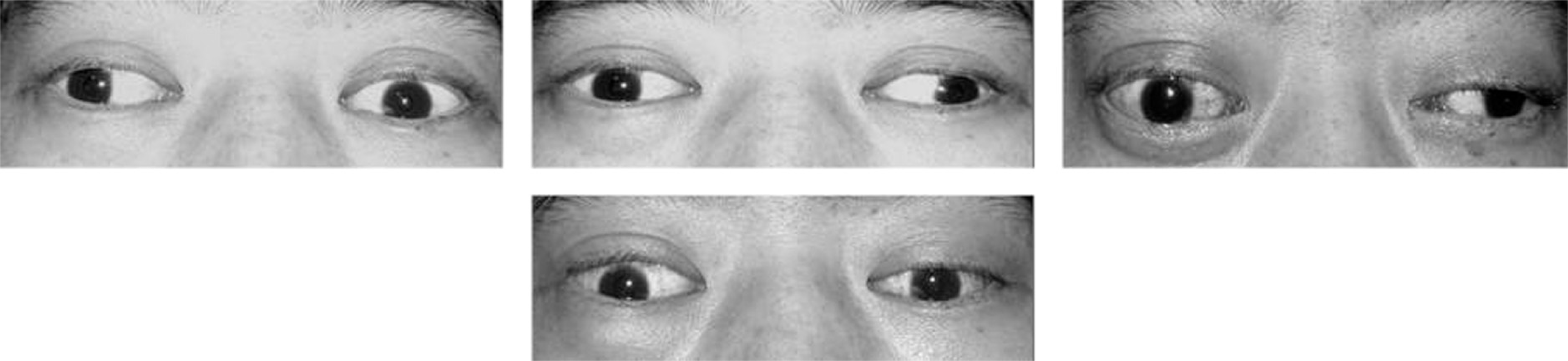
 | Figure 5.Preoperative (a), postoperative (b) and 2nd postoperative (c) photographs of patient 5. The patient showed 12ΔLHT at 2 months after 1st surgery and orthotropia at 1 month after the 2nd surgery in primary gaze. |
 | Figure 6.The patient 6 showed no definite extorsion in both eyes at 2 months after 1 st surgery. |
Table 1.
Patient profiles
| case | Sex/age | Duration (Mon) | Preop deviation | BCVA (OD/OS) | OP name | Postop deviation | Ptosis |
|---|---|---|---|---|---|---|---|
| 1 | F/8 | 33 | 30△ X(T)* 6△ RH‡ | 1.0/0.9 | RLR rec. 7 mm, LLR rec. 7 mm | 10△ X 4△ RH | + |
| 2 | F/45 | 180 | 60△ RXT† 5△ RH | 1.0/1.0 | LROU rec. 8.5 mm/8.5 mm | 6△ X | + |
| RMR res. 5 mm, LMR res. 3 mm | |||||||
| 3 | M/33 | 124 | 60△ LXT | 0.6/0.6 | LROU rec. 9 mm/9 mm | ortho | − |
| 4 | F/22 | 14 | 30△ LHOTΠ | 1.0/0.7 | LIR rec. 4 mm, RSR rec. 6 mm | ortho | + |
| 5 | M/50 | 10 | 30△ RHT§ 10△ X# | 1.0/1.0 | 1st: RSR rec. 6 mm, LIR rec. 4 mm | 18△ LHT | − |
| 18△ LHT | 2nd: LIR adv. 4 mm | ortho |
 XML Download