Abstract
Purpose
To report the case of a child with triple X syndrome presenting with exotropia and chorioretinal coloboma.
Case summary
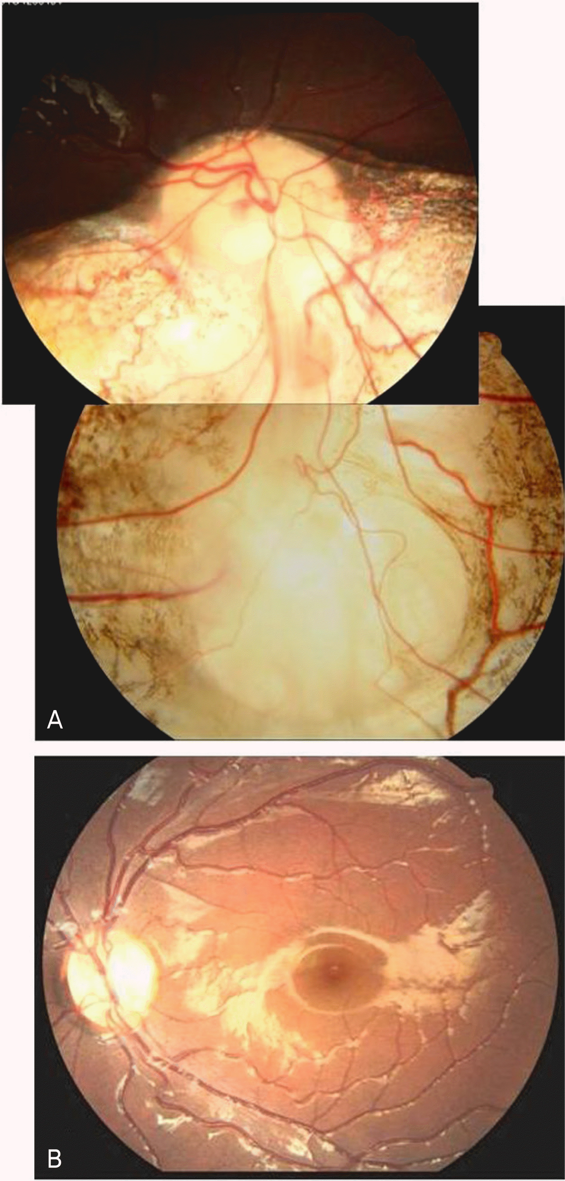
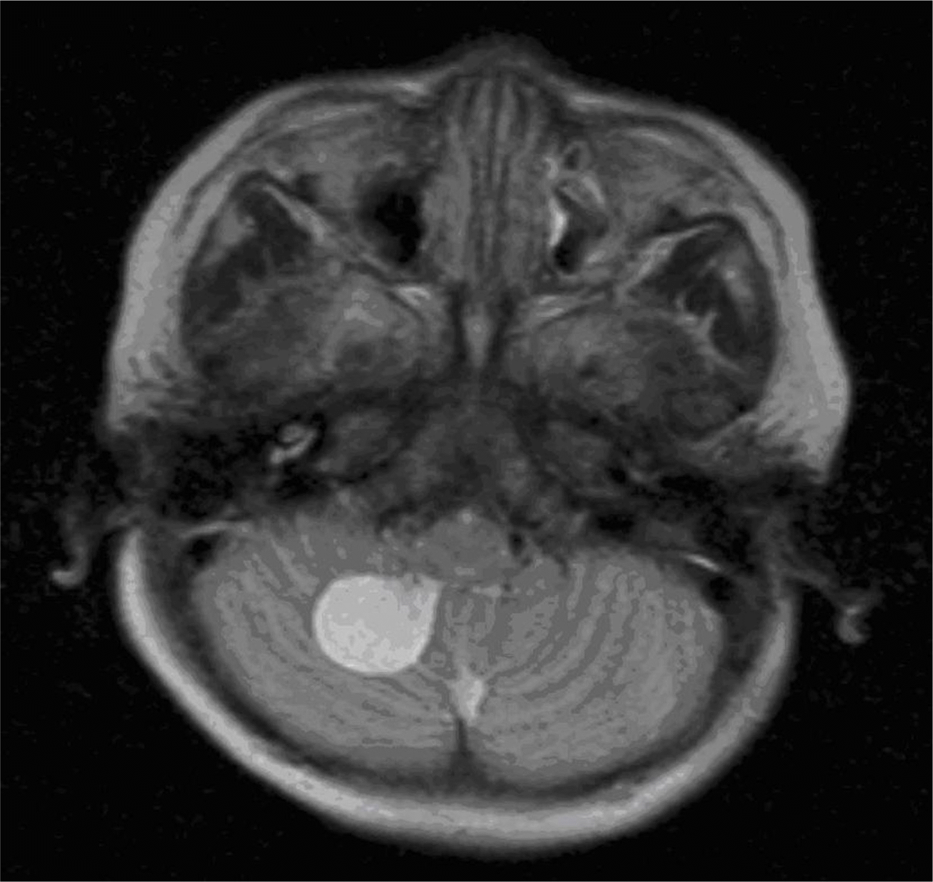
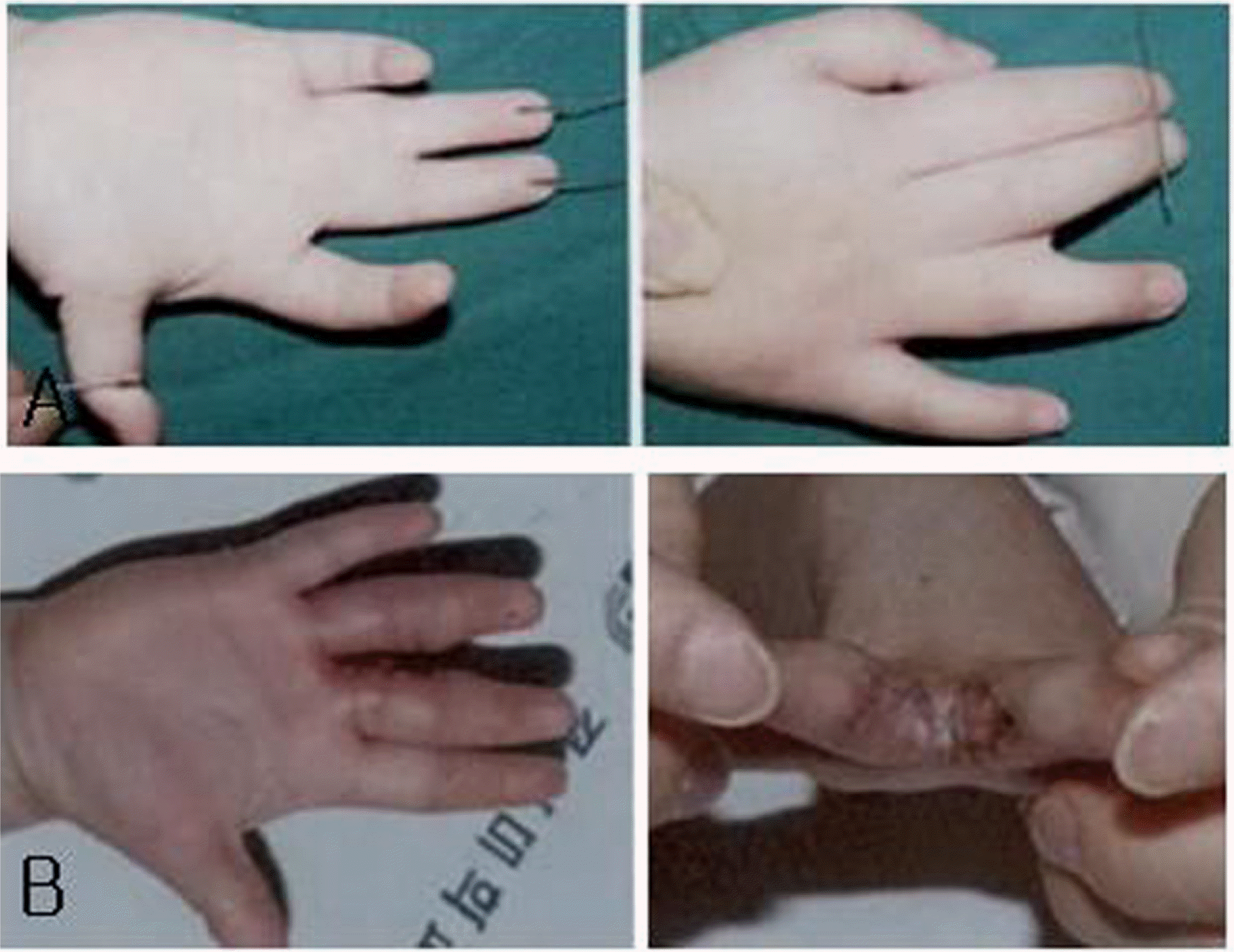
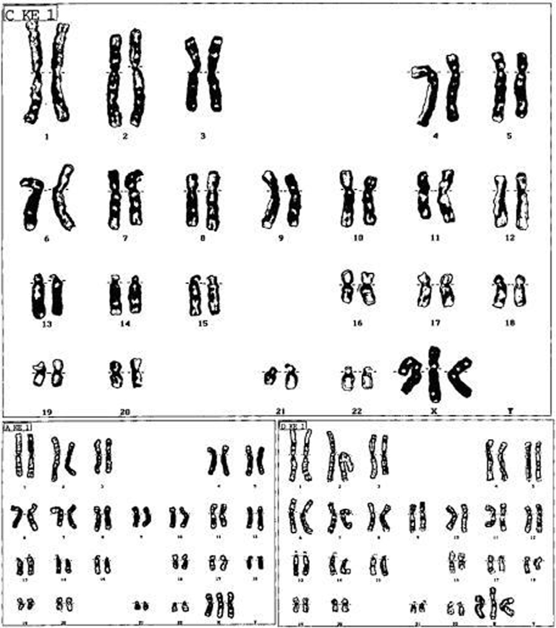
A one-year-old female infant presented with 35PD exotropia in the primary position. The patient had poor fixation of the right eye, and a fundus examination showed chorioretinal coloboma in the inferior region of her right eye. The patient also exhibited syndactyly of the right hand. Brain magnetic resonance imaging revealed a well-defined 2 cm cyst in the right cerebellum. Upon chromosomal study, the patient's karyotype was found to be 47, XXX.
References
1. Duke-Elder S. System of ophthalmology. London: Kimpton;1964. p. 456–87.
2. Kim SY, Lee YW, Koo HM, Chung SK. Chromosomal translocation occuring congenital coloboma in both eye. J Korean Ophthalmol Soc. 1992; 33:1233–7.
3. Moon AR, Moon NJ, Lee WK. A case of treatment of a retinal detachment associated with choroidal coloboma. J Korean Ophthalmol Soc. 1996; 37:1230–5.
4. Myron Y, Ben SF. Ocular pathology. A text and atlas. 2nd ed.Philadelphia: Harper & Row;1982. p. 1424.
5. Ginsberg J, Bove K, Nelson R, Englender GS. Ocular pathology of trisomy 18. Ann Ophthalmol. 1971; 3:273–9.
6. Jagadeesh S, Jabeen G, Bhat L, et al. Triple X Syndrome with Rare Phenotypic Presentation. Indian J Pediatr. 2008; 6:629–31.

7. Castane E, Peris E, Sanchez E. Ocular dysfunction associated with mental handicap. Ophthal Physiol Opt. 1995; 15:489–92.

9. Daufenbach DR, Ruttum MS, Pulido JS, Keech RV. Chorioretinal colobomas in a pediatric population. Ophthalmology. 1998; 8:1455–8.
10. Vaughan D, Asbury T, Tabbara K. General ophthalmology. 12th ed.Los Angels: Lange Medical Publications;1989. p. 13–4.
11. Johnston KM, Nevin NC, Park JM. Cloacal defect in a 23-year-old with 47, XXX karyotype and clinical features of Cat Eye syndrome. Obstet Gynaecol. 2002; 22:696.
12. Hero I, Farjah M, Scholtz CL. The prenatal development of the optic fissure in colobomatous microphthalmia. Invest Ophthalmol Vis Sci. 1991; 32:2622–35.
13. Sakurai E, Shirai S, Ozeki H, Majima A. A case of nonrhegmato-genous retinal detachment in Dandy-Walker Syndrome. Nippon Ganka Gakkai Zasshi. 1996; 10:832–6.
Figure 1.
Fundus photograghs show coloboma of choroids and retina involving optic disc in right eye(A) and normal fundus finging of left eye(B).

Figure 2.
Brain magnetic resonance imaging revealed a 2cm-sized well-defined cyst in the right cerebellum.


 PDF
PDF ePub
ePub Citation
 Print
Print


 XML Download