Abstract
Purpose
Anisometropia can lead to monocular deviation if the refractive error is not corrected. Therefore, the authors evaluated the change in angle deviation after visual acuity improvement by refractive correction in monocular deviated patients with anisometropia.
Methods
Changes in angle deviation were collated retrospectively for 9 patients with anisometric monocular deviation, 7 with monocular exotropia and 2 with monocular esotropia, according to medical records. The patients were admitted for strabismus surgery performed using cataract extraction or clear lens extraction (8 patients) or were treated nonsurgically using contact lenses for visual acuity recovery (1 patient).
Results
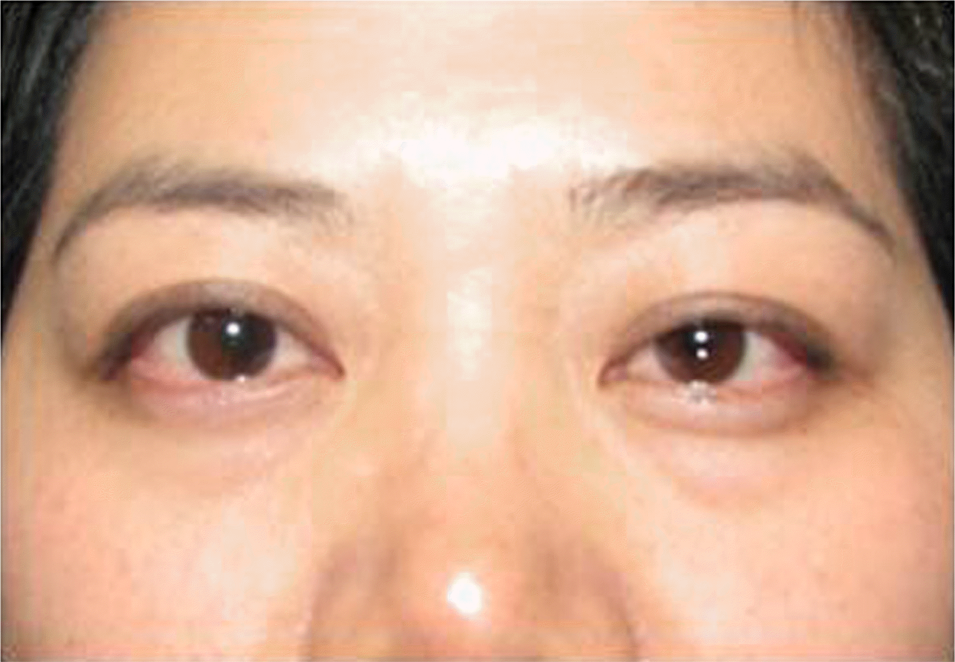
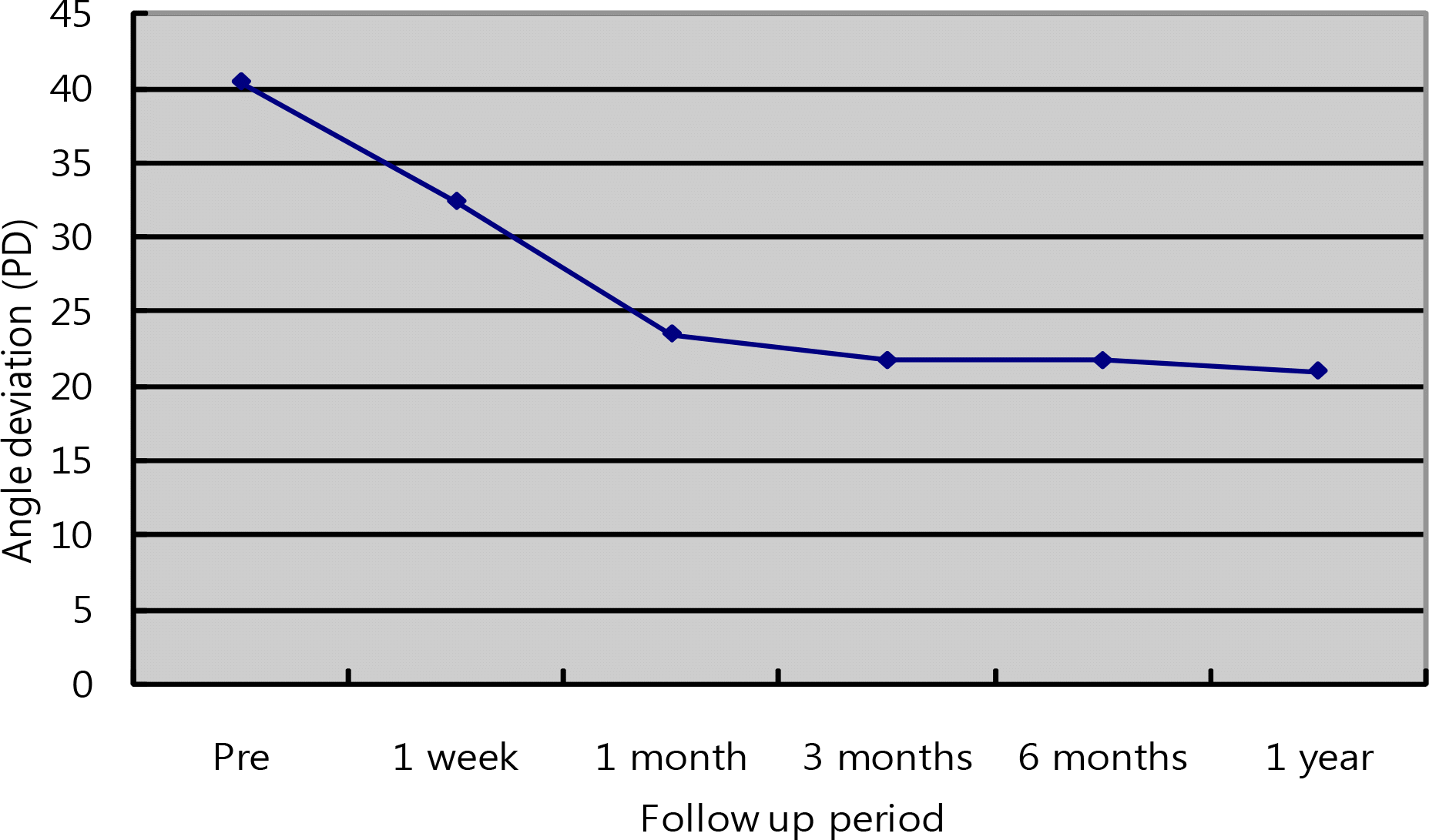
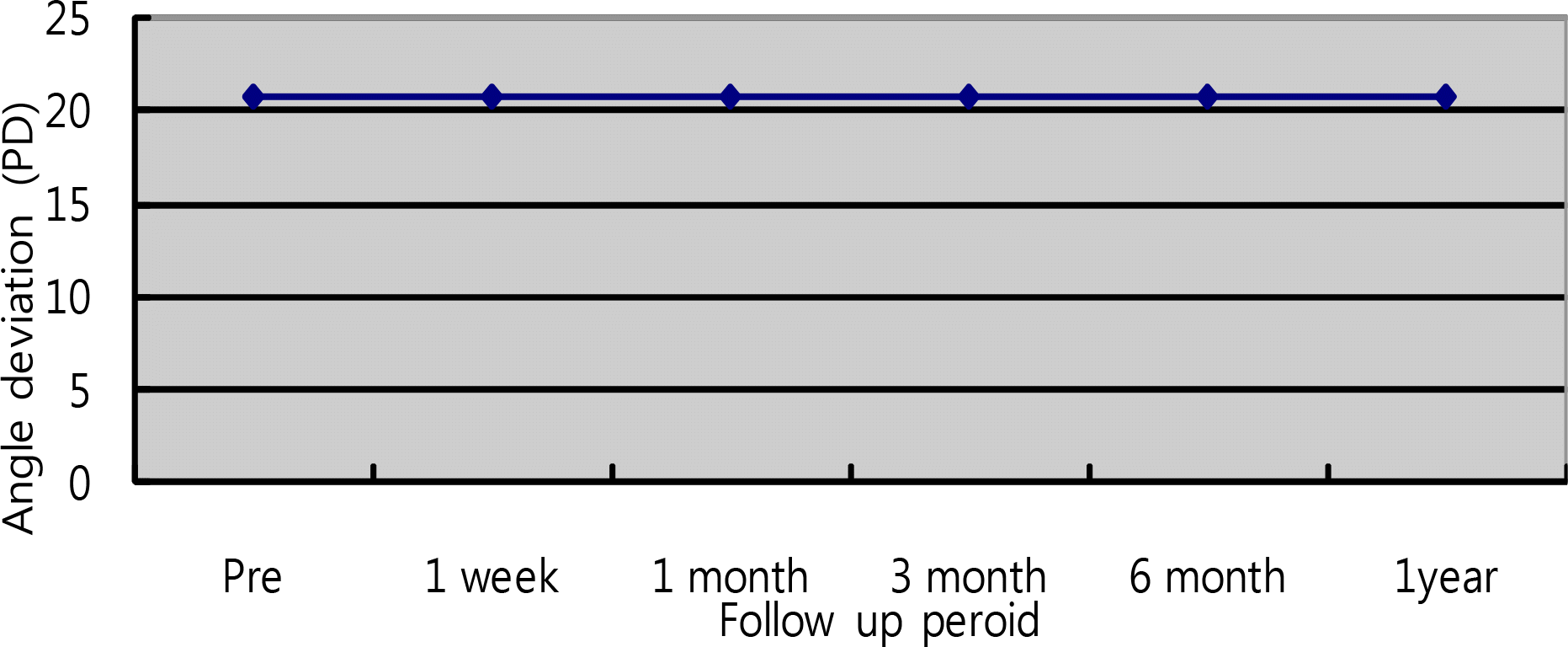
Prior to corrective measures, patients with exotropia had, on average, exodeviation of 40.43 PD, and those with esotropia had, on average, esodeviation of 27.50 PD. After corrective measures were taken, all 7 exotropia patients had decreased angle deviation, and, upon final evaluation, exodeviation had decreased to 21.71 PD, on average. In two exotropia patients, measures taken to correct refractive error shifted the exotropia to exophoria. There was no change in angle deviation after Corrective measures in 2 esotropia patients.
Go to : 
References
1. Abrahamsson M, Fabian G, Sjostrand J. A longitudianl study of a population based sample of astigmatic children, II. The changeability of anisometropia. Acta Ophthalmol. 1990; 68:435–40.
2. Abrahamsson M, Sjostrand J. Natural history of infantile anisometropia. Br J Ophthalmol. 1996; 80:860–3.

3. Sengpiel F, Jirmann KU, Vorobyov V, Eysel UT. Strabismic suppression is mediated by inhibitory interactions in the primary visual cortex. Cereb Cortex. 2006; 16:1750–8.

5. Nucci P, Drack AV. Refractive surgery for unilateral high myopia in children. J AAPOS. 2001; 5:348–51.

6. Autrata R, Rehurek J. Laser-assisted subepithelial keratectomy and photorefractive keratectomy versus conventional treatment of myopic anisometropic amblyopia in children. J Cataract Refract Surg. 2004; 30:74–84.

7. Ali A, Packwood E, Lueder G, Tychsen L. Unilateral lens extraction for high anisometropic myopia in children and adolescents. J AAPOS. 2007; 11:153–8.

8. Godts D, Trau R, Tassignon MJ. Effect of refractive surgery on binocular vision and ocular alignment in patients with manifest or intermittent strabismus. Br J Ophthalmol. 2006; 90:1410–3.

9. Nucci P, Drack AV. Refractive surgery for unilateral high myopia in children. J AAPOS. 2001; 5:348–51.

10. Sorsby A, Leary GA, Richards MJ. The optical components in anisometropia. Vis Res. 1962; 2:43–51.

11. Zadnik K, Satoriano WA, Mutti DO, et al. The effect of parental history of myopia on children's eye size. JAMA. 1994; 271:1323–7.

12. Young TL, Ronan SM, Alvear AB, et al. A second locus for familial high myopia maps to chromosome 12q. Am J Hum Genet. 1998; 63:1419–24.

13. Weiss AH. Unilateral high myopia: optical components, associated factors, and visual outcomes. Br J Ophthalmol. 2003; 87:1025–31.

14. Hussein MA, Coats DK, Muthialu A, et al. Risk factors for treatment failure of anisometropic amblyopia. J AAPOS. 2004; 8:429–34.

15. Helveston EM. Relationship between degree of anisometropia and depth of amblyopia. Am J Ophthalmol. 1966; 62:757–9.

16. Brooks SE, Johnson D, Fischer N. Anisometropia and binocularity. Ophthalmology. 1996; 103:1139–43.

17. McMullen WH. Some points in anisometropia in discussion on problem in refraction. Trans Ophthalmol Soc U K. 1939; 59:119.
18. Kivlin JD, Flynn JT. Therapy of anisometropic amblyopia. J Pediatr Ophthalmol Strabismus. 1981; 18:47–56.

19. Holmes JM, Kraker RT, Beck RW, et al. A randomized trial of prescribed patching regimens for treatment of severe amblyopia in children. Ophthalmology. 2003; 110:2075–87.

20. Nemet P, Levinger S, Nemet A. Refractive surgery for refractive errors which cause strabismus. Binocul Vis Strabismus Q. 2002; 17:187–90.
21. Hoyos JE, Cigales M, Maldonado-bas , et al. Hyperopic LASIK may work well in refractive accommodative esotropia. Ocular Surg News. 1998; 16:93–4.
22. Haw WW, Alcorn DM, Manche EE. Excimer laser refractive surgery in the pediatric population. J Pediatr Ophthalmol Strabismus. 1999; 36:173–7.

23. Sabetti L, Spadea L, D'Alessandri L, Balestrazzi E. Photorefractive keratectomy and laser in situ keratomileusis in refractive accommodative esotropia. J Cataract Refract Surg. 2005; 31:1899–903.

24. Hubel DH, Wiesel TN. Recptive fields of optic nerve fibers in th spinder monkey. J Physiol. 1960; 154:572–80.
25. Korean association for pediatric ophthalmology and strabismus, Current concepts in strabismus. 2nd ed.Seoul: Naewae Haksool;2008. p. 171–2.
Go to : 
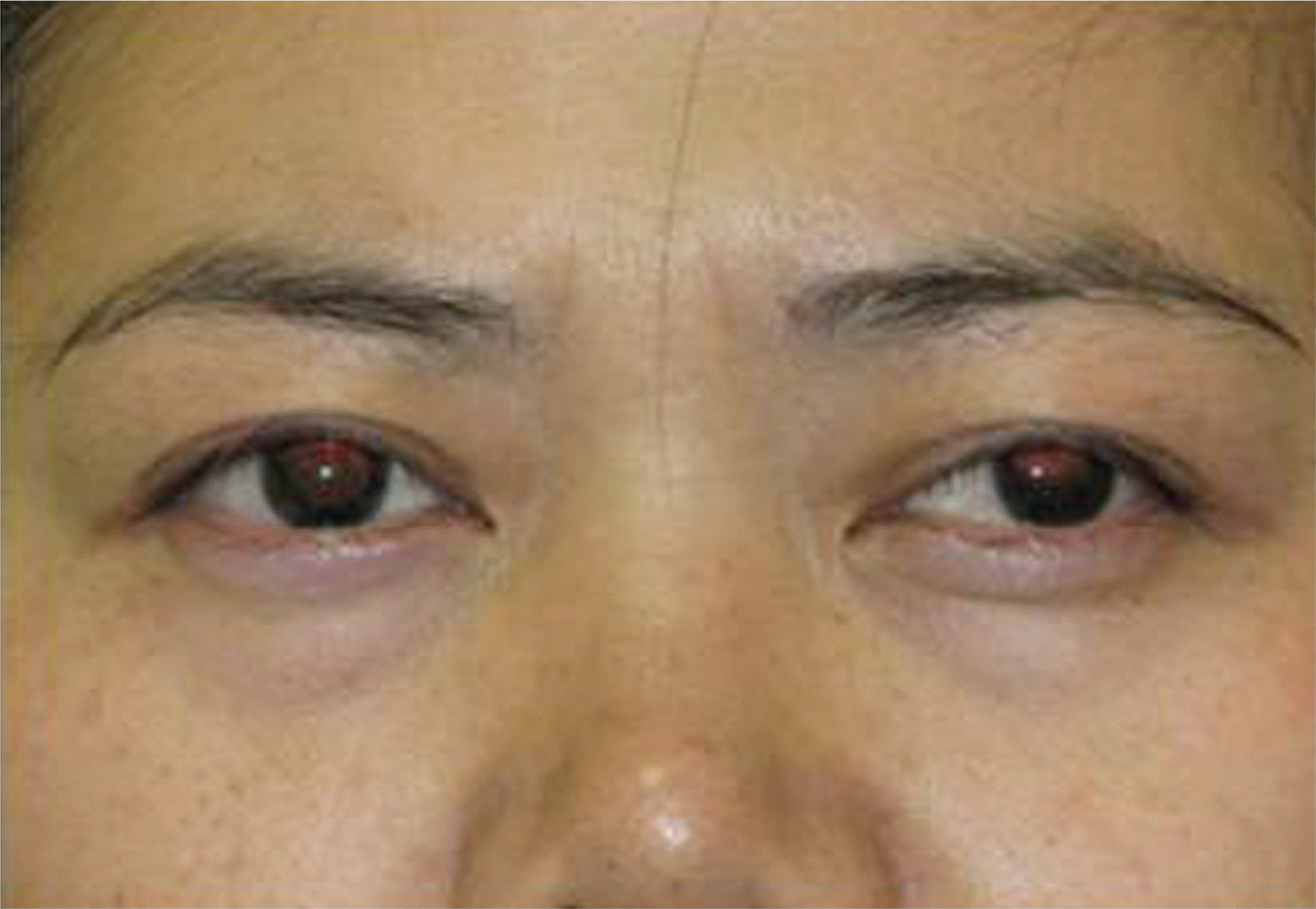
 | Figure 4.Digital photograph of recovered monocular deviation in anisometric patient after surgery. |
Table 1.
Clinical data and angle deviation outcomes in patients who underwent refractive correction


 PDF
PDF ePub
ePub Citation
 Print
Print


 XML Download