

 PDF
PDF ePub
ePub Citation
 Print
Print


Abstract
Purpose
To evaluate the clinical reliability of Pentacam® by comparing anterior chamber parameters measured by Oculus Pentacam system (Oculus Inc., Germany) and Hi-scan ultrasound biomicroscopy (OPTIKON 2000, Rome, Italy) in primary angle closure (PAC) and normal patients.
Methods
A prospective study was performed from June 2006 to January 2007. Fifty-one eyes in 26 primary angle-closure patients and 39 eyes in 20 normal control patients, for a total of 90 eyes of 46 patients were recruited from glaucoma outpatient clinics. The correlation and agreement of both measurements of anterior chamber depth and anterior chamber angle measured by UBM and Pentacam® were calculated. Sensitivity and specificity of each tool were also compared and the diagnostic value of angle closure was examined.
Results
Anterior chamber depth measured by UBM and Pentacam® showed strong correlation in the normal control group (r=0.821) and PAC group (r=0.957). Anterior chamber angle showed moderate correlation in the normal control group (r=0.523) and PAC group (r=0.456) while good agreement was also observed. In diagnosing angle closure, anterior chamber measurements appear similar in UBM and Pentacam® using the ROC curve (AUC of UBM, 0.942; AUC of Pentacam®, 0.931).
Go to : 
References
1. Kim YY, Jung HR. Clarifying the nomenclature for primary angle closure glaucoma. Surv Ophthalmol. 1997; 42:125–36.
2. Ang MH, Baskaran M, Kumar RS, et al. National survey of ophthalmologists in Singapore for the assessment and management of asymptomatic angle closure. J Glaucoma. 2008; 17:1–4.

3. Lavanya R, Wong TY, Friedman DS, et al. Determinants of angle closure in older Singaporeans. Arch Ophthalmol. 2008; 126:686–91.
4. Koranyi G, Lydahl E, Norrby S, Taube M. Anterior chamber depth measurement: A-scan versus optical methods. J Cataract Refract Surg. 2002; 28:243–7.
5. Reddy AR, Pande MV, Finn P, El-Gogary H. Comparative estimation of anterior chamber depth by ultrasonography, Orbscan II, and IOL master. J Cataract Refractive Surg. 2004; 30:1268–71.
6. Vetrugno M, Cardascia N, Cardia L. Anterior chamber depth measured by two methods in myopic and hyperopic phakic IOL implant. Br J Ophthalmol. 2000; 84:1113–6.
7. Foster PJ, Devereux JG, Alsbirk PH. Detection of gonioscopically occludable angles and primary angle closure glaucoma by estimation of limbal chamber depth in Asians: modified grading scheme. Br J Ophthalmol. 2000; 84:186–92.
8. Congdon NG, Spaeth GL, Augsburger J, et al. A proposed simple method for measurement in the anterior chamber angle: biometric gonioscopy. Ophthalmology. 1999; 106:2161–7.
9. Jang JW, Choe YJ, Hong YJ. Measurement of the depth and angle of the peripheral anterior chamber and iris thickness by ultrasonographic biomicroscopy. J Korean Ophthalmol Soc. 1995; 36:1179–84.
10. Lee SC, Jin KH. Changes in Ciliary Sulcus Size, Anterior chamber depth and angle during accommodation using ultrasound biomicroscopy. J Korean Ophthalmol Soc. 2005; 46:1809–14.
11. Elbaz U, Barkana Y, Gerber Y, et al. Comparison of different techniques of anterior chamber depth and keratometric measurements. Am J Ophthalmol. 2007; 143:48–53.
12. Emre S, Doganay S, Yologlu S. Evaluation of anterior segment parameters in keratoconic eyes measured with the Pentacam system. J Cataract Refract Surg. 2007; 33:1708–12.
13. Friedman DS, Gazzard G, Foster P, et al. Ultrasonographic biomicroscopy, Scheimpflug photography, and novel provocative tests in contralateral eyes of Chinese patients initially seen with acute angle closure. Arch Ophthalmol. 2003; 121:633–42.
14. Sihota R, Lakshmaiah NC, Agarwal HC, et al. Ocular parameters in the subgroups of angle closure glaucoma. Clin Exp Ophthalmol. 2000; 28:253–8.
15. Sihota R, Gupta V, Agarwal HC, et al. Comparison of symptomatic and asymptomatic, chronic, primary angle-closure glaucoma, open-angle glaucoma, and controls. J Glaucoma. 2000; 9:208–13.
16. Marchini G, Pagliarusco A, Toscano A, et al. Ultrasound biomicroscopic and conventional ultrasonographic study of ocular dimensions in primary angle-closure glaucoma. Ophthalmology. 1998; 105:2091–8.
17. Chen HB, Kashiwagi K, Yamabayashi S, et al. Anterior chamber angle biometry: quadrant variation, age change and sex difference. Curr Eye Res. 1998; 17:120–4.
18. Salmon JF. Predisposing factors for chronic angle-closure glaucoma. Prog Retin Eye Res. 1999; 18:121–32.
19. Reuland MS, Reuland AJ, Nishi Y, Auffarth GU. Corneal radii and anterior chamber depth measurements using the IOLmaster versus the Pentacam. J Refract Surg. 2007; 23:368–73.
20. Oka N, Otori Y, Okada M, et al. Clinical study of anterior ocular segment topography in angle-closure glaucoma using the three-dimensional anterior segment analyzer Pentacam. Nippon Ganka Gakkai Zasshi. 2006; 110:398–403.
21. Buehl W, Stojanac D, Sacu S, et al. Comparison of three methods of measuring corneal thickness and anterior chamber depth. Am J Ophthalmol. 2006; 141:7–12.
22. Lackner B, Schmidinger G, Skorpik C. Validity and repeatability of anterior chamber depth measurements with Pentacam and Orbscan. Optom Vis Sci. 2005; 82:858–61.
23. Yi JH, Hong S, Seong GJ, et al. Anterior chamber measurements by pentacam and AS-OCT in eyes with normal open angles. Korean J Ophthalmol. 2008; 22:242–5.
24. Yi J, Hong S, Seong GJ, et al. Detection of occludable angles with the Pentacam and the anterior segment optical coherence tomography. Yonsei Med J. (in press).
25. Kunimatsu S, Tomidokoro A, Mishima K, et al. Prevalence of appositional angle closure determined by ultrasonic biomicroscopy in eyes with shallow anterior chambers. Ophthalmology. 2005; 112:407–12.
Go to :
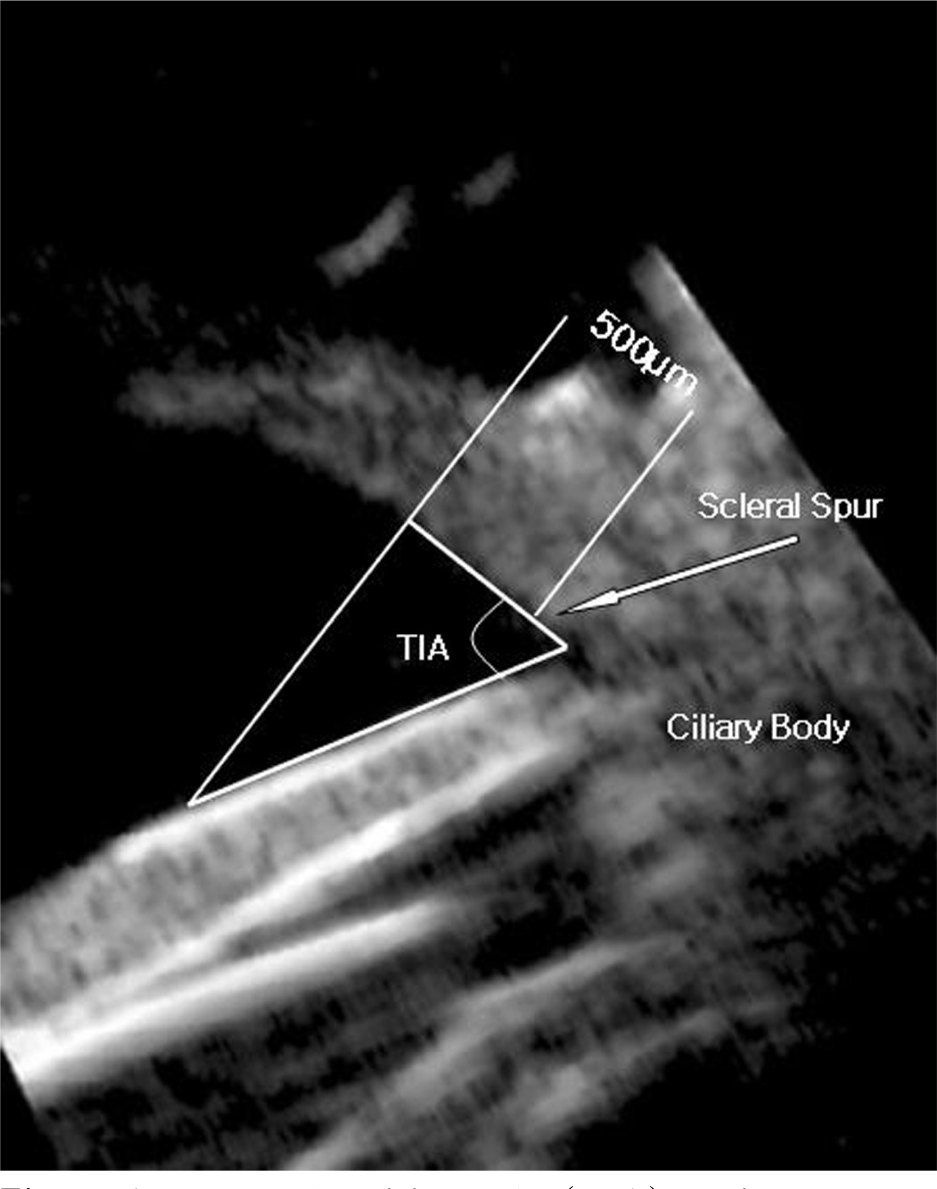 | Figure 1.Trabeculariris angle (TIA) depicted on an ultrasound biomicroscopic (UBM) image of a normal eye. |
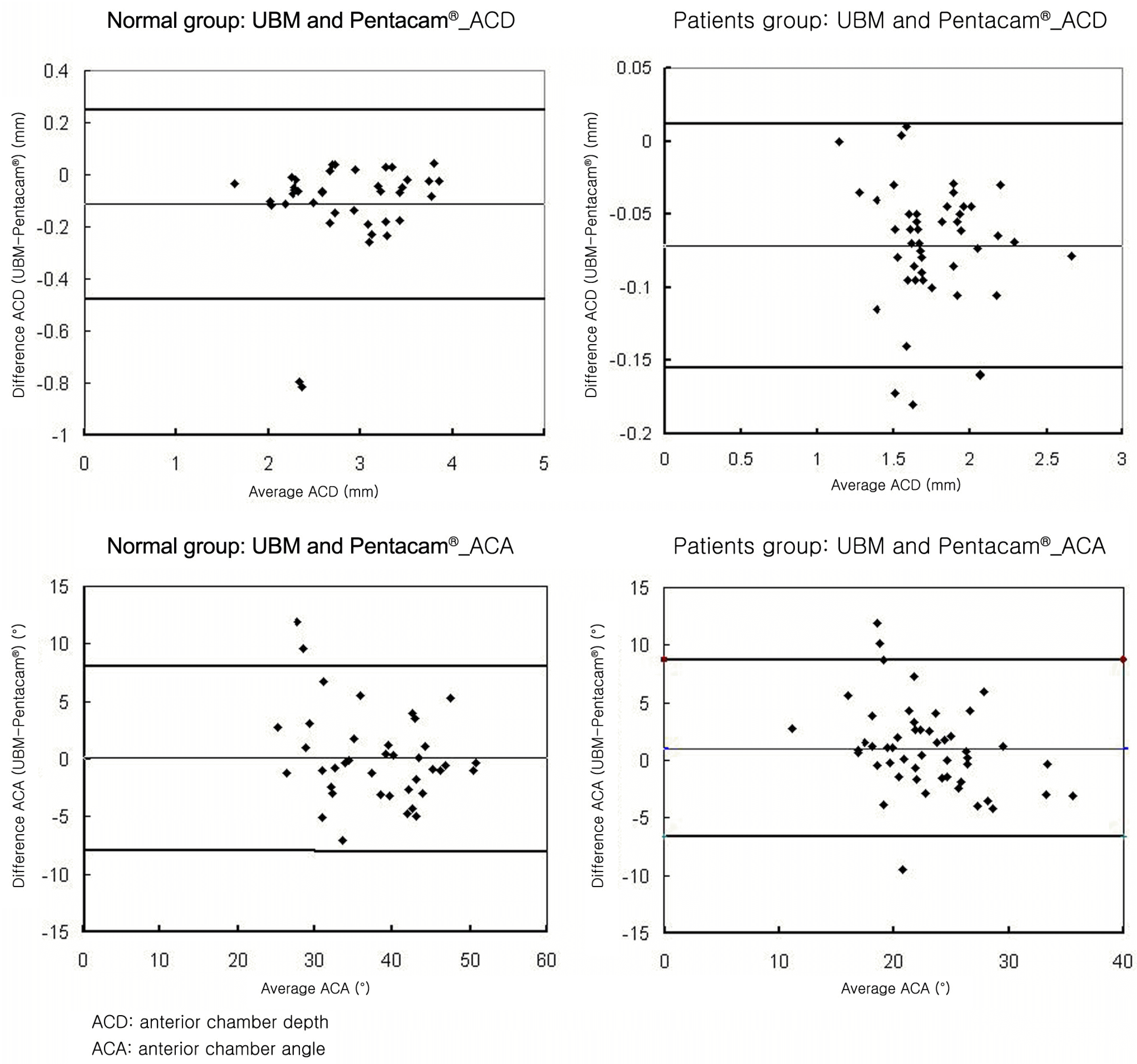 | Figure 2.Bland-Altmann plots of anterior biometry measurements between ultrasound biomicroscopy (UBM) and Pentacam®. Solid line: 95% confidence interval of the difference between the methods. |
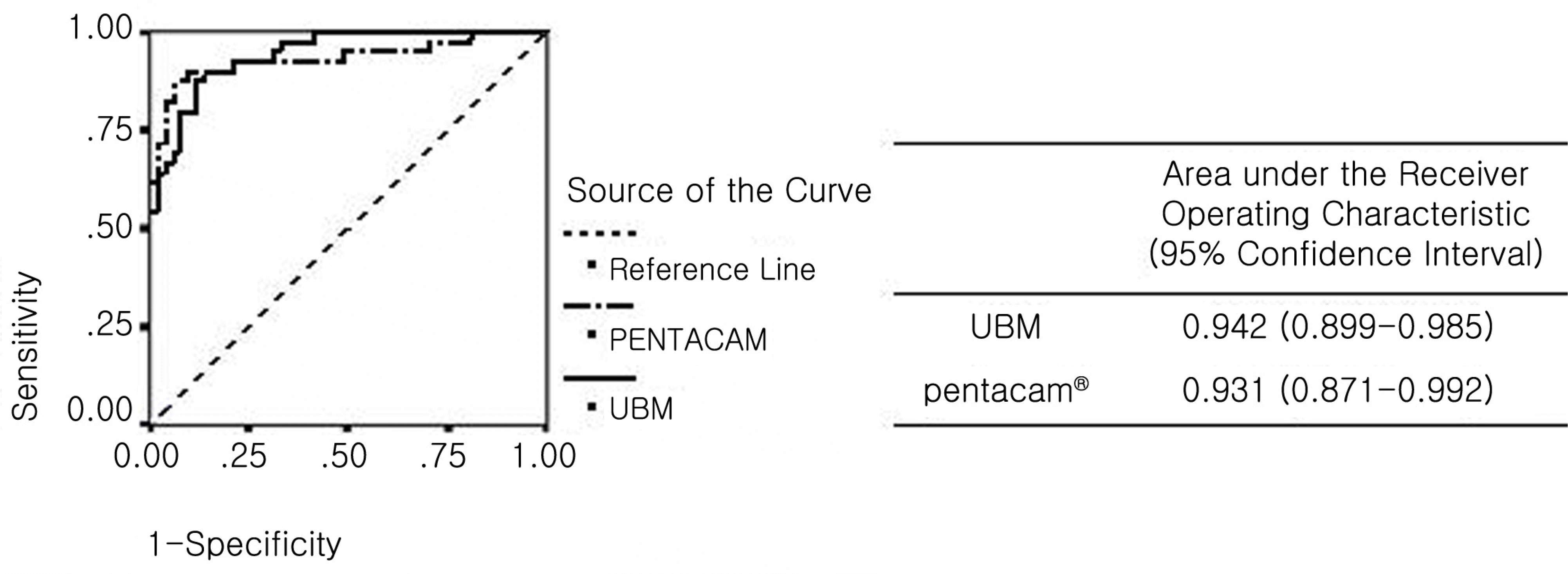 | Figure 3.Comparison of ultrasound biomicroscopy (UBM) and Pentacam® measurements of the anterior chamber angle for the diagnosis of primary angle-closure or primary angle-closure glaucoma. |
Table 1.
Characteristics of the study population
| Age (yrs) |
Sex |
||
|---|---|---|---|
| M | F | ||
| Normal (n=20) | 46.7±21.2 | 10 | 10 |
| Angle closure glaucoma (n=26) | 70.5±7.4 | 21 | 5 |
Table 2.
Correlation analysis between ultrasound biomicroscopy (UBM) and Pentacam® measurements of anterior chamber depth of normal and primary angle-closure patients
|
Anterior chamber depth (mm) |
Pearson's correlation coefficient |
|||
|---|---|---|---|---|
| UBM | Pentacam® | Coefficeint | p-value* | |
| Normal | 2.74±0.63 | 3.00±0.60 | 0.821 | <0.01 |
| Patients | 1.70±0.27 | 1.80±0.30 | 0.957 | <0.01 |
Table 3.
Correlation analysis between ultrasound biomicroscopy (UBM) and Pentacam® measurements of anterior chamber angle of normal and primary angle-closure patients
|
Anterior chamber angle (°) |
Pearson's correlation coefficient |
|||
|---|---|---|---|---|
| UBM | Pentacam® | Coefficeint | p-value* | |
| Normal | 38.00±7.00 | 37.80±8.82 | 0.523 | 0.01 |
| Patients | 24.10±5.50 | 21.38±5.01 | 0.456 | 0.01 |
 XML Download