Abstract
Purpose
To establish the accuracy of the newly released biometer Ocuscan RxP® (Alcon, USA) by comparison with the established Ultrasonic Biometer Model 820® (Allergan Humphrey, USA), and to compare the accuracy of contact and immersion biometries.
Methods
This is a prospective study involving 27 patients (40 eyes) who were scheduled for cataract surgery and had axial lengths measured with an Ocuscan RxP® biometer using both contact (Method 1) and immersion (Method 2) techniques. As a reference, a contact type Ultrasonic biometer 820® (Method 3) was also used. IOL(Intraocular Lens) power for the cataract surgery was calculated
using this result. An axial length which would have caused no post-operative refractive error was reversely calculated from the difference of target diopter and post-operative refractive error. This length was compared with the axial lengths obtained via Methods 1, 2 and 3.
Results
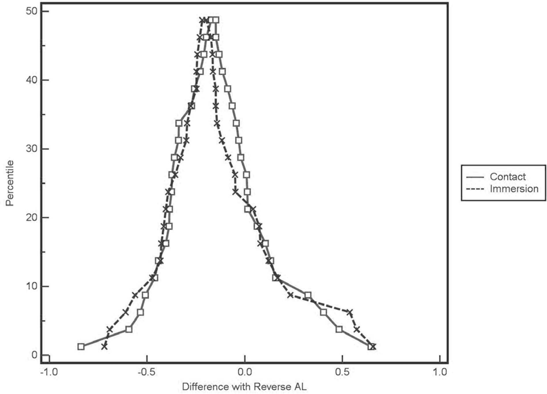
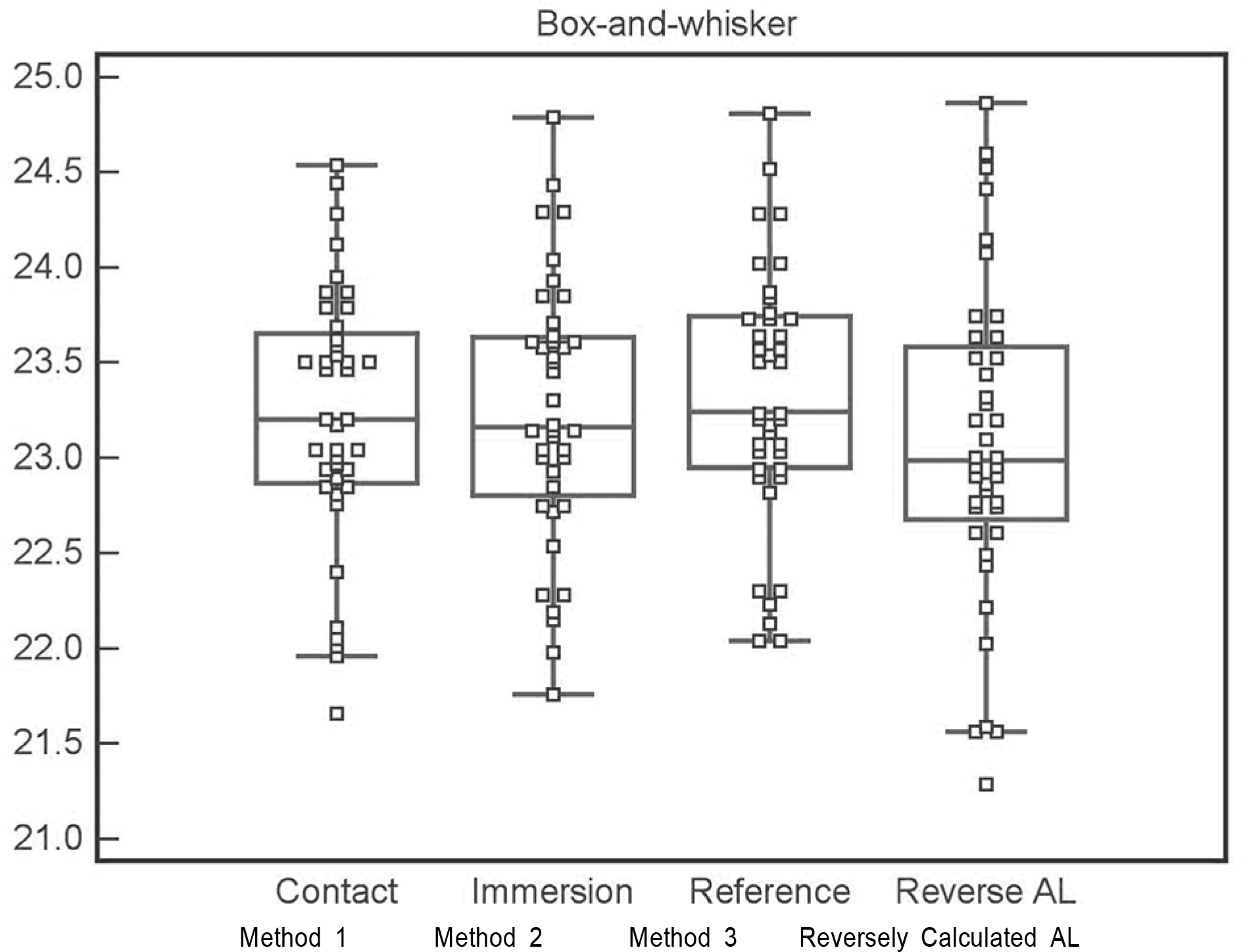
The means and standard deviations for the measurement sets were compared. Methods 1 and 2 showed no significant difference (23.22±0.68, 23.24±0.69 mm, p=0.55). The axial length measured by Method 3 was 23.32±0.67 mm. The difference between the target refraction and post-operative refractive error was 0.29±0.60D. The axial length was reversely calculated from the difference (23.07±0.84 mm). The differences between the reversely calculated axial lengths and those of Methods 1, 2 and 3 were 0.15± 0.31, 0.17±0.31 and 0.24±0.28 mm, respectively.
Go to : 
References
1. Olsen T. Sources of error in intraocular lens power calculation. J Cataract Refract Surg. 1992; 18:125–9.

2. Olsen T. Theoretical approach to intraocular lens calculation using Gaussian optics. J Cataract Refract Surg. 1987; 13:141–5.

3. Leaming DV. Practice styles and preferences of ASCRS members-1999 survey. J Cataract Refract Surg. 2000; 26:913–21.

4. Binkhorst RD. The accuracy of ultrasonic measurement of the axial length of the eye. Ophthalmic Surg. 1981; 12:363–5.

5. Olsen T. The accuracy of ultrasonic determination of axial length in pseudophakic eyes. Acta Ophthalmol. 1989; 67:141–4.

6. Olsen T, Nielsen PJ. Immersion versus contact technique in the measurement of axial length by ultrasound. Acta Ophthalmol. 1989; 67:101–2.

7. Hitzenberger CK, Drexler W, Dolezal C. Measurement of the axial length of cataract eyes by laser Doppler interferometry. Invest Ophthalmol Vis Sci. 1993; 34:1886–93.
8. Findl O, Drexler W, Menapace R, et al. Improved prediction of intraocular lens power using partial coherence interferometry. J Cataract Refract Surg. 2001; 27:861–7.

9. Giers U, Epple C. Comparison of A-scan device accuracy. J Cataract Refract Surg. 1990; 16:235–42.

10. Binkhorst RD. The accuracy of ultrasonic measurement of the axial length of the eye. Ophthalmic Surg. 1981; 12:363–5.

11. Hennessy MP, Franzco , Chan DG. Contact versus immersion biometry of axial length before cataract surgery. J Cataract Refract Surg. 2003; 29:2195–8.

12. Retzlaff JA, Sanders DR, Kraff MC. Development of the SRK /T intraocular lens implant power calculation formula. J Cataract Refract Surg. 1990; 16:333–40.
13. Hoffer KJ. The Hoffer Q Formula: a comparison of theoretic and regression formulas. J Cataract Refract Surg. 1993; 19:700–12.

14. Holladay JT. Standardizing constants for ultrasonic biometry, keratometry, and intraocular lens power calculations. J Cataract Refract Surg. 1997; 23:1356–70.

15. Hwang JS, Lee JH. Comparison of the IOL master(R) and A-scan ultrasound: refractive results of 96 consecutive cases. J Korean Ophthalmol Soc. 2007; 48:27–32.
16. Song BY, Yang KJ, Yoon KC. Accuracy of partial coherence interferometry in intraocular lens power calculation. J Korean Ophthalmol Soc. 2005; 46:775–80.
17. Chung JK, Choe CM, You YS, Lee SJ. Biometry with partial coherence interferometry and ultrasonography in high myopes. J Korean Ophthalmol Soc. 2006; 47:355–61.
18. Cho YA, Lee RH, Kim KS. The axial length of normal emmetropic eyes by ultrasonic biometry. J Korean Ophthalmol Soc. 1983; 24:27–33.
19. Kim WJ, Park SH, Shin HH. The change of axial length according to age in the eyeball of premature infants by ultrasonic biometry. J Korean Ophthalmol Soc. 1983; 34:667–71.
Go to : 
 | Figure 3.Comparison of method 1 (contact) and method 2 (immersion) with reversely calculated axial length. |
Table 2.
Mean axial length and difference with the reversely calculated axial length
| Mean± SD | Difference with the reversely calculated axial length | |
|---|---|---|
| Method 1 (Contact) | 23.22±0.68 | 0.15±0.31 |
| Method 2 (Immersion) | 23.24±0.69 | 0.17±0.31 |
| Method 3 (Reference) | 23.32±0.67 | 0.24±0.28 |


 PDF
PDF ePub
ePub Citation
 Print
Print


 XML Download