Abstract
Purpose
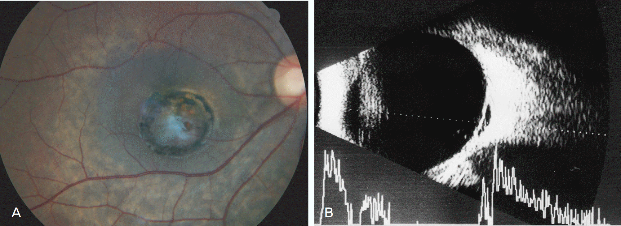
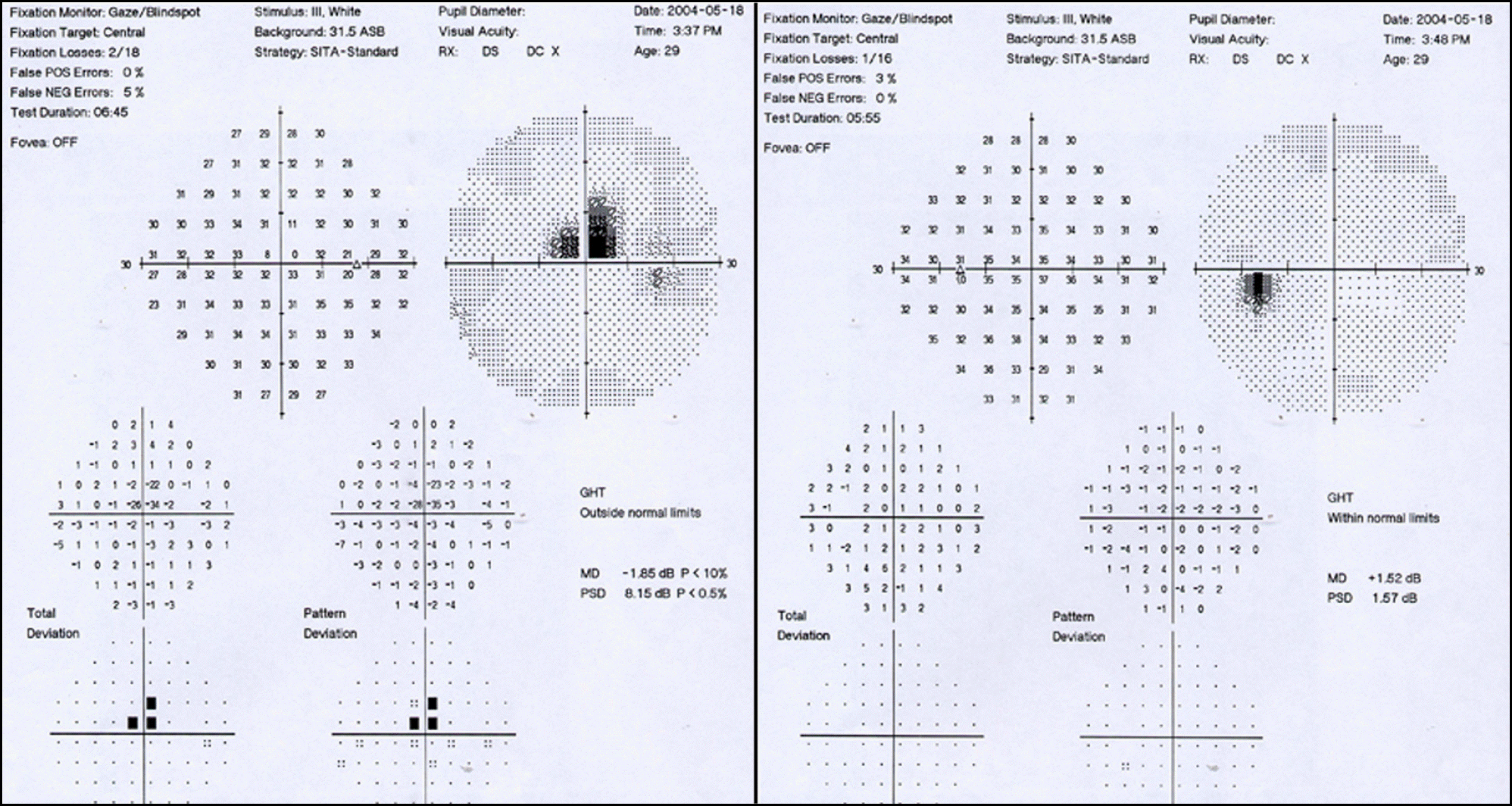
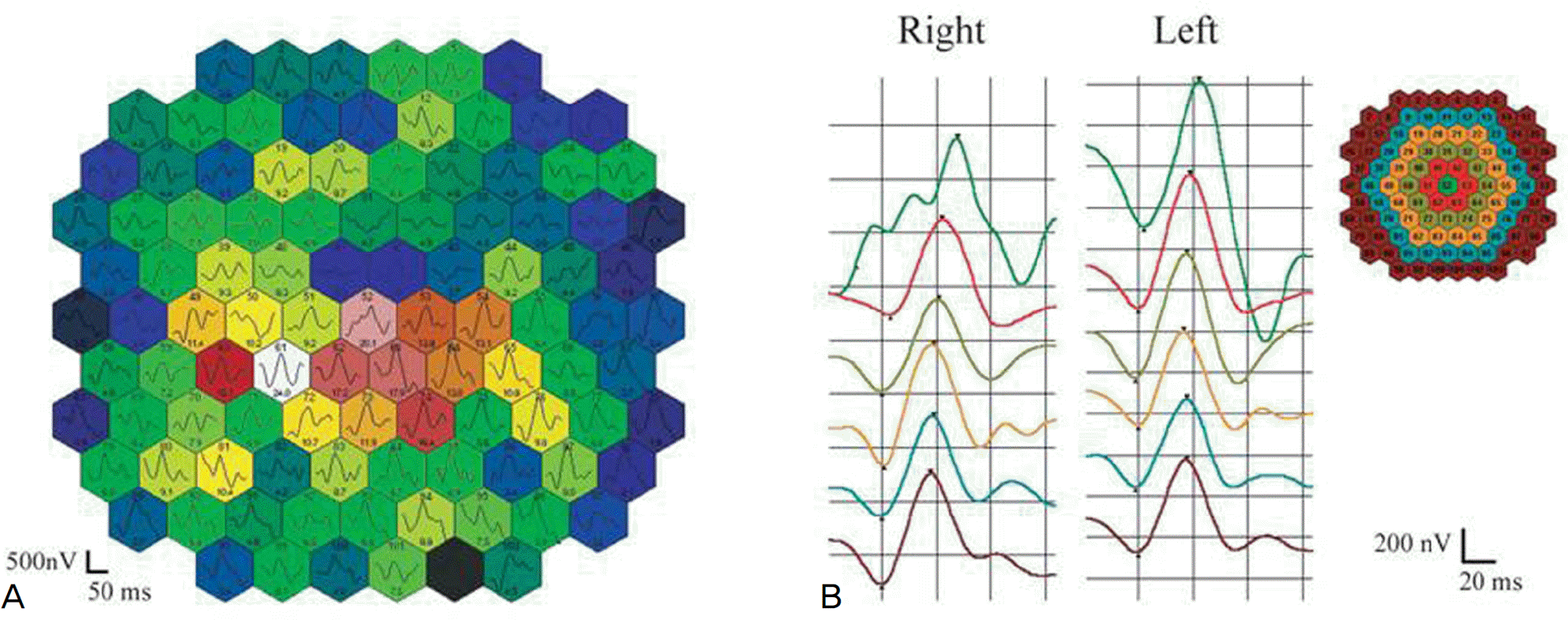
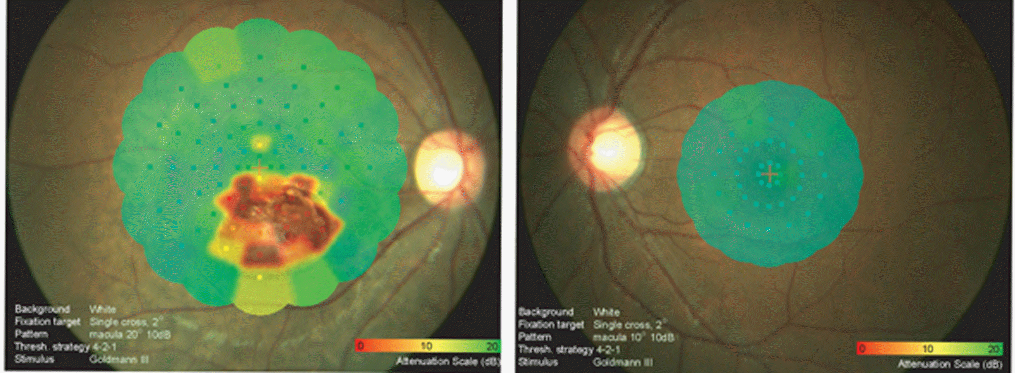
To report a case of isolated choroidal cystic lesion in the macula with no interval change for two years. Case summary: A 29-year-old woman who had suspicious maculopathy was referred to our clinic. Her best corrected visual acuity was 20/20 in the affected right eye, which showed a choroidal cystic lesion on a fundus exam, fluorescein angiography, USG and OCT. The multifocal ERG showed reduced amplitudes of the cystic area in the right eye, and SLO microperimetry revealed reduced retinal sensitivity in the cystic lesion as well as a stable fixation and spared foveal function. There was no evidence of underlying ocular disease in clinical assessment, and the lesion had not undergone interval change for the past two years.
Go to : 
References
1. Espinoza G, Rosenblatt B, Harbour JW. Optical coherence tomography in the evaluation of retinal changes associated with suspicious choroidal melanocytic tumors. Am J Ophthalmol. 2004; 137:90–5.

2. Benhamou N, Massin P, Hauchine B, et al. Macular retinoschisis in highly myopic eyes. Am J Ophthalmol. 2002; 133:794–800.

3. Theodossiadis GP, Theodossiadis PG, Ladas ID, et al. Cyst formation in optic disc pit maculopathy. Doc Ophthalmol. 1999; 97:329–35.

4. Espinoza G, Rosenblatt B, Harbour JW. Optical coherence tomography in the evaluation of retinal changes associated with suspicious choroidal melanocytic tumors. Am J Ophthalmol. 2004; 137:90–5.

5. Higashide T, Akao N, Shirao E, Shirao Y. Optical coherence tomo-graphic and angiographic findings of a case with subretinal toxocara granuloma. Am J Ophthalmol. 2003; 136:188–90.

Go to : 
 | Figure 1.(A) A 1.5 disc area-sized round mass lesion with surrounding hyperpigmentation was shown in the macula of the right eye. White glistening glial tissues overlaid the mass lesion. The peripheral retina of the right eye was normal. (B) Ultrasonography revealed that the mass was a cystic lesion with minimal internal echo signals. |
 | Figure 2.Fluorescein angiography showed hypofluorescence due to macular pigmentation throughout the early and late period, but there was no leakage from the lesion. |
 | Figure 4.Initial Humphrey visual field test (central 30-2 threshold) shows paracentral scotoma in the right eye. |
 | Figure 5.(A) Multifocal electroretinogram (mfERG) responses from the cystic mass were reduced in a 103 response density plot. However, the responses beyond the lesion were within normal range. (B) Averaged responses from the first three rings of the mfERG were reduced compared with those of the left eye. |


 PDF
PDF ePub
ePub Citation
 Print
Print


 XML Download