Abstract
Purpose
To evaluate the long-term (12 to 30 months) effect of L-dopa with part-time occlusion in patients in which occlusion therapy failed.
Methods
Seventeen eyes of 12 amblyopic children who failed with part-time occlusion (4 to 8 hours/day) treatment for a minimum period of 6 months were studied. The follow-up period was 12 to 30 months. The average best corrected visual acuity (BCVA) before treatment was 0.28±0.20 (0.05-0.5). After full informed consent was obtained from their parents, the children received levodopa (2 to 4 mg/kg) for 8 weeks combined with part-time occlusion and spectacles.
Results
The average age of the subjects was 7.0±2.7 years and the mean follow-up period was 23.7±7.7 (12 to 30) months. After the administration of levodopa for 8 weeks, 9 eyes (53%) showed improvement in BCVA, and only 4 eyes showing a mean regression of 0.20±0.11 logMAR visual acuities. The BCVA reached the maximum value after a mean of 8.47 months. After 8 weeks from baseline, 13 eyes (76%) reached the maximum BCVA. After 12 to 30 months of follow-up, 12 out of 17 eyes (70.6%) showed a BCVA improvement of 0.14±0.19 logMAR.
Go to : 
References
1. von Noorden GK, Campos EC. Binocular Vision and Ocular Mo-tility. 6th ed.St Louis: Mosby;2002. p. 246.
2. Beardsell R, Clarke S, Hill M. Outcome of occlusion treatment for amblyopia. J Pediatric Ophthalmol Strabismus. 1999; 36:19–24.

3. Campos EC, Fresina M. Medical treatment of amblyopia: present state and perspectives. Strabismus. 2006; 14:71–3.

4. Kim SH, Shin HH, Koh SB, Cho YA. The Effect of L-dopa in Amblyopic Children for Whom Occlusion Therapy Failed. J Korean Ophthalmol Soc. 2006; 47:443–8.
5. Fredrick JM, Rayborn ME, Laties AM, et al. Dopaminergic neurons in human retina. J Comp Neurol. 1982; 210–65.
6. Daw NW, Rader RK, Robertson TW, Ariel M. Effect of 6-hydroxy-dopamine on visual deprivation in the kitten striate cortex. J Neurosci. 1983; 3:907–14.
7. Kasamatsu T, Pettigrew JD. Depletion of brain catecholamines: fai-lure of ocular dominance shift after monocular occlusion in kittens. Science. 1976; 8194:206–9.

8. Pettigrew JD, Kasamatsu T. Local perfusion of noradrenaline main-tains visual cortical plasticity. Nature. 1979; 271:761–3.

9. Kasamatsu T. Enhancement of neuronal plasticity by activating the norepinephrine system in the brain: a remedy for amblyopia. Hum Neurobiol. 1982; 1:49–54.
10. Imamura K, Kasamatsu T. Interaction of noradrenergic and cholinergic systems in regulation of ocular dominance plasticity. Neurosci Res. 1989; 6:519–36.

11. Kasamatsu T, Heggelund P. Single el responses in cat visual cortex to visual stimulation during iontophoresis of noradrenaline. Exp Brain Res. 1982; 45:317–27.
12. Gottlob I, Stangler-Zuschrott E. Effect of levodopa on contrast sensitivity and scotomas in human amblyopia. Invest Ophthalmol Vis Sci. 1990; 31:776–80.
13. Leguire LE, Rogers GL, Bremer DL, et al. Levodopa and childhood amblyopia. J Pediatr Ophthalmol Strabismus. 1992; 29:290–8.

14. Leguire LE, Rogers GL, Bremer DL, et al. Levodopa/carbidopa for childhood amblyopia. Invest Ophthalmol Vis Sci. 1993; 34:3090–5.
15. Leguire LE, Walson PD, Rogers GL, et al. Longitudinal study of levodopa/carbidopa for childhood amblyopia. J Pediatr Opbthalmol Strabismus. 1993; 30:354–60.

16. Gottlob I, Charlier J, Reinecke RD. Visual acuities and scotomas after one week levodopa administration in human amblyopia. Invest Ophthalmol Vis Sci. 1992; 33:2722–8.
17. Leguire LE, Walson PD, Rogers GL, et al. Levodopa/carbidopa treatment for amblyopia in older children. J Pediatr Ophthalmol Strabismus. 1995; 32:143–51.

18. Gottlob I, Wizov SS, Reinecke RD. Visual acuities and scotomas after 3 weeks' levodopa administration in adult amblyopia. Graefes Arch Clin Exp Ophthalmol. 1995; 233:407–13.

19. Leguire LE, Rogers GL, Walson PD, et al. Occlusion and levodopa-carbidopa treatment for childhood amblyopia. J AAPOS. 1998; 2:257–64.

20. Leguire LE, Komaromy KL, Nairus TM, Rogers GL. Long-term follow-up of L-dopa treatment in children with amblyopia. J Pediatr Ophthalmol Strabismus. 2002; 39:326–30.

21. Ferris FL III, Kassoff A, Bresnick GH, Bailey I. New visual acuity charts for clinical research. Am J Ophthalmol. 1982; 94:91–6.
22. Lee JH, Choi DG. Effect of levodopa on visual function in amblyopia. J Korean Ophthalmol Soc. 1996; 37:1354–9.
Go to : 
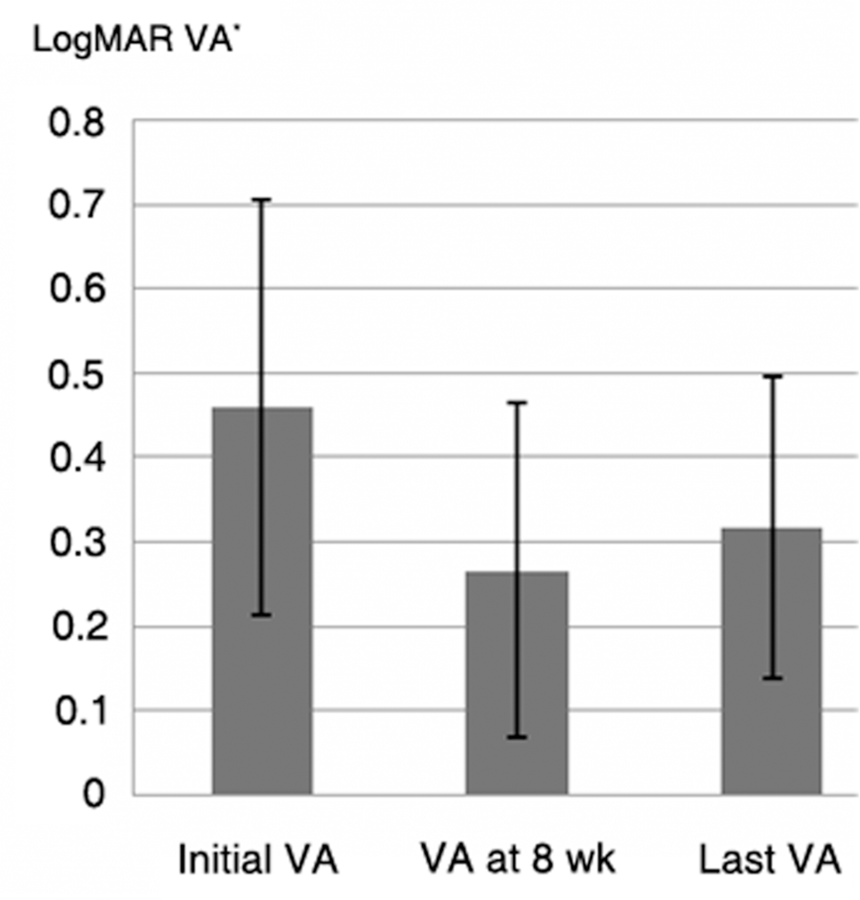
 | Figure 1.Average best corrected visual acuity (BCVA) of patients with improved visual acuity after administration of levodopa for 8 weeks (9 eyes).(* the log of minimum angle of resolution visual acuity) |
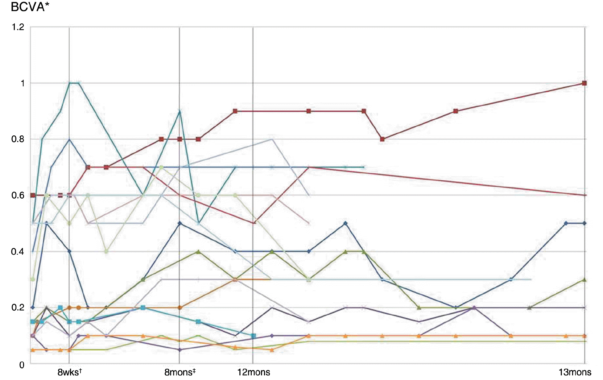
 | Figure 2.Changes in best corrected visual acuity (BCVA) in all patients after dopamine administration. (* BCVA=best corrected visual acuity; † wks=weeks; ‡ mons=months) |
Table 1.
Patient profiles and characteristics
| Patient | Age (years) | Sex | Diagnosis | Follow-up periods (months) | Initial VA* | VA at 8 wks | Maximum VA | Last VA | Side effect |
|---|---|---|---|---|---|---|---|---|---|
| 1 | 5.2 | M† | AA§ | 30 | 0.2 | 0.4 | 0.5 | 0.5 | None |
| 2 | 10.8 | F‡ | AA | 18 | 0.5 | 1.0 | 1.0 | 1.0 | None |
| 3 | 5.3 | M | Mixed(AA+SA∏) | 14 | 0.1 | 0.2 | 0.3 | 0.3 | None |
| 4 | 5.8 | F | DA# | 15 | 0.5 | 0.6 | 0.8 | 0.6 | Nervousness |
| 0.5 | 0.6 | 0.6 | 0.5 | ||||||
| 5 | 4.5 | F | DA | 30 | 0.05 | 0.05 | 0.1 | 0.08 | Loss of |
| 0.6 | 0.6 | 1.0 | 1.0 | appetite | |||||
| 6 | 5.1 | M | DA | 30 | 0.15 | 0.15 | 0.4 | 0.3 | Nervousness |
| 0.1 | 0.1 | 0.3 | 0.2 | ||||||
| 7 | 3.8 | M | Mixed(DA+SA) | 30 | 0.05 | 0.05 | 0.1 | 0.1 | None |
| 8 | 6.3 | F | Mixed(AA+OA**) | 12 | 0.15 | 0.15 | 0.2 | 0.1 | None |
| 9 | 6.5 | M | Mixed(AA+OA) | 30 | 0.1 | 0.1 | 0.2 | 0.1 | None |
| 10 | 11.1 | M | AA | 14 | 0.3 | 0.5 | 0.7 | 0.3 | None |
| 0.1 | 0.1 | 0.3 | 0.15 | ||||||
| 11 | 12 | F | SA | 27 | 0.5 | 0.6 | 0.6 | 0.3 | None |
| 12 | 8.5 | M | DA | 30 | 0.4 | 0.8 | 0.8 | 0.6 | None |
| 0.5 | 0.6 | 0.7 | 0.6 | ||||||
| Mean | 7.0±2.70 | 23.7±7.7 |
Table 2.
Patients with improved visual acuity at 8 week
| Patient | Initial VA* | VA at 8 wks | Maximum VA | Periods† (months) | Last VA |
|---|---|---|---|---|---|
| 1 | 0.2 | 0.4 | 0.5 | 2 | 0.5 |
| 2 | 0.5 | 1.0 | 1.0 | 2.5 | 1.0 |
| 3 | 0.1 | 0.2 | 0.3 | 11 | 0.3 |
| 4 | 0.5 | 0.6 | 0.8 | 13 | 0.6 |
| 0.5 | 0.6 | 0.6 | 6 | 0.5 | |
| 10 | 0.3 | 0.5 | 0.7 | 7 | 0.3 |
| 11 | 0.5 | 0.6 | 0.6 | 2.5 | 0.3 |
| 12 | 0.4 | 0.8 | 0.8 | 2 | 0.6 |
| 0.5 | 0.6 | 0.7 | 3 | 0.6 | |
| ‡Mean(decimal) | 0.39±0.15 | 0.59±0.23 | 0.67±0.20 | 0.52±0.22 | |
| § Mean(logMAR) | 0.46±0.25 | 0.26±0.20 | 0.20±0.15 | 0.33±0.15 |


 PDF
PDF ePub
ePub Citation
 Print
Print


 XML Download