

 PDF
PDF ePub
ePub Citation
 Print
Print


Abstract
Purpose
To define and measure macular thickness and volume using Fourier domain OCT (FD OCT) and compare the values with Stratus OCT in normal eyes and eyes with macular disease.
Methods
On the same day, macular thicknesses of the ETDRS 9 subfield and total macular volumes were measured in 35 eyes of 23 normal subjects and 19 diseased eyes of 24 patients with FD OCT and Stratus OCT. The macular cube scan protocol for FD OCT and the fast macular thickness map protocol for Stratus OCT were used to measure macular thicknesses.
Results
Foveal thickness of the central subfield in FD OCT (251.49±79.45 μ m) was thicker than the value of Stratus OCT (210.26 ±60.57 μ m) (p<0.001) in all eyes. Total macular volume was 7.72±1.06 mm3 and 7.04±0.96 mm3 for FD OCT and Stratus OCT, respectively (p<0.001). Retina thickness of the ETDRS 9 subfields in FD OCT was thicker than the value obtained using Stratus OCT. In addition, foveal thickness differences were statistically significant in both the normal and diseased eye groups.
Conclusions
Macular thickness and total macular volume as measured by the FD OCT were larger than the values obtained using the Stratus OCT in both the normal and the diseased eye groups. The measuring algorithm of FD OCT defines the top of RPE as the outer retinal boundary, but Stratus OCT defines the outer retinal boundary as the IS/OS junction of the photoreceptor. Therefore, macular thicknesses of FD OCT are thicker than those of Stratus OCT. This difference should be considered when comparing the results of FD OCT with those of Stratus OCT.
Go to : 
References
1. Jaffe GJ, Caprioli J. Optical coherence tomography to detect and manage retinal disease and glaucoma. Am J Ophthalmol. 2004; 137:156–69.

2. Otani T, Kishi S, Maruyama Y. Patterns of diabetic macular edema with optical coherence tomography. Am J Ophthalmol. 1999; 127:688–93.
3. Haouchine B, Massin P, Tadayoni R, et al. Diagnosis of macular pseu-doholes and lamellar macular holes by optical coherence tomography. Am J Ophthalmol. 2004; 138:732–9.
4. Sanchez-Tocino H, Valdez-Vidal A, Maldonado M, et al. Retinal thickness study with optical coherence tomography in patients with diabetes. Invest Ophthalmol Visc Sci. 2002; 43:1588–94.
5. Dacosta S, Rajendran B, Janakiraman P. Spectral domain optical co-herence tomography a practical guide. 1st ed.New Delhi: Jaypee Bro-thers Medical Publisheres;2008. p. 3–7.
6. Zeimer RC, Shahidi M, Mori M, et al. A new method for rapid mapping of the retinal thickness at the posterior pole. Invest Ophthalmol Vis Sci. 1996; 37:1994–2001.
7. Legarreta JE, Gregori G, Punjabi OS, et al. Macular thickness measurements in normal eyes using spectral domain optical coherence tomography. Ophthalmic Surg Lasers Imaging. 2008; 39:S43–9.
8. Forooghian F, Cukras C, Meyerle CB, et al. Evaluation of Time Domain and Spectral Domain Optical Coherence Tomography in the Measurement of Diabetic Macular Edema. Invest Ophthalmol Vis Sci. 2008; 49:4290–6.
9. Kiernan D, Hariprasad S, Chin E, et al. Prospective Comparison of Cirrus and Stratus Optical Coherence Tomography for Quantifying Retinal Thickness. Am J Ophthalmol. 2009; 147:267–75.
10. Leung CK, Cheung CY, Weinreb RN, et al. Comparison of Macular Thickness Measurements between Time Domain and Spectral Domain Optical Coherence Tomography. Invest Ophthalmol Vis Sci. 2008; 49:4893–7.
11. Forte R, Cennamo GL, Finelli ML, de Crecchio G. Comparison of time domain Stratus OCT and spectral domain SLO/OCT for assessment of macular thickness and volume. Eye 2008 Dec 12. [Epub ahead of print].
12. Srinivasan VJ, Monson BK, Wojtkowski M, et al. Characterization of outer retinal morphology with high-speed, ultra-high resolution optical coherence tomography. Invest Ophthalmol Vis Sci. 2008; 49:1571–9.
13. Sayanagi K, Sharma S, Yamamoto T, Kaiser PK. Comparison of Spectral-Domain versus Time-Domain Optical Coherence Tomography in Management of Age-Related Macular Degeneration with Ranibizumab. Ophthalmology. 2009; 116:947–55.
14. Han IC, Jaffe GJ. Comparison of Spectral- and Time-Domain Optical Coherence Tomography for Retinal Thickness Measure-ments in Healthy and Diseased Eyes. Am J Ophthalmol. 2009; 147:847–58.
Go to :
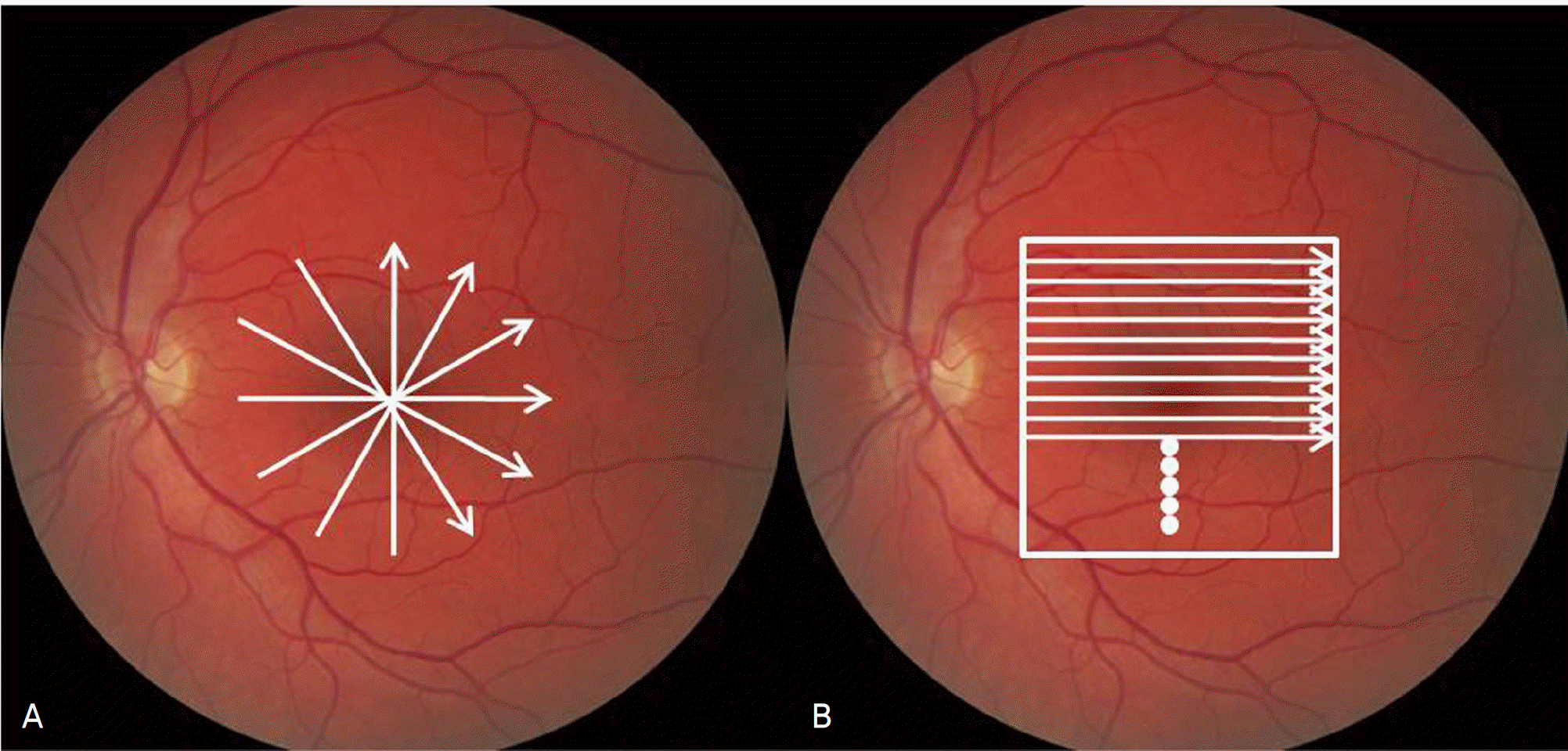 | Figure 1.Six radial scans of Stratus OCT (A) and macular cube scans of Fourier domain OCT (B). |
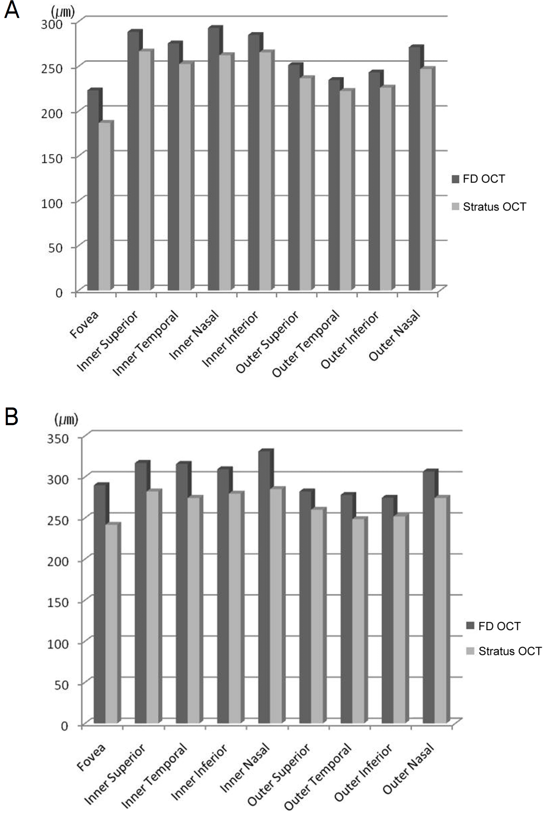 | Figure 2.Comparison of macular thickness measurements in 35 normal eyes (A) and 19 diseased eyes (B) between Fourier domain OCT (FD OCT) and Stratus OCT. |
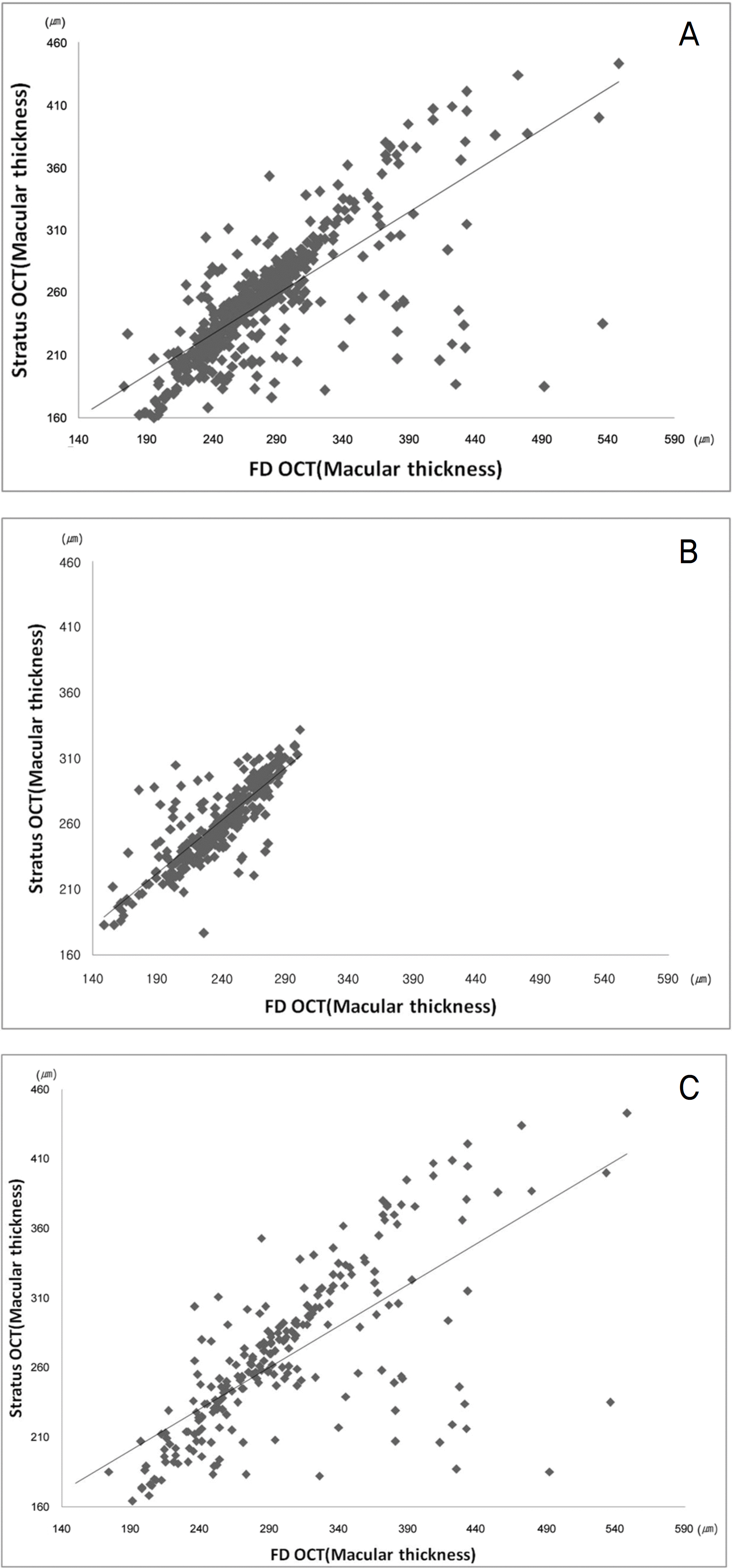 | Figure 3.Fourier domain OCT (FD OCT) and Stratus OCT measurements in all patients (A), normal eyes (B), and diseased eyes (C) are graphed together. |
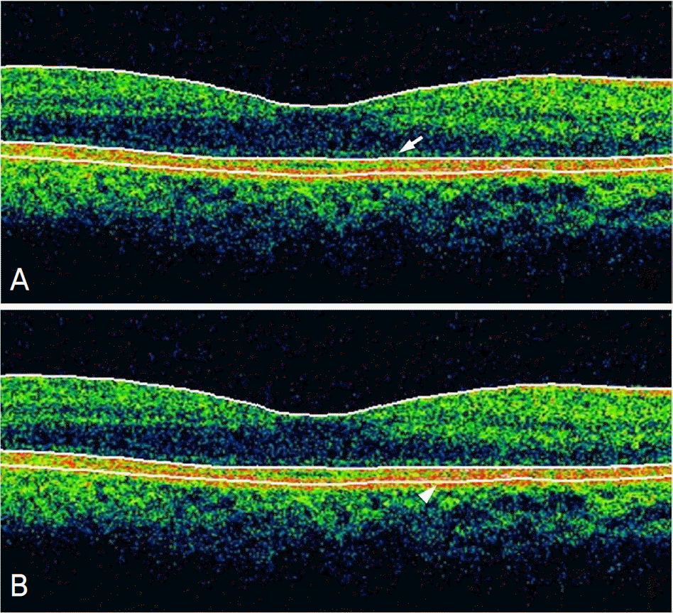 | Figure 4.Outer retinal boundary in Stratus OCT (arrow) and Fourier domain OCT (arrow head). The arrow indicates the inner segment/outer segment photoreceptor junction and the arrow head indicates the inner border of the retinal pigment epithelium. |
Table 1.
Comparison of macular thickness measurement in all patients between Fourier domain OCT (FD OCT) and Stratus OCT
| ETDRS* area | FD OCT (μm) | Stratus OCT (μm) | Difference | p-value | r† |
|---|---|---|---|---|---|
| Fovea (500 μm radius) | 251.49±79.45 | 210.26±60.57 | 41.23±53.87 | <0.001 | 0.74 |
| Inner ring (1.5 mm radius) | |||||
| Superior | 300.54±46.58 | 273.23±40.79 | 27.31±30.23 | <0.001 | 0.77 |
| Temporal | 292.59±50.41 | 261.82±50.84 | 30.77±33.11 | <0.001 | 0.79 |
| Inferior | 295.28±44.58 | 271.56±41.35 | 23.72±39.60 | <0.001 | 0.58 |
| Nasal | 309.08±55.43 | 272.03±42.60 | 37.05±43.32 | <0.001 | 0.64 |
| Outer ring (3.0 mm radius) | |||||
| Superior | 264.51±39.94 | 246.66±36.01 | 17.85±23.08 | <0.001 | 0.82 |
| Temporal | 253.11±47.87 | 233.56±40.31 | 19.56±35.22 | <0.001 | 0.69 |
| Inferior | 256.51±35.78 | 237.05±35.25 | 19.46±33.37 | <0.001 | 0.56 |
| Nasal | 286.25±42.65 | 258.64±39.89 | 27.61±25.79 | <0.001 | 0.81 |
| Total macular volume (mm3) | 7.72±1.06 | 7.44±1.31 | 0.67±0.63 | <0.001 | 0.81 |
Table 2.
Comparison of macular thickness measurement in 35 normal eyes and 26 diseased eyes between Fourier domain OCT (FD OCT) and Stratus OCT
| ETDRS* area | FD OCT (μm) | Stratus OCT (μm) | Difference | p-value | r† |
|---|---|---|---|---|---|
| Normal eyes | |||||
| Fovea (500 μm radius) | 223.00±27.49 | 186.86±21.47 | 36.14±22.08 | <0.001 | 0.62 |
| Inner ring (1.5 mm radius) | |||||
| Superior | 288.31±17.65 | 266.49±24.54 | 21.83±8.74 | <0.001 | 0.89 |
| Temporal | 275.31±18.80 | 252.40±23.22 | 22.91±18.41 | <0.001 | 0.63 |
| Inferior | 284.80±20.24 | 265.49±20.03 | 19.31±6.75 | <0.001 | 0.94 |
| Nasal | 292.66±20.92 | 262.29±24.54 | 30.37±17.88 | <0.001 | 0.70 |
| Outer ring (3.0 mm radius) | |||||
| Superior | 251.31±16.25 | 236.60±15.60 | 14.71±9.11 | <0.001 | 0.84 |
| Temporal | 234.51±13.88 | 222.37±23.63 | 12.14±22.16 | <0.001 | 0.40 |
| Inferior | 243.03±15.61 | 226.06±16.10 | 16.97±13.17 | <0.001 | 0.66 |
| Nasal | 271.11±15.83 | 246.86±21.17 | 24.26±18.70 | <0.001 | 0.52 |
| Total macular volume (mm3) | 7.26±0.35 | 6.75±0.40 | 0.50±0.20 | <0.001 | 0.87 |
| Diseased eyes | |||||
| Fovea (500 μm radius) | 289.85±107.03 | 241.77±79.86 | 48.08±78.84 | <0.001 | 0.68 |
| Inner ring (1.5 mm radius) | |||||
| Superior | 317.00±65.52 | 282.31±57.74 | 34.69±44.62 | <0.001 | 0.75 |
| Temporal | 315.85±68.11 | 274.50±71.97 | 41.35±44.35 | <0.001 | 0.80 |
| Inferior | 309.38±62.06 | 279.73±58.63 | 29.65±60.31 | 0.004 | 0.50 |
| Nasal | 331.19±76.76 | 285.15±56.78 | 46.04±62.63 | <0.001 | 0.60 |
| Outer ring (3.0 mm radius) | |||||
| Superior | 282.27±53.82 | 260.19±49.48 | 22.08±33.66 | 0.003 | 0.79 |
| Temporal | 278.15±64.04 | 248.62±52.24 | 29.54±46.15 | 0.001 | 0.70 |
| Inferior | 274.65±46.31 | 251.85±47.26 | 22.81±49.15 | 0.003 | 0.45 |
| Nasal | 306.62±57.20 | 274.50±52.47 | 32.12±32.92 | <0.001 | 0.82 |
| Total macular volume (mm3) | 8.32±1.37 | 7.44±1.31 | 0.87±0.90 | <0.001 | 0.77 |
Table 3.
Comparison of macular thickness differences between Fourier domain OCT and Stratus OCT in normal eyes and diseased eyes
| ETDRS* area | Normal eyes | Diseased Eye | p-value |
|---|---|---|---|
| Fovea (500 μm radius) | 36.14±22.08 | 48.08±78.84 | 0.46 |
| Inner ring (1.5 mm radius) | |||
| Superior | 21.83±8.74 | 34.69±44.62 | 0.16 |
| Temporal | 22.91±18.41 | 41.35±44.35 | 0.06 |
| Inferior | 19.31±6.75 | 29.65±60.31 | 0.39 |
| Nasal | 30.37±17.88 | 46.04±62.63 | 0.23 |
| Outer ring (3.0 mm radius) | |||
| Superior | 14.71±9.11 | 22.08±33.66 | 0.29 |
| Temporal | 12.14±22.16 | 29.54±46.15 | 0.09 |
| Inferior | 16.97±13.17 | 22.81±49.15 | 0.56 |
| Nasal | 24.26±18.70 | 30.12±32.92 | 0.28 |
| Total macular volume (mm3) | 0.50±0.20 | 0.44±1.31 | 0.06 |
Table 4.
Comparison of macular thickness differences between Fourier domain OCT and Stratus OCT for DME and wet AMD compared with normal eyes
| ETDRS* area | Normal eyes | DME† | p-value | Wet AMD‡ | p-value |
|---|---|---|---|---|---|
| Fovea (500 μm radius) | 36.14±22.08 | 55.33±68.42 | 0.08 | 38.18±93.80 | 0.90 |
| Inner ring (1.5 mm radius) | |||||
| Superior | 21.83±8.74 | 31.80±24.78 | 0.07 | 38.64±63.94 | 0.07 |
| Temporal | 22.91±18.41 | 43.33±49.99 | 0.06 | 38.64±37.46 | 0.13 |
| Inferior | 19.31±6.75 | 26.87±53.33 | 0.41 | 33.45±71.30 | 0.07 |
| Nasal | 30.37±17.88 | 35.73±40.63 | 0.51 | 60.09±84.38 | 0.24 |
| Outer ring (3.0 mm radius) | |||||
| Superior | 14.71±9.11 | 19.47±30.84 | 0.40 | 18.08±33.66 | 0.56 |
| Temporal | 12.14±22.16 | 32.60±59.44 | 0.08 | 19.54±46.15 | 0.48 |
| Inferior | 16.97±13.17 | 25.07±51.52 | 0.39 | 19.73±48.02 | 0.76 |
| Nasal | 24.26±18.70 | 25.87±30.08 | 0.82 | 33.64±36.11 | 0.36 |
| Total macular volume (mm3) | 0.50±0.20 | 0.88±0.83 | 0.06 | 0.87±1.04 | 0.06 |
 XML Download