Abstract
Purpose
To investigate the clinical features of patients with macular edema persisting for three months after a successful vitrectomy and removal of the epiretinal membrane.
Methods
The authors retrospectively reviewed the records of 32 patients (32 eyes) with epiretinal membranes who underwent pars plana vitrectomies and membranectomies from January 2005 to September 2008. The patients selected for the study underwent optical coherence tomography (OCT) which revealed macular edema. Macular edema is defined as central macular thickness measuring over 300 m. Several factors including age, presence of systemic disorders, preoperative visual acuity, surgical technique, preoperative state of the macula, and the response to treatment for macular edema were reviewed.
Results
Eight out of 32 eyes (25%) had no improvement of visual acuity after surgery of the epiretinal membrane. Six out of 32 eyes (18.75%) persisted in their macular edema. No common features were detected. All patients showed no improvement of visual acuity after their vitrectomies. Two of the eyes were treated with intravitreal or subtenon triamcinolone injection or non-steroidal anti-inflammatory eye drops. Only one eye (16.7%) achieved visual improvement as of the last follow-up.
Go to : 
References
2. Margherio RR, Cox MS, Trese MT, et al. Removal of epimacular membranes. Ophthalmology. 1985; 92:1075–83.

3. McDonald HR, Verre WP, Aaberg TM. Surgical management of idiopathic epiretinal membranes. Ophthalmology. 1986; 93:978–83.

4. Rice TA, De Bustros S, Michels RG, et al. Prognostic factors in vitrectomy for epiretinal membranes of the macula. Ophthalmology. 1986; 93:602–10.

5. Abdelkader E, Lois N. Internal Limiting Membrane Peeling in Vitreoretinal Surgery. Surv Ophthalmol. 2008; 53:368–96.

6. Massin P, Allouch C, Haouchine B, et al. Optical coherence tomography of idiopathic macular epiretinal membranes before and after surgery. Am J Ophthalmol. 2003; 130:732–9.

7. Choi YK, Yoo JS, Kim MH. Result of Surgery for Epiretinal Membrane and Their Recrrence. J Korean Ophthlamol Soc. 2000; 41:2357–62.
8. Lee HJ, Kim HC. The Clinical Outcome and Prognostic Factors of Vitrectomy for Macular Epiretinal Membranes. J Korean Ophthlamol Soc. 2003; 44:857–64.
9. Foos RY. Vitreoretinal junction: topographical variations. Invest Ophthalmol. 1972; 11:801–8.
11. Sebag J, Balazs EA. Pathogenesis of cystoid macular edema:an ana-tomic consideration of vitreoretinal adhesion. Surv Ophthalmol. 1984; 28:493–8.
12. Gomes NL, Corcostegui I, Fine HF, Chang S. Subfoveal pigment changes in patients with longstanding epiretinal membrane. Am J Ophthalmol. 2009; 147:865–8.
13. Kwok AK, Lai TY, Li WW, et al. Indocyanine green-assisted internal limiting membrane removal in epiretinal membrane surgery. Am J Ophthalmol. 2004; 138:194–9.
14. Sivalingam A, Eagle RC, Duker JS, et al. Visual prognosis correlated with the presence of internal limiting membrane peeling in histo-pathologic specimens obtained from epiretinal membrane surgery. Ophthalmology. 1990; 97:1549–52.
15. Gandorfer A, Haritoglou C, Gass CA, et al. Indocyanine Green-Assisted Peeling of the Internal Limiting Membrane may cause retinal damage. Am J Ophthalmol. 2001; 132:431–3.

16. Kim DS, Moon SW, Yu SY, Kwak HW. The structure of the in-termal limiting membrane removed by vitrectomy using tissue plas-minogen activator. J Korean Ophthalmol Soc. 2008; 49:917–24.
17. Stefabsson E, MAchemer R, de Juan E Jr, et al. Retinaloxygenation and laser treatment in patients with diabetic retinopathy. Am J Ophthalmol. 1992; 113:36–8.
18. Martidis A, Duker JS, Greenberg PB, et al. Intravitreal Triamcinolone for Refractory Diabetic Macular Edema. Ophthalmology. 2002; 109:920–7.

19. Ikeda T, Sato K, Katano T, Hayashi Y. Improved visual acuity following pars plana vitrectomy for diabetic cystoid macular edema and detached posterior hyaloid. Retina. 2000; 20:220–2.

20. Suckling RD, Maslin KF. Pseudophakic cystoid macular oedema and its treatment with local steroids. Aust N Z J Ophthalmol. 1988; 16:353–9.

21. Jonas JB, Kreissig I, Söfker A, Degenring RF. Intravitreal Injection of Triamcinolone for Diffuse Diabetic Macular Edema. Arch Ophthalmol. 2003; 121:57–61.

22. Lee DG, Cho NC. Combination of laser treatment and intravitreal triamcinolone injection for macular edema with branch retinal vein occlusion. J Korean Ophthalmol Soc. 2005; 46:287–96.
Go to : 
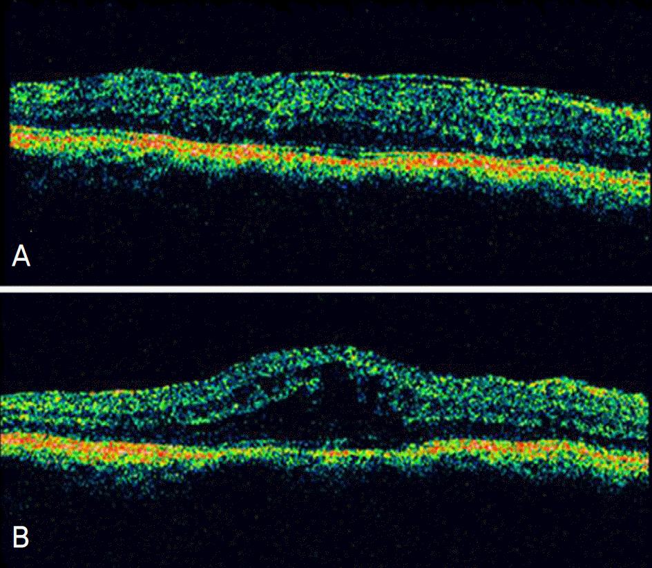
 | Figure 1.OCT images of No.6 patient shows that the macular edema aggravated after surgical treatment. (A) Preoperative OCT image, central macular thickness was about 565 µm. (B) Postoperative OCT image at 3 months after surgery revealed that the macular thickness was measured about 655 µm. |
Table 1.
Characteristics of the patients who had idiopathic epiretinal membrane and remained macular edema after surgical treatment
| No | Sex | Age | ILM§ | ICG‡ | Gas† | Preop. VA | Postop. VA | Preop. CMT* | Postop. CMT | Cystoid | Tx. |
|---|---|---|---|---|---|---|---|---|---|---|---|
| 1 | M | 74 | No | No | SF6 | 20/40 | 20/40 | 506 | 387 | − | NSAID# |
| 2 | F | 77 | No | No | − | 20/80 | 20/32 | 406 | 549 | − | STTA** |
| 3 | M | 72 | No | No | SF6 | 20/20 | 20/40 | 431 | 439 | − | NSAID |
| 4 | F | 60 | Yes | Yes | − | 20/200 | 20/200 | 723 | 556 | + | IVTAП |
| 5 | F | 61 | No | No | SF6 | 20/125 | 20/100 | 564 | 375 | − | IVTA |
| 6 | F | 65 | Yes | Yes | − | 20/50 | 20/63 | 565 | 655 | + | STTA |


 PDF
PDF ePub
ePub Citation
 Print
Print


 XML Download