

 PDF
PDF ePub
ePub Citation
 Print
Print


Abstract
Purpose
To report on 2 cases of Eales’ disease that were successfully regressed with laser photocoagulation and intravitreal bevacizumab (Avastin; Genetech, Inc, San Francisco, California, USA) injection.
Case summary
Two male patients (30 years and, 40 years of age,) with a history of recurrent vitreous hemorrhage were diagnosed with Eales’ disease. The 2 patients had peripheral retina neovascularization and active phlebitis in fundus fluorescein angiography. No other findings were observed in their eyes in the general examination. Scatter laser photocoagulation and intravitreal bevacizumab injection were performed. After 1 month follow-up, neovascularization completely regressed. There was no complication or recurrent vitreous hemorrhage at the 1 year follow up.
Go to : 
References
1. Biswas J, Sharma T, Gopal L, et al. Eales disease-an update. Surv Ophthalmol. 2002; 47:197–214.

2. El-asrar AM, Al-kharashi SA. Full panretinal photocoagulation and early vitrectomy improve prognosis of retinal vasculitis associated with tuberculoprotein hypersensitivity (Eales' disease). Br J Ophthalmol. 2002; 86:1248–51.
3. Kurup S, Lew J, Byrnes G, et al. Therapeutic efficacy of intravitreal bevacizumab on posterior uveitis complicated by neovascularization. Acta Ophthalmol. 2000; 87:349–52.
4. Tatar O, Yoeruek E, Szurman P, et al. Effect of Bevacizumab on Inflammation and Proliferation in Human Choroidal Neovascularization. Arch Ophthalmol. 2008; 126:782–90.
5. Moradian S, Ahmadieh H, Malihi M, et al. Intravitreal bevacizumab in active progressive proliferative diabetic retinopathy. Graefes Arch Clin Exp Ophthalmol. 2008; 246:1699–705.
6. Murugeswari P, Shukla D, Rajendran A. Proinflammatory cytokines and angiogenic and anti-angiogenic factors in vitreous of patients with proliferative diabetic retinopathy and eales' disease. Retina. 2008; 28:817–24.
7. Küçükerdönmez C, Akova YA, Yilmaz G. Intravitreal injection of Bevacizumab in Eales disease. Ocul Immunol Inflamm. 2008; 16:63–5.
8. Chanana B, Azad RV, Patwardhan S. Role of intravitreal bevacizumab in the management of Eales' disease. Int Ophthalmol 2009 Jan 23. [Epub ahead of print].
Go to :
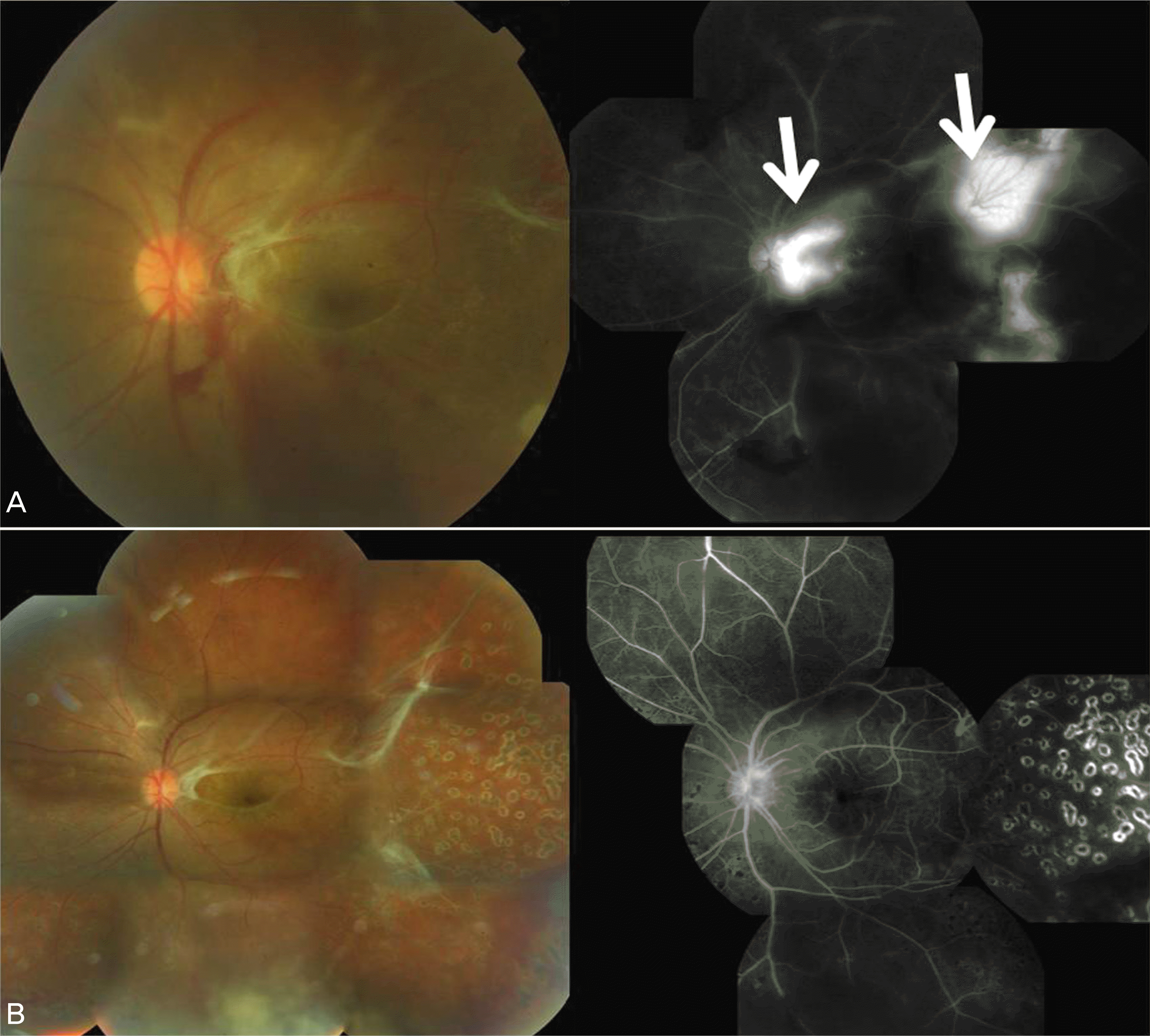 | Figure 1.Fundus photographs and fluorescein angiographs of Case 1. (A) Fundus photograph showing active periphlebitis and slcerosed vein with subhylaoid hemorrhage. Fibrovascular prolife-ration causing tractional retinal membrane seen also at juxtafoveolar area. There was late fluorescein leakage due to neovascularization at the optic disc and peripheral retina (white arrows). (B) Montage fundus photography shows successfully regressed neovascularization of the optic disc and peripheral retina six weeks after panretinal photocoagulation and intravitreal bevacizumab injection. There was no progression of fibrovascular membrane. |
 | Figure 2.Fundus photographs and fluorescein angiographs of Case 2. (A) There were vascular obliterance and sheath in the nasal retina. And fibrovascluar membrane was observed in the peripheral inferior nasal retina (white arrow). Fundus fluorecein angiogram shows large non-perfusion area and late leakage at the neovascularization site. (B) One month after scatter laser photocoagulation and intravitreal bevacizumab injection, neovascularization regressed and vitreous hemorrhage did not recur. |
 XML Download