Abstract
Purpose
To report 2 cases of bilateral endogenous endophthalmitis in patients with Klebsiella pneumoniae primary liver abscess.
Case summary
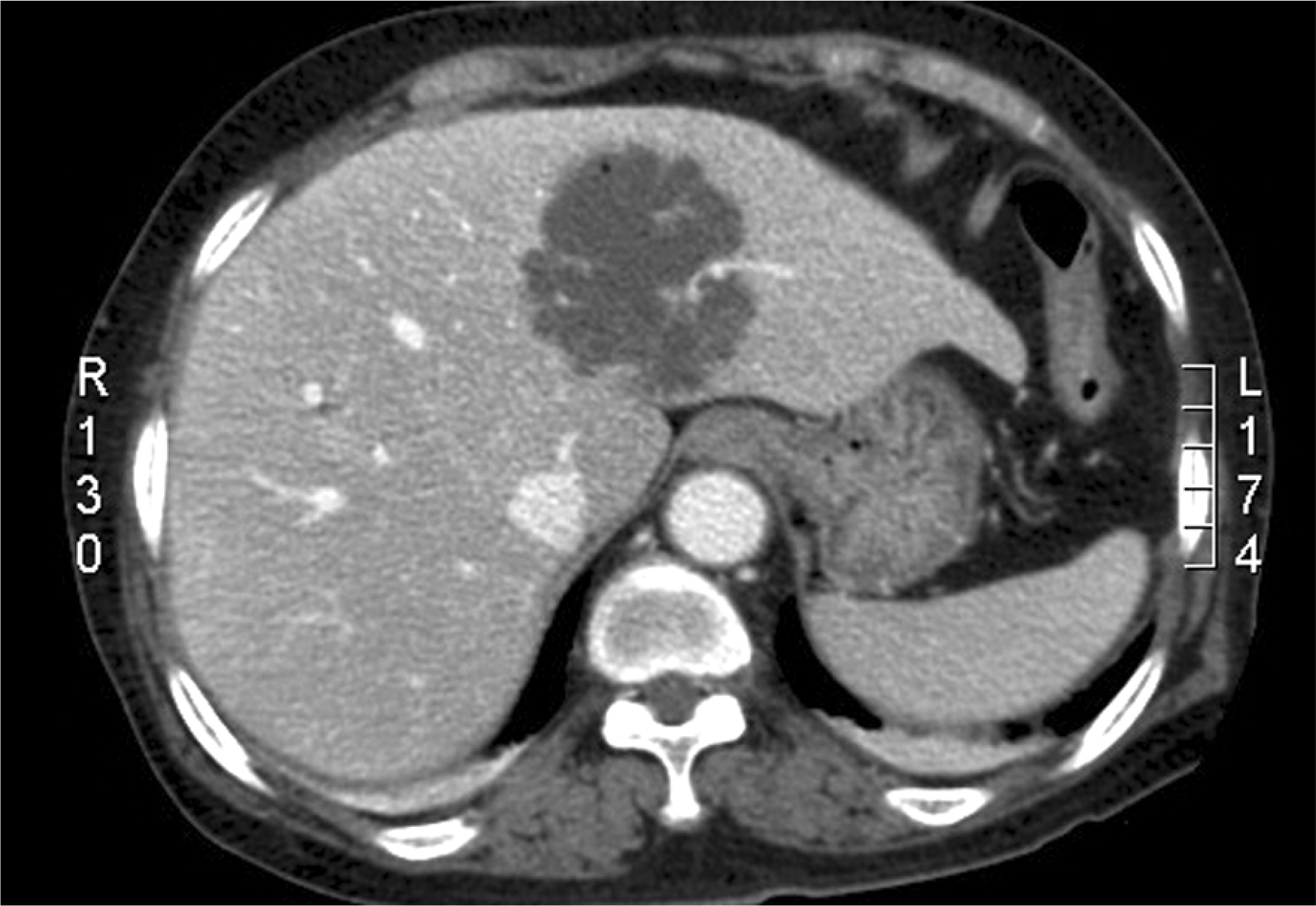
A 78-year-old woman and a 76-year-old woman presented with bilateral blurred vision for 2 to 3 days. The women had fever, cough, and chills for 5 to 10 days. Abdominal computed tomography revealed liver abscess and ophthalmologic examination results suggested bilateral endophthalmitis. In both patients, Klebsiella pneumoniae was identified from the liver abscess aspirate. In one patient, Klebsiella pneumoniae was also identified from a vitreous sample. Both patients lost their vision despite intravitreal and intravenous antibiotics injection.
References
1. Lee YH, Choi SJ, Kim IC, Chung YT. A case of bilateral metastatic endophthalmitis. J Korean Ophthalmol Soc. 1995; 36:2048–53.
2. Wang JH, Liu YC, Lee SS, et al. Primary liver abscess due to Klebsiella pneumoniae in Taiwan. Clin Infect Dis. 1998; 26:1434–8.
3. Yang CC, Yen CH, Ho MW, Wang JH. Comparison of pyogenic liver abscess caused by non-Klebsiella pneumoniae and Klebsiella pneumoniae. J Microbiol Immunol Infect. 2004; 37:176–84.
4. Tsay RW, Siu LK, Func CP, Chang FY. Characteristics of bacteremia between community-acquired and nosocomial Klebsiella pneu– moniaeinfection: risk factor for mortality and the impact of capsular serotypes as a herald for community-acquired infection. Arch Intern Med. 2002; 162:1021–7.
5. Ko WC, Paterson DL, Sagnimeni AJ, et al. Community-acquired Klebsiella pneumoniae bacteremia: global differences in clinical patterns. Emerg Infect Dis. 2002; 8:160–6.

6. Chung DR, Lee SS, Lee HR, et al. Emerging invasive liver abscess caused by K1 serotype Klebsiella pneumoniae in Korea. J Infect. 2007; 54:578–83.

7. Kang CI, Kim SH, Bang JW, et al. Community-acquired versus nosocomial Klebsiella pneumoniae bacteremia: clinical features, treatment outcomes, and clinical implication of antimicrobial resistance. J Korean Med Sci. 2006; 21:816–22.

8. Fang CT, Lai SY, Yi WC, et al. Klebsiella pneumoniae genotype K1: An emerging pathogen that causes septic ocular or central nervous system complications from pyogenic liver abscess. Clin Infect Dis. 2007; 45:284–93.

9. Cheng DL, Liu YC, Yen MY, et al. Septic metastatic lesions of pyogenic liver abscess. Their association with Klebsiella pneumoniae bacteremia in diabetic patients. Arch Intern Med. 1991; 151:1557–9.

10. Chen YJ, Kuo HK, Wu PC, et al. A 10-year comparison of endogenous endophthalmitis outcomes: an East Asian experience with Klebsiella pneumoniae infection. Retina. 2004; 24:383–90.

11. Tan YM, Chee SP, Soo KC, Chow P. Ocular manifestations and complications of pyogenic liver abscess. World J Surg. 2004; 28:38–42.

12. Wong JS, Chan TK, Lee HM, Chee SP. Endogenous bacterial endophthalmitis: an East Asian experience and a reappraisal of a severe ocular affliction. Ophthalmology. 2000; 107:1483–91.

13. La TY, Kim CW, Lee JS. A case of endogenous endophthalmitis accompanying orbital cellulitis caused by Klebsiella pneumoniae from liver abscess. J Korean Ophthalmol Soc. 2000; 41:1000–5.
14. Tae KS, Kim TH, Moon YS, Chin HS. A case of Klebsiella endogenous endophthalmitis accompanied by necrotizing scleral perforation. J Korean Ophthalmol Soc. 2003; 44:2680–6.
Figure 1.
Photographs of patient 1. (A) Photograph of the right eye on the day of admission. Note severe corneal edema and hypopyon. (B) Ultrasonography revealed vitreous opacity and choroidal thickening. (C) On abdominal computed tomography (CT), multiseptated cystic mass (about 6.7×3.9×3.8 cm in size) with adjacent parenchymal enhancement was found in the right lobe of the liver, which confirmed the diagnosis of liver abscess. (D) Photograph of the left eye on the 4th day of admission. Note the corneal perforation and uveal prolapse.


 PDF
PDF ePub
ePub Citation
 Print
Print


 XML Download