

 PDF
PDF ePub
ePub Citation
 Print
Print


Abstract
Purpose
To report a rare case of primary mucinous adenocarcinoma arising from a sweat gland in the eyelid.
Case summary
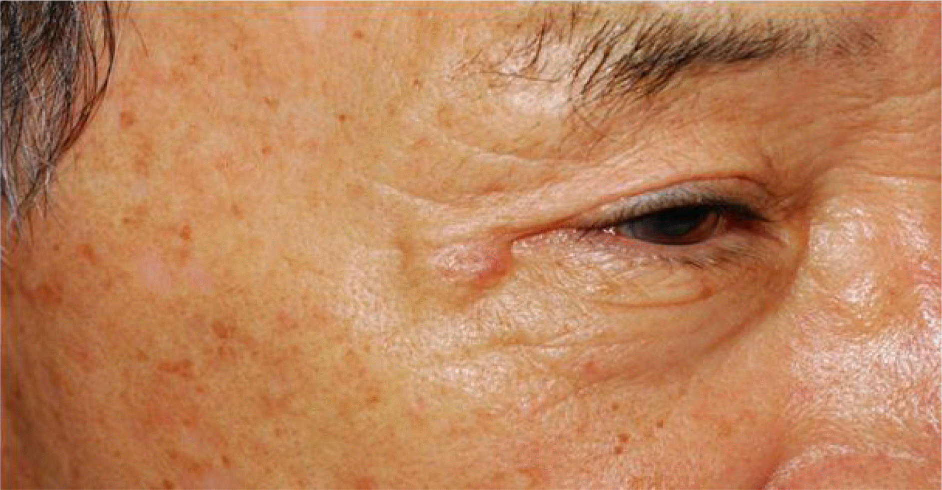
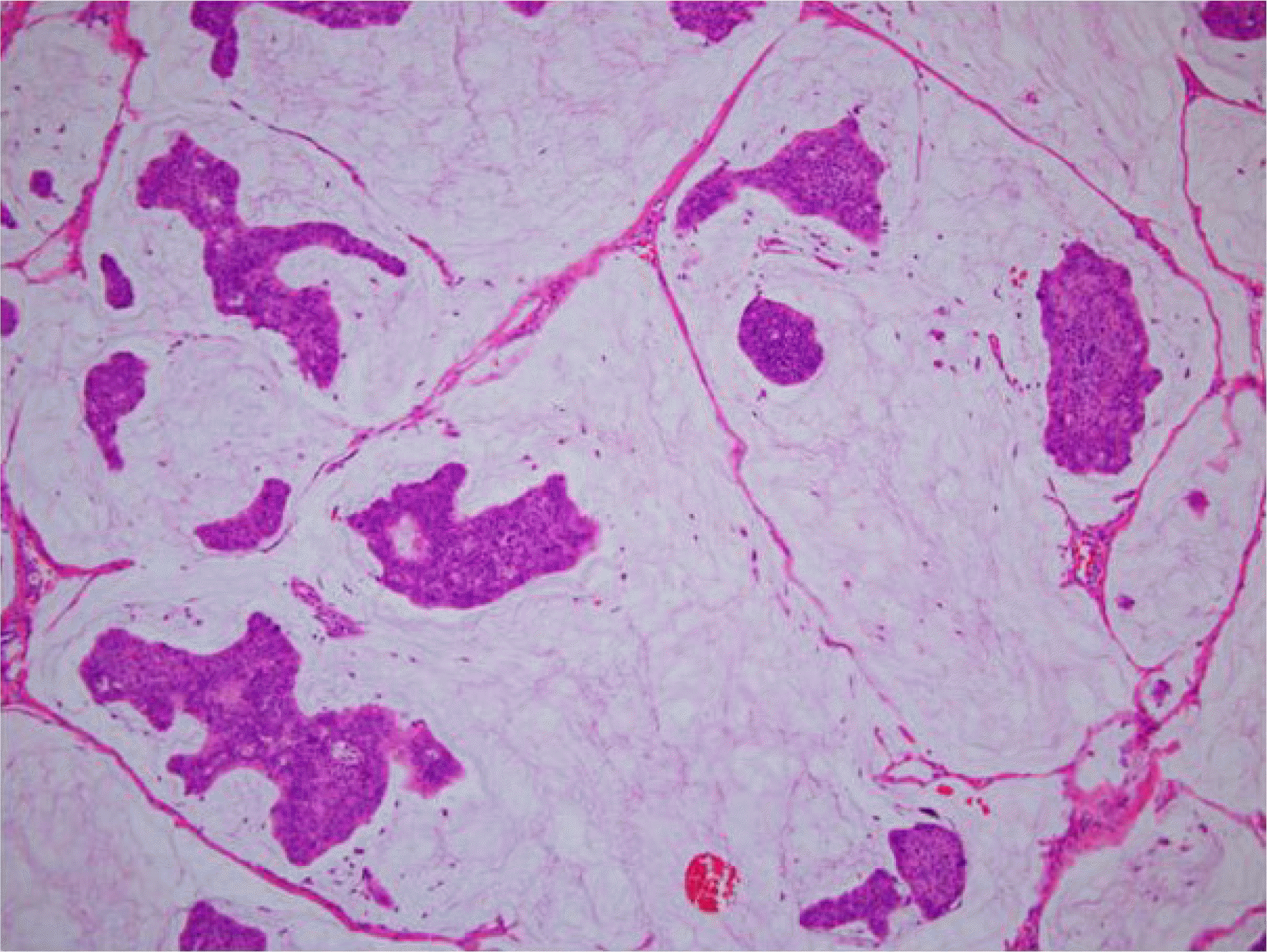
A 68-year-old male presented to our hospital with a painless, superficial nodular lesion over the skin of the right lateral canthus that had slowly grown over the past two years. The patient had a history of surgical excision for three nodular lesions at the same site 5 years ago, and an excisional biopsy was mucinous adenocarcinoma with a positive margin. A systemic evaluation, including whole-body Positron Emission Tomography scan (PET), chest computerized tomography, gastro-intestinal endoscopy, and colonoscopy, revealed no other abnormal lesions. Therefore, the eyelid lesion was considered a primary mucinous adenocarcinoma of the skin.
References
1. Lennox B, Pearse AG, Richards HG. Mucin-secreting tumours of the skin with special reference to the so-called mixed-salivary tumour of the skin and its relation to hidradenoma. J Pathol Bacteriol. 1952; 64:865–80.

2. Seo HC, Ahn M, Cho NC, Lee DO. A case of primary mucinous adenocarcinoma of the upper eyelid. J Korean Ophthalmol Soc. 2007; 48:599–603.
3. Kazakov DV, Suster S, LeBoit PE, et al. Mucinous carcinoma of the skin, primary, and secondary: a clinicopathologic study of 63 cases with emphasis on the morphologic spectrum of primary cutaneous forms: homologies with mucinous lesions in the breast. Am J Surg Pathol. 2005; 29:764–82.
4. Carson HJ, Gattuso P, Raslan WF, Reddy V. Mucinous carcinoma of the eyelid: An immunohistochemical study. Am J Dermato-pathol. 1995; 17:494–8.
5. Breiting L, Christensen L, Dahlstrøm K, et al. Primary mucinous carcinoma of the skin: a population-based study. Int J Dermatol. 2008; 47:242–5.
6. Wright JD, Font RL. Mucinous sweat gland adenocarcinoma of eyelid: a clinicopathologic study of 21 cases with histochemical and electron microscopic observations. Cancer. 1979; 44:1757–68.
7. el-Domeiri AA, Brasfield RD, Huvos AG, Strong EW. Sweat gland carcinoma: a clinicopathologic study of 83 patients. Ann Surg. 1971; 173:270–4.
8. Durairaj VD, Hink EM, Kahook MY, et al. Mucinous eccrine adenocarcinoma of the periocular region. Ophthal Plast Reconstr Surg. 2006; 22:30–5.
9. Martinez SR, Young SE. Primary mucinous carcinoma of the skin: a review. Int J Oncol. 2005; 2:2.
10. Hanby AM, McKee P, Jeffery M, et al. Primary mucinous carcinomas of the skin express TFF1, TFF3, estrogen receptor, and progesterone receptors. Am J Surg Pathol. 1998; 22:1125–31.
11. Rdrigues MM, Font RL, Shannon GM. Metastatic mucussecreting mammary carcinoma in the eyelid. Report of two cases. Br J Ophthalmol. 1974; 58:877–81.
12. Chauhan A, Ganguly M, Takkar P, Dutta V. Primary mucinous carcinoma of eyelid: a rare clinical entity. Indian J Ophthalmol. 2009; 57:150–2.
13. Krishnakumar S, Rambhatla S, Subramanian N, et al. Recurrent mucinous carcinoma of the eyelid. Indian J Opthalmol. 2004; 52:156–7.
14. Snow SN, Reizner GT. Mucinous eccrine carcinoma of the eyelid. Cancer. 1992; 70:2099–104.
15. Ortiz KJ, Gaughan MD, Bang RH, et al. A case of primary mucinous carcinoma of the scalp treated with mohs surgery. Dermatol Surg. 2002; 28:751–4.
16. Andrews TM, Gluckman JL, Weiss MA. Primary mucinous adenocarcinoma of the eyelid. Head Neck. 1992; 14:303–7.
17. Breier F, Calbian M, Pokiser W, et al. Primary mucinous carcinoma of the scalp. Dermatology. 2000; 200:250–3.
18. Gizzard WS, Torczynski E, Edwards WC. Adenocarcinoma of eccrine sweat glands. Arch Ophthalmol. 1976; 94:2119–23.
 XML Download