Abstract
Purpose
The aim of this study was to understand the pathogenesis of the enophthalmos of Horner’s syndrome.
Methods
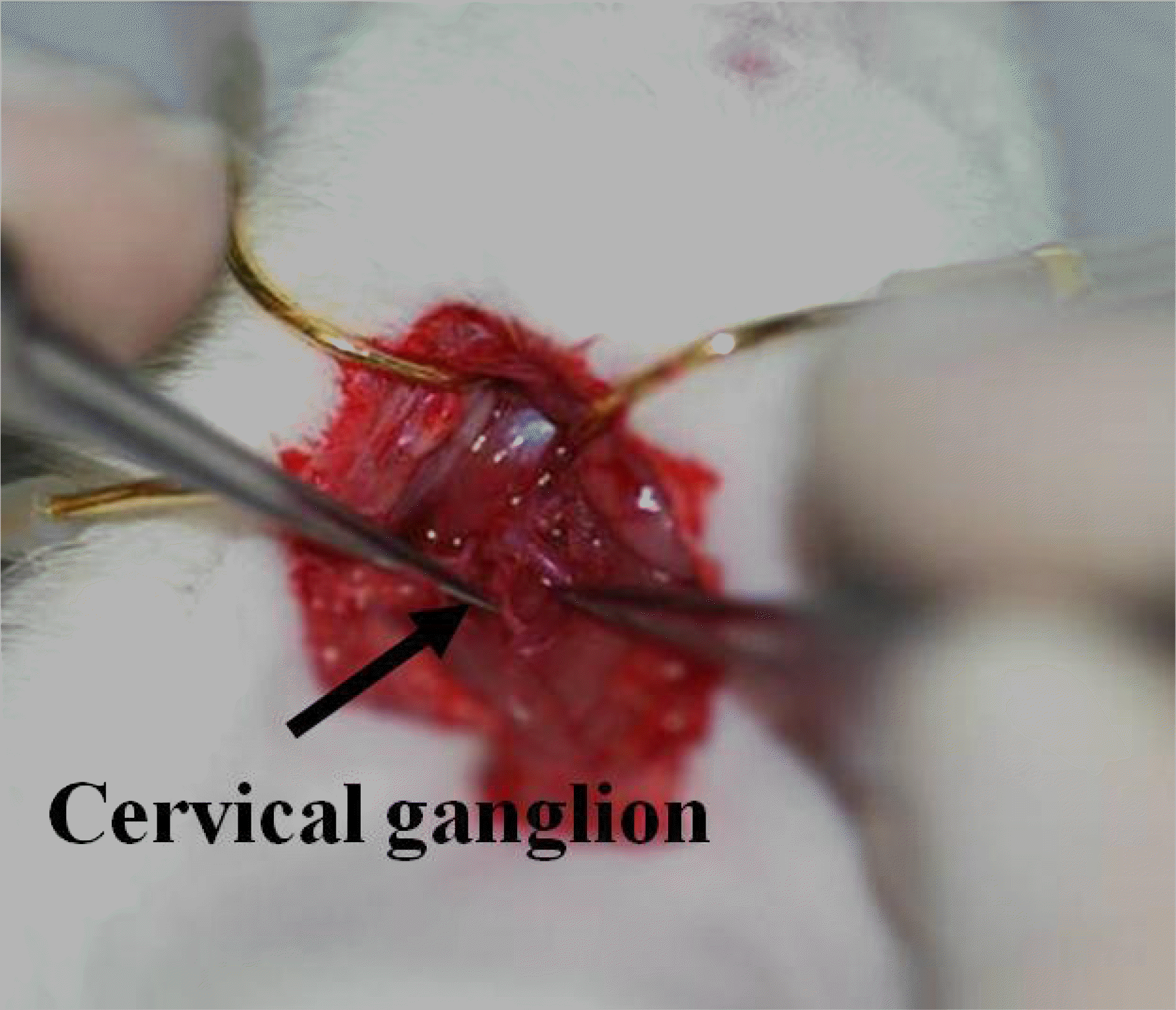
We performed right cervical sympathectomy in 10 Sprague-Dawley rats weighing 200∼300 grams at 8 weeks of age. We obtained bilateral extraocular muscle and fat from all 10 rats. These tissues were observed under light and electron microscopy.
Results
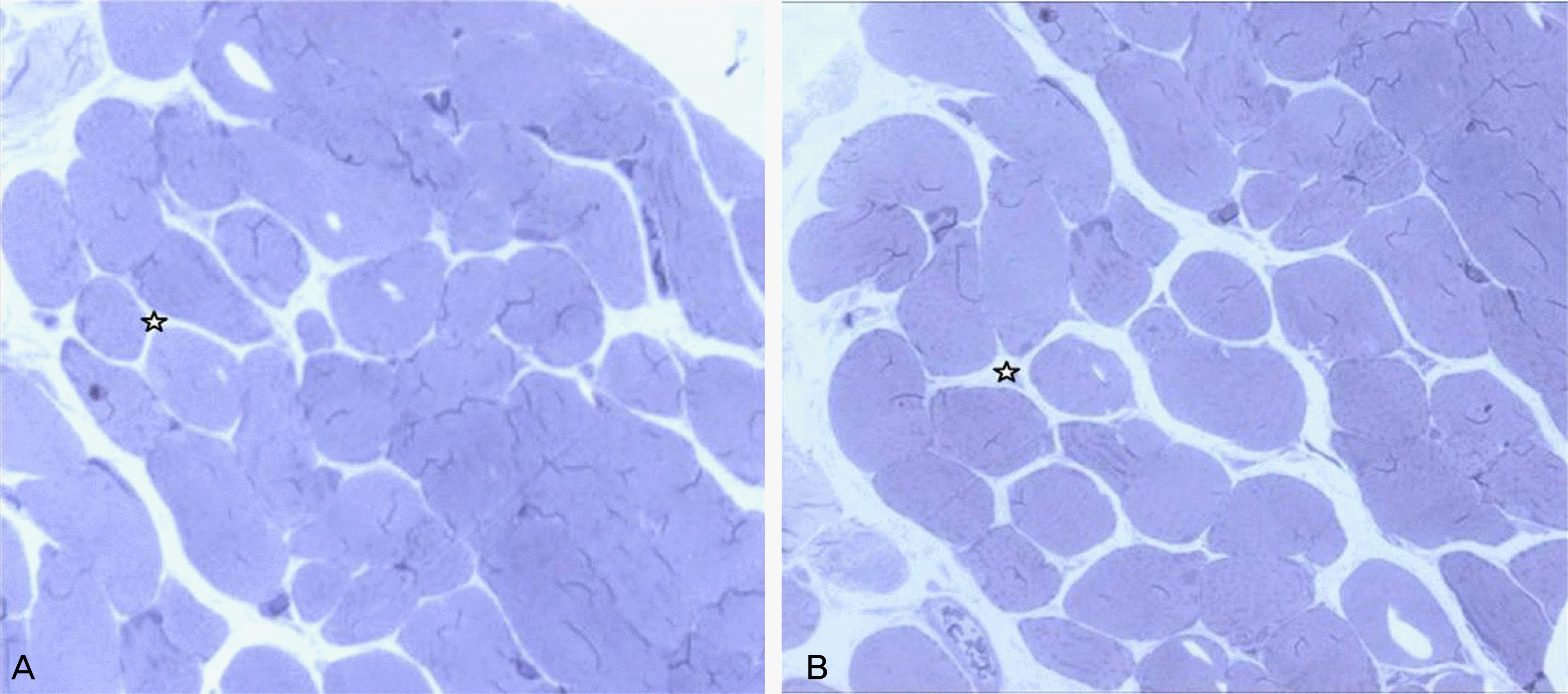
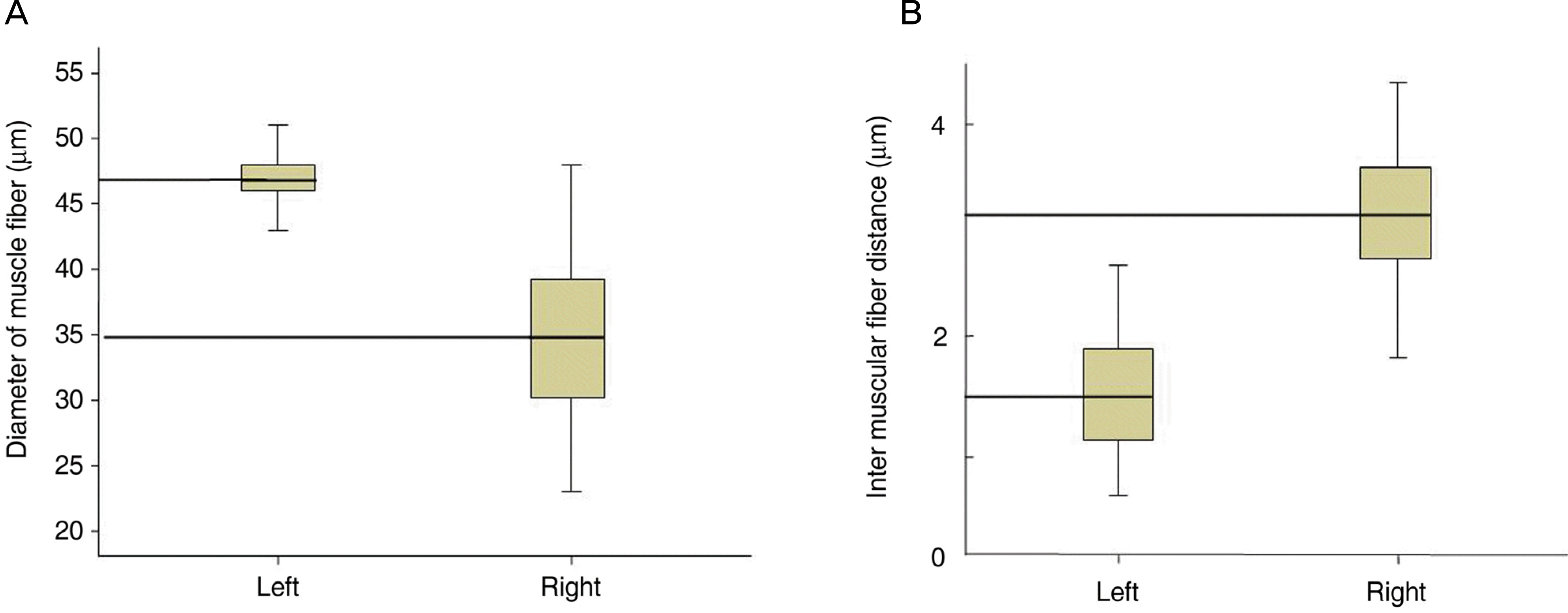
After 8 weeks, ptosis and enophthalmos were seen in the right eye in all rats. We found no change bilateral fat, but the right extraocular muscle fibers had smaller diameters than the left. The right intermuscular distance was longer than the left upon light microscopy and the right extraocular muscle contained fewer and smaller mitochondria than the left extraocular muscle upon electron microscopy.
Go to : 
References
1. Simoens P, Lauwers H, De Muelenare C, et al. Horner's syndrome in the horse: a clinical, experimental and morphological study. Equine Vet J Suppl. 1990; 10:62–5.

3. Firth EC. Horner's syndrome in the horse: experimental induction and a case report. Equine Vet J. 1978; 10:9–13.

4. Jones BR, Studdert VP. Horner's syndrome in the dog and cat as an aid to diagnosis. Aust Vet J. 1975; 51:329–32.

5. Bang YH, Park SH, Kim JH, et al. The role of Müller's muscle reconsidered. Plast Reconstr Surg. 1999; 103:1788–91.
6. Rubin LF. Diagnosis and treatment. Canine neurology. 2nd ed.Philadelphia: WB Saunders;1971. p. 502.
7. Lembo TM, Wright KC, Cromeens DM, et al. Iatrogenic Horner's syndrome in an experimental pig. Contemp Top Lab Anim Sci. 2001; 40:33–5.
9. Van der Wiel HL, van Gijn J. No enophthalmos in Horner's syndrome. J Neurol Neurosurg Psychiatry. 1987; 50:498–9.

10. Cavazza S, Bocciolini C, Gasparrini E, Tassinari G. Iatrogenic Horner's syndrome. Eur J Ophthalmol. 2005; 15:504–6.

11. Ozel SK, Kazez A. Horner's syndrome secondary to tube thoracostomy. Turk J Pediatr. 2004; 46:189–90.
12. Cozzaglio L, Coladonato M, Doci R, et al. Horner's syndrome as a complication of thyroidectomy: Report of a case. Surg Today. 2008; 38:1114–6.

13. Weinreb I, Goldstein D, Perez-Ordoñez B. Primary extraskeletal Ewing family tumor with complex epithelial differentiation: a unique case arising in the lateral neck presenting with Horner syndrome. Am J Surg Pathol. 2008; 32:1742–8.

14. Clauser L, Galiè M, Pagliaro F, et al. Posttraumatic enophthalmos: etiology, principles of reconstruction, and correction. J Craniofac Surg. 2008; 19:351–9.
15. Manson PN, Clifford CM, Su CT, et al. Mechanisms of global support and posttraumatic enophthalmos: The Anatomy of the ligament sling and its relation to intramuscular cone orbital fat. Plast Reconstr Surg. 1986; 77:193–202.
16. Kim YK, Park CS, Kim HK, et al. Correlation between changes of medial rectus muscle section and enophthalmos in patients with medial orbital wall fracture. J Plast Reconstr Aesthet Surg 2008 Oct 17.

17. Devoe AG. Experiences with the surgery of the anophthalmic orbit. Am J Ophthalmol. 1945; 28:1346–51.

19. Kronish JW, Gonnering RS, Dortzbach RK, et al. The pathophysiology of the anophthalmic socket. Part II. Analysis of orbital fat. Ophthal Plast Reconstr Surg. 1990; 6:88–95.

20. Lim NK, Kim TH, Song JK, Yoo JM. The effect of Sympath-ectomy on Circardian Rhythm of Intraocular Pressure in Rat. J Korean Ophthalmol Soc. 2008; 49:1839–44.

21. Brecher GA, Mitchell WG. Studies on the role of sympathectic nervous stimulation in extraocular muscle movements. Am J Ophthalmo. 1957; 44:144–9.
22. Blodi FC, Van allen MW. The effect of paralysis of the cervical sympathetic system on the electromyogram of extraocular muscles in the human. With particular reference to the straited levator in Horner's syndrome. Am J Ophthalmol. 1960; 49:679–83.
Go to : 
 | Figure 2.Pre-operative (A) and post-operative 8 weeks (B) of case 1 showed drooping of the upper lid and enophthalmos in the right eye. |
 | Figure 3.Light microscopy of the left rectus muscle (A) and right rectus muscle (B) at postoperative 8 weeks. It found that right extraocular muscle fibers had smaller diameter and wider interval than left. (Toluidine blue ×200) (☆ : intermuscular distance) |
 | Figure 4.Difference of the right and left muscle fiber diameters (A) and of the right and left intermuscular fiber distance (B). |


 PDF
PDF ePub
ePub Citation
 Print
Print


 XML Download