Abstract
Purpose
To investigate the relationship between intraocular pressure (IOP) and visual field defect progression (VFP) in normal- tension glaucoma (NTG).
Methods
We reviewed the records of patients who were enrolled according to the following inclusion criteria: at least one IOP measurement at every section, which was divided into four sections (90 minutes) by IOP measurement time and a follow-up for 2 years or more. Patients were divided into VFP (n=9) and non-visual field defect progression (NVFP, n=28) groups. The baseline IOP was defined as an average IOP measured five times with 90-minute intervals before treatment. The maximal, minimal and mean IOPs were defined as the highest, lowest and average IOPs among all checked IOPs during follow-up. IOP fluctuation was defined as the difference between the maximal and minimal IOPs. The section IOP was defined as an average IOP among all checked IOPs in each section, and section IOP fluctuation was the difference between the highest and lowest section IOPs. We reviewed and compared the IOP indices of the two groups and the risk factors, including hypertension, diabetes, migraine, familial history of glaucoma, disc hemorrhage, and number of eyedrops.
Results
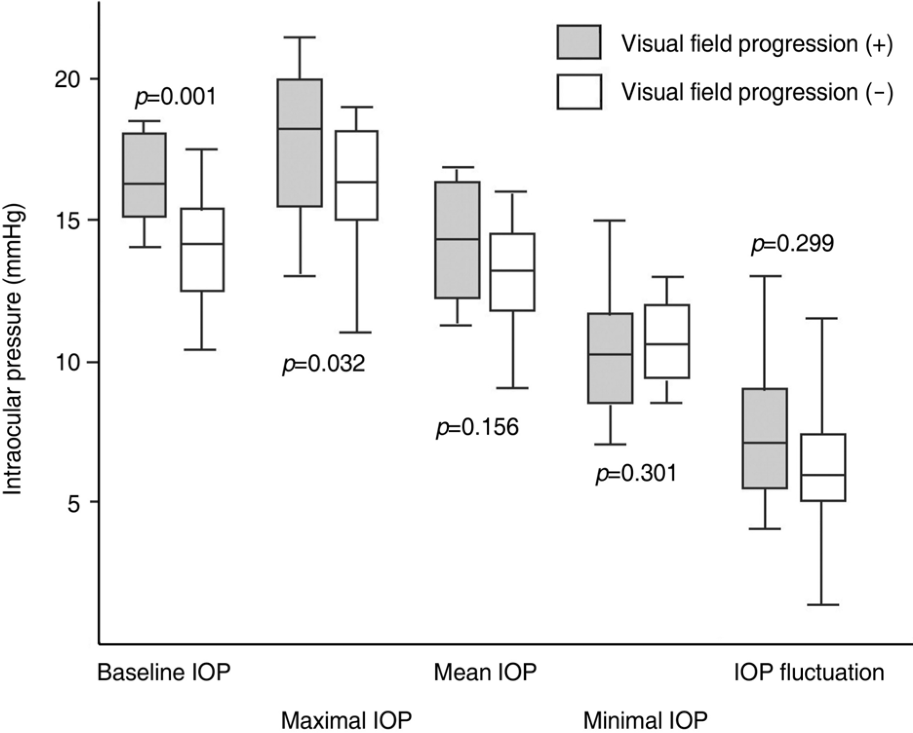
Thirty-seven eyes with an average follow-up of 50.4±18.9 months were included. The baseline and the maximal IOPs were higher than those of the NVFP group (p=0.001 and 0.032, respectively), but the mean, minimal and IOP fluctuations were not different (all, p >0.05). All section IOPs, section IOP fluctuations and other risk factors were not different (all, p >0.05).
Go to : 
References
1. Choe YJ, Hong YJ. The prevalence of glaucoma in Korean careermen. J Korean Ophthalmol Soc. 1993; 34:153–8.
2. Kwak HW, Joo MJ, Yoo JH. The significance of fundus photography without mydriasis during health mass screening. J Korean Ophthalmol Soc. 1997; 38:1585–9.
3. Cartwright MJ, Anderson DR. Correlation of asymmetric damage with asymmetric intraocular pressure in normal-tension glaucoma. Arch Ophthalmol. 1998; 106:898–900.
4. Araie M, Sekine M, Suzuki Y, Koseki N. Factors contributing to the progression of visual field damage in eyes with normal tension glaucoma. Ophthalmology. 1994; 101:1440–4.
5. Ishida K, Yamamoto T, Sugiyama K, Kitazawa Y. Disk hemorrhage is a significantly negative prognostic factor in normal-tension glaucoma. Am J Ophthalmol. 2000; 129:707–14.

6. Drance S, Anderson DR, Schulzer M. Risk factors for progression of visual field abnormalities in normal tension glaucoma. Am J Ophthalmol. 2001; 131:699–708.
7. Daugeliene L, Yamamoto T, Kitazawa Y. Risk factors for visual field damage progression in normal-tension glaucoma eyes. Graefes Arch Clin Exp Ophthalmol. 1999; 237:105–8.

8. Crichton A, Drance SM, Douglas GR, Schulzer M. Unequal intraocular pressure and its relation to asymmetric visual field defects in low-tension glaucoma. Ophthalmology. 1989; 96:1312–4.

9. Zeimer RC, Wilensky JT, Gieser DK, Viana MA. Association between intraocular pressure peaks and progression of visual field loss. Ophthalmology. 1991; 98:64–9.

10. Hughes E, Spry P, Diamond J. 24-hour monitoring of intraocular pressure in glaucoma management: a retrospective review. J Glaucoma. 2003; 12:232–6.

11. Sacca SC, Rolando M, Marletta A, et al. Fluctuations of intraocular pressure during the day in open-angle glaucoma, normal-tension glaucoma and normal subjects. Ophthalmologica. 1998; 212:115–9.

12. Caprioli J, Coleman AL. Intraocular pressure fluctuation a risk factor for visual field progression at low intraocular pressures in the advanced glaucoma intervention study. Ophthalmology. 2008; 115:1123–9.
13. Bengtsson B, Leske MC, Hyman L, Heijl A. Fluctuation of intraocular pressure and glaucoma progression in the early manifest glaucoma trial. Ophthalmology. 2007; 114:205–9.

14. Singh K, Shrivastava A. Intraocular pressure fluctuations: how much do they matter? Curr Opin Ophthalmol. 2009; 20:84–7.

15. Collaborative normal-tension glaucoma study group. Natural history of normal-tension glaucoma. Ophthalmology. 2001; 108:247–53.
16. Kim SH, Park KH. The relationship between recurrent optic disc hemorrhage and glaucoma progression. Ophthalmology. 2006; 113:598–602.

17. Collaborative normal-tension glaucoma study group. Comparison of glaucomatous progression between untreated patients with normal-tension glaucoma and patients with therapeutically reduced intraocular pressures. Am J Ophthalmol. 1998; 126:487–97.
18. Krieglstein G, Langham ME. Influence of body position on the intraocular pressure of normal and glaucomatous eyes. Ophthalmologica. 1975; 171:132–45.

19. Jain MR, Marmion VJ. Rapid pneumatic and Mackey-Marg applanation tonometry to evaluate the postural effect on intraocular pressure. Br J Ophthalmol. 1976; 60:687–93.

20. Tsukahara S, Sasaki T. Postural change of IOP in normalpersons and in patients with primary wide open-angle glaucoma and low-tension glaucoma. Br J Ophthalmol. 1984; 68:389–92.
21. De Vivero C, O'Brien C, Lanigan L, Hitchings R. Diurnal intraocular pressure variation in low-tension glaucoma. Eye. 1994; 8:521–3.

22. David R, Zangwill L, Briscoe D, et al. Diurnal intraocular pressure variationsan analysis of 690 diurnal curves. Br J Ophthalmol. 1992; 76:280–3.
23. Kano K, Kuwayama Y. Diurnal variation of intraocular pressure in normal-tension glaucoma. Nippon Ganka Gakkai Zasshi. 2003; 107:375–9.

24. Wilensky JT, Gieser DK, Dietsche ML, et al. Individual variability in the diurnal intraocular pressure curve. Ophthalmology. 1993; 100:940–4.

25. Asrani S, Zeimer R, Wilensky J, et al. Large diurnal fluctuations in intraocular pressure are an independent risk factor in patients with glaucoma. J Glaucoma. 2000; 9:134–42.

26. Ishida K, Yamamoto T, Kitazawa Y. Clinical factors associated with progression of normal-tension glaucoma. J Glaucoma. 1998; 7:372–7.

27. Kim NJ, Lee SM, Park KH, Kim DM. Factors associated with progression of visual field defect in normal tension glaucoma. J Korean Ophthalmol Soc. 2003; 44:1351–5.
Go to : 
 | Figure 1.The comparison of intraocular pressure indices of 37 eyes with and without visual field defect progression. IOP=intraocular pressure. Mann-Whitney U test. Boxes (25∼75%), bars (the highest and lowest intraocular pressure), and bars in the box (mean intraocular pressure) were displayed. |
Table 1.
Results of intraocular pressure (IOP, mmHg) indices of 37 eyes
| Parameters | Mean± standard deviation of IOP (range) |
|---|---|
| Baseline IOP | 14.62±1.98 (10.40∼18.40) |
| Minimal IOP | 10.51±1.69 (7.00∼15.00) |
| Mean IOP | 13.39±1.89 (9.00∼16.83) |
| Maximal IOP | 16.77±2.48 (11.00∼21.50) |
| IOP fluctuation* | 6.21±2.12 (1.33∼13.00) |
| 1st section IOP† | 13.39±1.89 (9.00∼16.83) |
| 2nd section IOP‡ | 13.59±1.73 (10.00∼16.73) |
| 3rd section IOP§ | 13.35±2.05 (9.00∼17.75) |
| 4th section IOP∏ | 13.62±2.33 (9.50∼18.00) |
| Section IOP fluctuation# | 1.95±0.77 (0.80∼3.67) |
Table 2.
The comparison of intraocular pressure (IOP, mmHg) indices, which were divided by IOP-measuring time, of 37 eyes with and without visual field defect progression (VFP)
| VEP (+) (n=9) Mean± SD (range) | VEP (-) (n=28) Mean± SD (range) | p value* | |
|---|---|---|---|
| 1st section IOP† | 14.24±1.94 | 12.88±1.90 | 0.173 |
| (11.2∼16.8) | (9.5∼16.4) | ||
| 2nd section IOP‡ | 13.92±1.83 | 13.43±1.65 | 0.387 |
| (11.3∼16.3) | (10.0∼16.5) | ||
| 3rd section IOP§ | 14.09±1.97 | 13.18±1.92 | 0.378 |
| (11.2∼16.7) | (9.0∼17.4) | ||
| 4th section IOP∏ | 13.91±2.45 | 13.30±2.19 | 0.151 |
| (10.5∼17.8) | (9.5∼18.0) | ||
| Section IOP fluctuation# | 2.13±0.91 | 1.90±0.74 | 0.469 |
| (0.9∼3.4) | (0.8∼3.7) |
Table 3.
The clinical data except intraocular pressure of 37 eyes with and without visual field defect progression (VFP)
| VFP (+) (n=9) | VFP (-) (n=28) | p value* | |
|---|---|---|---|
| Age (years) | 56.4±12.0 | 49.7±15.3 | 0.290 |
| Sex (M:F) | 2:7 | 9:19 | 0.695 |
| Follow-up (months) | 58.5±24.9 | 47.8±16.3 | 0.186 |
| Hypertension | 0 (0%) | 9 (32.1%) | 0.079 |
| Diabetes | 2 (22.2%) | 3 (10.7%) | 0.577 |
| Migraine | 0 (0%) | 2 (7.7%)† | 1.000 |
| Family history of glaucoma | 1 (11.1%) | 2 (7.1%) | 1.000 |
| Disc hemorrhage | 3 (33.3%) | 7 (25.0%) | 0.472 |
| LogMAR V/A‡ | 0.037±0.047 | 0.048±0.088 | 0.739 |
| Refractive error (SE§, diopters) | -1.51±3.52 | -1.56±3.26 | 0.909 |
| Initial MD∏ (dB) | -4.27±3.20 | -3.91±3.49 | 0.664 |
| Final MD (dB) | -5.05±3.24 | -3.28±3.66 | 0.138 |
| Number of eye drops | 1.55±0.46 | 1.29±0.50 | 0.180 |


 PDF
PDF ePub
ePub Citation
 Print
Print


 XML Download