

 PDF
PDF ePub
ePub Citation
 Print
Print


Abstract
Purpose
To compare the effects of early and late intravitreal injection of bevacizumab in patients with macular edema (ME) due to branch retinal vein occlusion (BRVO).
Methods
The study sample included 56 eyes of 56 patients who received intravitreal bevacizumab injection for ME due to BRVO and were followed up with at least six months of observation. We retrospectively divided eyes into two classes that included 36 eyes with a disease duration of ≤ 3 months (early treatment group) and 20 eyes with a disease duration of > 3 months (late treatment group). We assessed the effects of injection on the best corrected visual acuity (BCVA), central retinal thickness and IOP at one, three, and six months after treatment.
Results
There was no statistically significant differences in terms of sex, age, number of injections, ischemia, pre-injection visual acuity and central retinal thickness between the two treatment groups. Improvements in central retinal thickness were observed in both groups but were not significantly different between the groups. Visual acuity improved in both groups after treatment; the improvement was significantly better in the early treatment group observed three and six months after treatment. IOP did not change after treatment in either group.
References
1. Kim SY. Retina. 2nd ed. Seoul: Jinprint;2004. p. 551–2.
2. The Branch Vein Occlusion Study Group. Argon laserphotocoa-gulation for macular edema in branch vein occlusion. Am J Ophthalmol. 1984; 98:271–82.
3. Jonas JB, Akkoyun I, Kamppeter B, et al. Branch retinal vein occlusion treated by intravitreal triamcinolone acetonide. Eye. 2005; 19:65–71.

4. Rabena MD, Pieramici DJ, Castellarin AA, et al. Intravitreal bevacizumab (Avastin) in the treatment of macular edema secondary to branch retinal vein occlusion. Retina. 2007; 27:419–25.
5. Lardenoye CW, Probst K, DeLint PJ, Rothova A. Photoreceptor function in eyes with macular edema. Invest Ophthalmol Vis Sci. 2000; 41:4048–53.
6. Oh JY, Seo JH, Ahn JK, et al. Early versus late intravitreal triamcinolone acetonide for macular edema associated with branch retinal vein occlusion. Korean J Ophthalmol. 2007; 21:18–20.
7. Moon SW, Kim ES, Kim YG, et al. The Comparison of macular thickness measurements and repeatabilities between time domain and spectral domain OCT. J Korean Ophthalmol Soc. 2009; 50:1050–9.
8. Silva RM, Faria de Abreu JR, Cunha-Vaz JG. Blood-retina barrier in acute retinal branch vein occlusion. Graefes Arch Clin Exp Ophthalmol. 1995; 233:721–6.
9. Arnarsson A, Stefansson E. Laser treatment and the mechanism of edema reduction in branch retinal vein occlusion. Invest Ophthalmol Vis Sci. 2000; 41:877–9.
10. Noma H, Minamoto A, Funatsu H, et al. Intravitreal levels of vascular endothelial growth factor and interleukin-6 are correlated with macular edema in branch retinal vein occlusion. Graefes Arch Clin Exp Ophthalmol. 2006; 244:309–15.
11. Rosenfeld PJ, Fung AE, Puliafito CA. Optical coherence tomography findings after an intravitreal injection of bevacizumab (Avastin) for macular edema from central retinal vein occlusion. Ophthalmic Surg Lasers Imaging. 2005; 36:336–9.
13. Tso MO. Pathology of cystoid macular edema. Ophthalmology. 1982; 89:902–15.
14. Ota M, Tsujikawa A, Murakami T, et al. Foveal photoreceptor layer in eyes with persistent cystoid macular edema associated with branch retinal vein occlusion. Am J Ophthalmol. 2008; 145:273–80.
15. Rensch F, Jonas JB, Spandau UH. Early intravitreal bevacizumab for non-ischaemic branch retinal vein occlusion. Ophthalmologica. 2009; 223:124–7.
Figure 1.
Graph showing changes in central macular thickness after intravitreal bevacizumab injection. The error bars denote the standard errors of the means. * Statistically significant change from baseline at each visit within group (p<0.05).
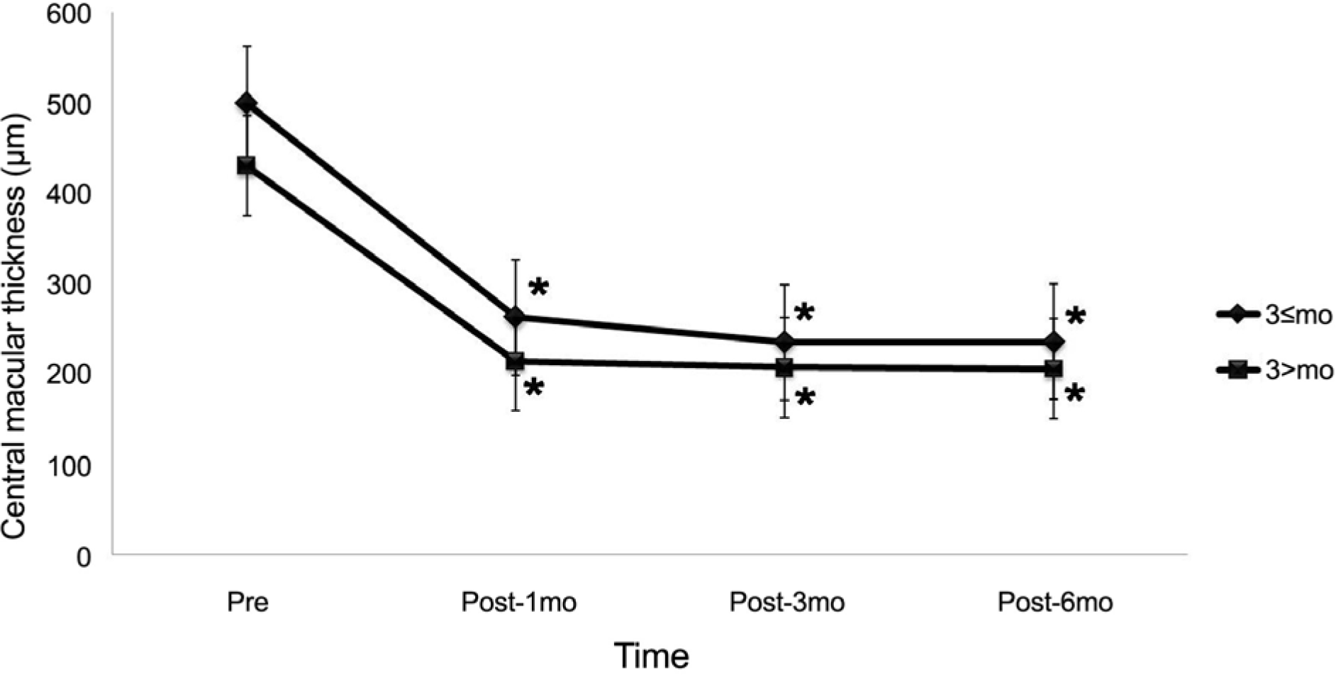
Figure 2.
Graph showing changes in visual acuity after intravitreal bevacizumab injection. The error bars denote the standard errors of the means.
* Statistically significant change from baseline at each visit within group (p<0.05); † Statistically significant difference between two groups at each follow-up visit (p<0.05).
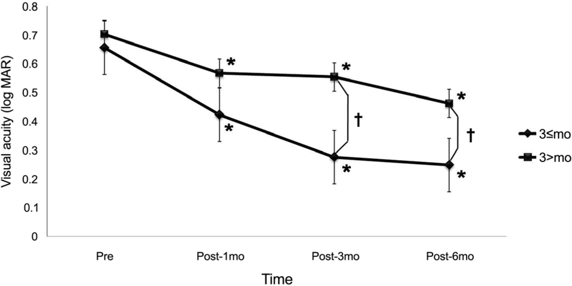
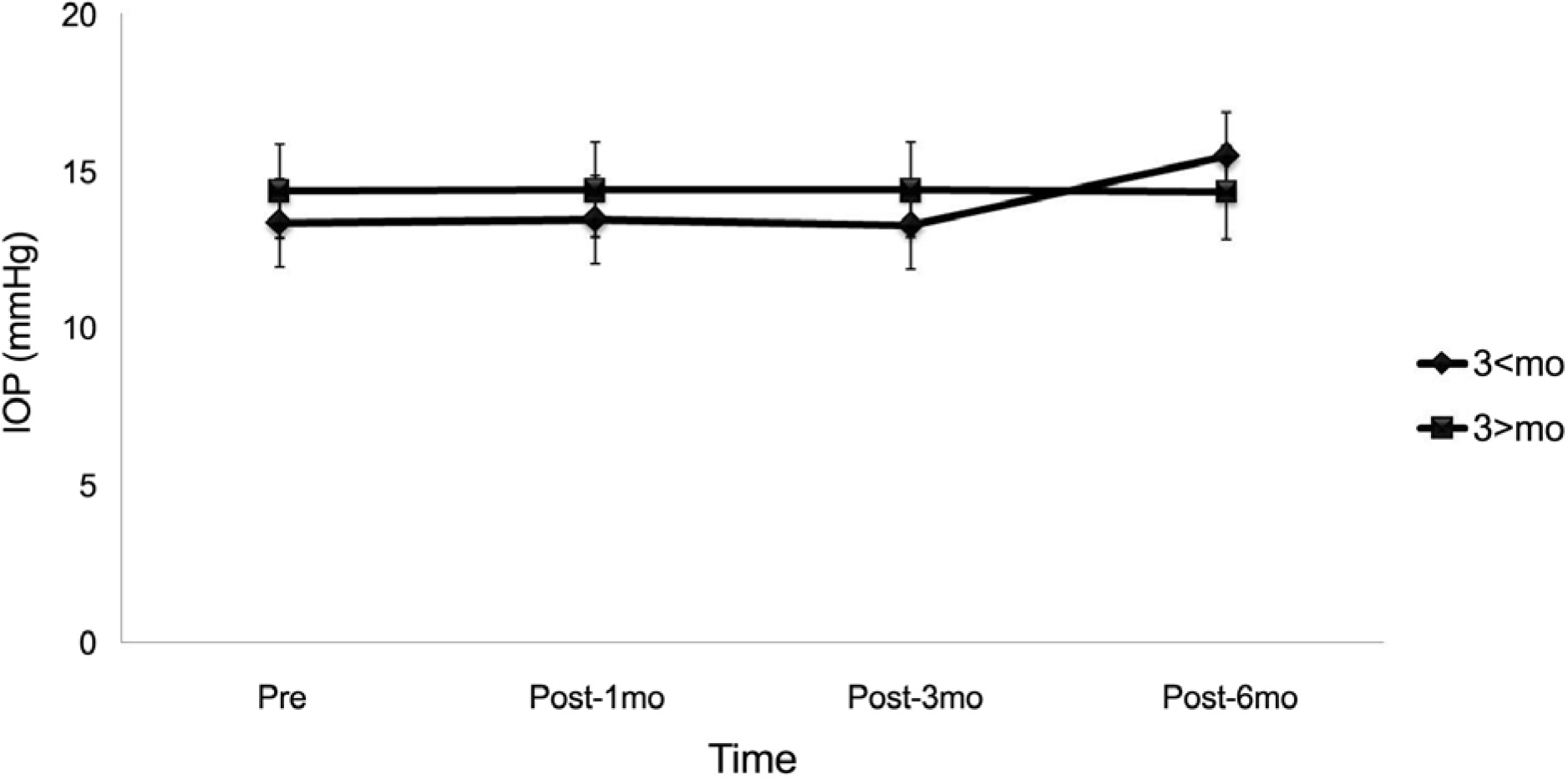
Table 1.
Patient demographics
| Characteristics | Early treatment (≤ 3mo) | Late treatment (> 3mo) | p-value |
|---|---|---|---|
| Numbers of eyes | 36 | 20 | |
| Sex (male/female) | 13/23 | 4/16 | 0.209 |
| Right/Left | 20/16 | 12/8 | 0.747 |
| Age | 58.83±7.8 | 56.3±8.6 | 0.268 |
| Symptom duration (week) | 6.4±2.7 | 26.4±18.0 | 0.011* |
| Numbers of injection | 3.19±0.8 | 3.25±1.3 | 0.848 |
| Ischemic/non-ischemic | 18/18 | 6/14 | 0.147 |
| Visual acuity (ETDRS) | 0.65±0.3 | 0.70±0.4 | 0.666 |
| Central macular thickness (um) | 498.7±207 | 430.0±199 | 0.234 |
 XML Download