Abstract
Purpose
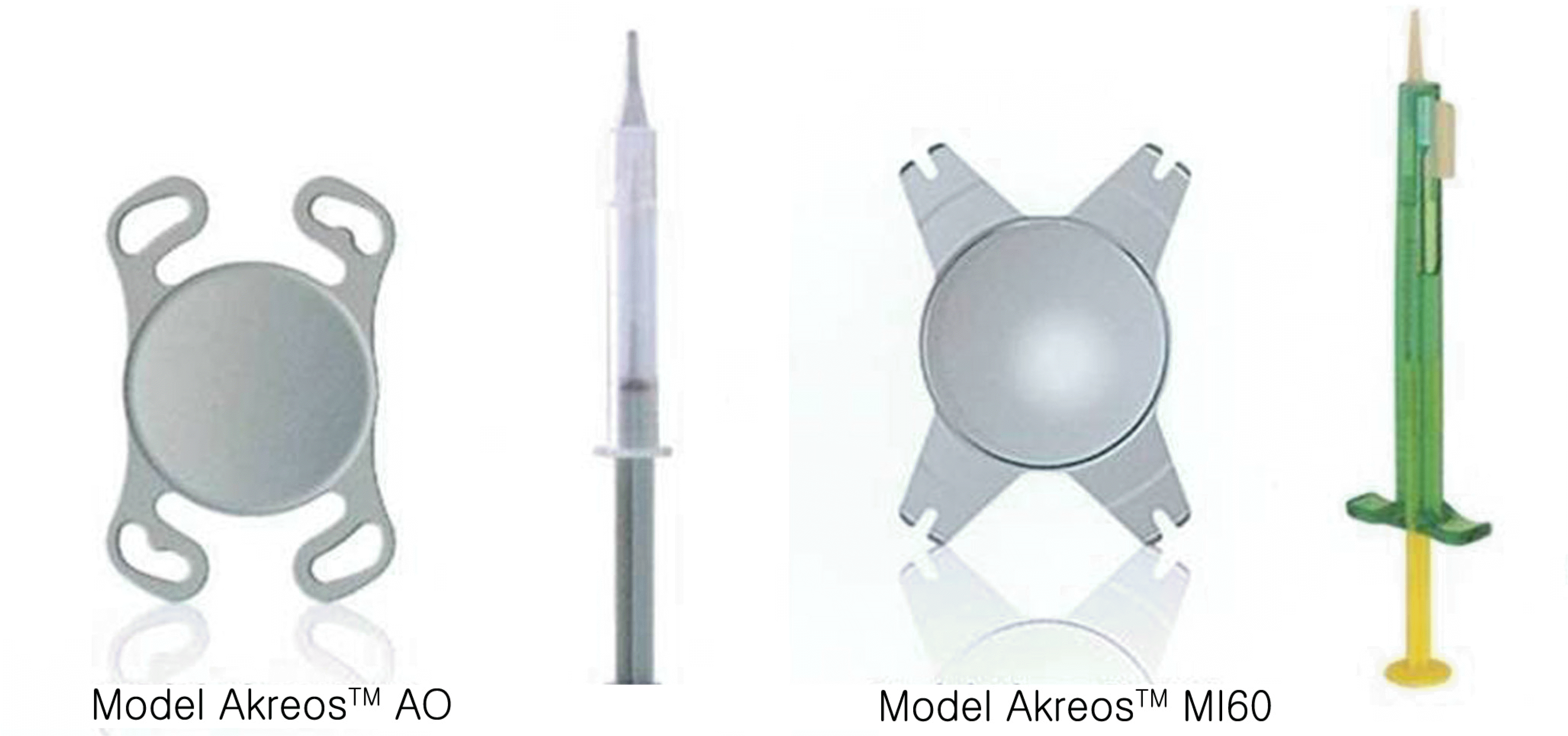
To compare clinical outcomes of aberration-free MI60 intraocular lens (IOL) in microincision cataract surgery and an aberration-free intraocular lens, Akreos AO in conventional cataract surgery.
Methods
Patients were randomly assigned to two IOL groups, and were examined preoperatively, and at one and three months postoperatively. The performed ophthalmologic evaluation included best corrected visual acuity (BCVA), measurement of refractive error, corneal astigmatism, and surgically induced astigmatism. The spherical, total, and higher-order aberration analysis of the two groups were assessed at one month and three months after operation.
Results
MI60 IOL group provided significantly less corneal astigmatism (p=0.020) one month after operation, compared to Akreos AO group. The MI60 group also showed significantly less surgically-induced astigmatism at one month (p=0.021) and three months postoperatively (p=0.043). There was no statistically significant difference in the spherical, higher-order, and total aberration between the two groups at one and three months after operation.
Go to : 
References
1. Grabow HB. Topical anesthesia for cataract surgery. Eur J Implant Refractive Surg. 1993; 5:20–4.
2. Gimbel HV, Neuhann T. Development advantages and methods of the continuous circular capsulorhexis technique. J Cataract Refract Surg. 1990; 16:31–7.

3. Dogru M, Honda R, Omoto M, et al. Early visual results with the rollable ThinOptx intraocular lens. J Cataract Refract Surg. 2004; 30:558–65.

4. Pandey SK, Werner L, Agarwal A, et al. Phakonit: cataract removal through a sub 1.0 mm incision and implantation of the Thin-Optx rollable intraocular lens. J Cataract Refract Surg. 2002; 28:1710–3.
5. Long DA, Monica ML. A prospective evaluation of corneal curva-ture change with 3.0 to 3.5mm corneal tunnel phacoemulsification. Ophthalmology. 1996; 103:226–32.
6. Alio' JL, Rodfiguez-Prats JL, Gala A, Ramzy M. Outcomes of microincision cataract surgery versus coaxial phacoemulsification. Ophthalmology. 2005; 112:1997–2003.
7. Mester U, Dillinger P, Anterist N. Impact of a modified optic design on visual function: clinical comparative study. J Cataract Refract Surg. 2003; 29:652–60.

8. Rawer R, Stork W, Spraul CW, Lingenfelder C. Imaging quality of intraocular lenses. J Cataract Refract Surg. 2005; 31:1618–31.

9. Artal P, Berrio E, Guirao A, Piers P. Contribution of the cornea and internal surface to the change of ocular aberrations with age. J Opt Soc Am A Opt Image Sci. 2002; 19:137–43.
10. Guirao A, Redondo M, Geraghty E, et al. Corneal optical aberrations and retinal image quality in patients in whom monofocal intraocular lenses were implanted. Arch Ophthalmol. 2002; 120:1143–51.

11. Applegate RA, Howland HC, Sharp RP, et al. Corneal aberrations and visual performance after keratotomy. J Refract Surg. 1998; 14:397–407.
12. Chalita MR, Chavala S, Xu M, Krueger RR. Wavefront analysis in post-LASIK eyes and its correlation with visual symptoms, refrac-tion, and topography. Ophthalmology. 2004; 111:447–53.

13. Altmann GE, Nichamin LD, Lane SS, Pepose JS. Optical performance of 3 intraocular lens designs in the presence of decentration. J Cataract Refract Surg. 2005; 31:574–85.

15. Jiang Y, Le Q, Yang J, Lu Y. Change in corneal astigmatism and higher order aberrations after clear corneal tunnel phacoemulsification guided by corneal topography. J Refract Surg. 2006; 22:S1083–8.

16. Steiner GA, Binder PS, Parker WT, Perl T. The natural and modified course of post-cataract astigmatism. Ophthalmic Surg. 1982; 13:822–7.
17. Hu YJ, Lee KH, Joo CK. Comparison of surgically induced astigmatism between superior and temporal clear corneal incision in sutureless cataract surgery. J Korean Ophthalmol Soc. 1998; 39:495–500.
18. Simsek S, Yasar T, Demirok A, et al. Effect of superior and temporal clear corneal incision on astigmatism after sutureless phacoemulsification. J Cataract Refract Surg. 1998; 24:515–8.
19. Hwang SJ, Choi SK, Oh SH, et al. Surgically induced astigmatism and corneal higher order aberrations in microcoaxial and conventional cataract surgery. J Korean Ophthalmol Soc. 2008; 49:1597–602.

20. Ku HC, Kim HJ, Joo CK. The comparison of astigmatism according to the incision size in small incision cataract surgery. J Korean Ophthalmol Soc. 2005; 46:416–21.
21. Cavallini GM, Campi L, Masini C, et al. Bimanual micro phacoemulsification versus coaxial mini phacoemulsification: prospective study. J Cataract Refract Surg. 2007; 33:387–92.
22. Kurz S, Krummenauer F, Gabriel P, et al. Biaxial microincision versus coaxial small-incision clear corneal cataract surgery. Ophthalmology. 2006; 113:1818–26.
23. Oshika T, Klyce SD, Applegate RA, Howland HC. Changes in corneal wavefront aberrations with aging. Invest Ophthalmol Vis Sci. 1999; 40:1351–5.
24. Artal P, Guirao A, Berrio E, Williams DR. Compensation of corneal aberrations by the internal optics in the human eye. J Vis. 2001; 1:1–8.

25. Applegate RA, Howland HC, Sharp RP, et al. Corneal aberrations and visual performance after radial keratotomy. J Refract Surg. 1998; 14:397–407.

26. Rocha K, Soriano E, Chalita M, et al. Wavefront analysis and contrast sensitivity of aspheric and spherical intraocular lenses. Am J Ophthalmol. 2006; 142:750–6.
27. Tzelikis P, Akaishi L, Trindade F, Boteon JE. Ocular aberration and contrast sensitivity after cataract surgery with AcrySof IQ intraocular lens implantation. J Cataract Refract Surg. 2007; 33:1918–24.
28. Caporossi A, Martone G, Casprini F, Rapisarda L. Prospective randomized study of clinical performanceof 3 aspheric and 2 spherical intraocular lenses in 250 eyes. J Refract Surg. 2007; 23:639–48.
29. Kim HS, Kim SW, Ha BJ, et al. Ocular aberrations and contrast sensitivity in eyes implanted with aspheric and spherical intraocular lenses. J Korean Ophthalmol Soc. 2008; 49:1256–62.

30. Rocha K, Soriano E, Chamon W, et al. Spherical aberration and depth of focus in eyes implanted with aspherical and spherical intraocular lenses. Ophthalmology. 2007; 114:2050–4.
Go to : 
Table 1.
Characteristics of the two IOL in the study
| IOL* Characteristics | Akreos AO | MI60 |
|---|---|---|
| Type | 1 piece | 1 piece |
| Optic material | 26% Acrylic material | 26% Hydrophilic acrylic |
| Refractive index | 1.458 | 1.458 |
| Optic shape | Biconvex aspheric anterior and posterior | Biconvex aspheric anterior and posterior |
| Haptics shape | One-piece | One-piece |
| 10˚ average angulation | 0˚ average angulation |
Table 2.
Preoperative and postoperative visual acuity and refraction, corneal astigmatism, surgically induced astigmatism of Akreos AO
| UCVA* | BCVA† | SE‡ | Cor. Astig.§ | SIAП | |
|---|---|---|---|---|---|
| Pre op. | 0.44±0.27 | 0.56±0.33 | 0.13±1.87 | 0.62±0.28 | |
| Post op. 1M | 0.66±0.28 | 0.86±0.22 | -0.28±0.43 | 1.05±0.36 | 1.06±0.75 |
| Post op. 3M | 0.78±0.25 | 0.94±0.15 | -0.33±0.76 | 0.87±0.44 | 0.93±0.55 |
| p-value | 0.006 | 0.004 | 0.262 | 0.010 | 0.620 |
Table 3.
Preoperative and postoperative visual acuity and refraction, corneal astigmatism, surgically induced astigmatism of MI60
| UCVA* | BCVA† | SE‡ | Cor. Astig.§ | SIAП | |
|---|---|---|---|---|---|
| Pre op. | 0.36±0.25 | 0.57±0.26 | -0.15±1.72 | 0.67±0.39 | |
| Post op. 1M | 0.65±0.16 | 0.91±0.10 | -0.85±0.53 | 0.63±1.85 | 0.58±0.33 |
| Post op. 3M | 0.58±0.18 | 0.93±0.08 | -0.92±0.64 | 0.71±0.47 | 0.59±0.37 |
| p-value | 0.000# | 0.000# | 0.028 | 0.921 | 0.970 |
Table 4.
Comparison of postoperative corneal astigmatism, surgically induced astigmatism, difference of corneal astigmatism
| Akreos AO | MI60 | p-value | |
|---|---|---|---|
| Cor. Astig* | |||
| Post op 1M | 1.05±0.36 | 0.63±0.36 | 0.020 |
| Post op 3M | 0.87±0.44 | 0.71±0.47 | 0.254 |
| Cor. Astig Diff.† | |||
| Post op 1M | 0.43±0.44 | 0.06±0.36 | 0.005 |
| Post op 3M | 0.25±0.54 | -0.02±0.51 | 0.314 |
| SIA‡ | |||
| Post op 1M | 1.06±0.75 | 0.58±0.33 | 0.021 |
| Post op 3M | 0.93±0.55 | 0.59±0.37 | 0.043 |
Table 5.
Comparison of aberrations in each study group
| Akreos AO 1M | MI60 1M | Akreos AO 3M | MI60 3M | p-value (1M/3M) | |
|---|---|---|---|---|---|
| RMS total* | 1.73±0.65 | 2.41±1.33 | 1.43±0.62 | 2.91±1.82 | 0.117/0.012 |
| Sph A† | 0.25±0.21 | 0.09±0.28 | 0.17±0.27 | -0.07±0.75 | 0.067/0.117 |
| (entire) | |||||
| SphA† | -0.02±0.2 | -0.06±0.35 | -0.02±0.24 | -0.27±0.75 | 0.789/0.079 |
| (int. optics§) | |||||
| SphA† | 0.27±0.09 | 0.25±0.19 | 0.22±0.11 | 0.21±0.14 | 0.509/0.682 |
| (Cornea) | |||||
| HOtotal‡ | 1.00±0.42 | 1.05±0.84 | 0.96±0.51 | 1.60±1.56 | 0.682/0.762 |
| Int. optics§ | |||||
| Trefoil6 | 0.05±0.49 | 0.33±0.51 | 0.15±0.57 | 0.40±0.56 | 0.155/0.215 |
| Trefoil9 | -0.48±0.47 | -0.01±0.37 | -0.18±0.47 | -0.04±0.56 | 0.020/0.307 |
| Coma7 | 0.10±0.45 | 0.15±0.75 | 0.02±0.52 | -0.16±1.17 | 0.343/0.929 |
| Coma8 | 0.08±0.39 | 0.01±0.20 | 0.05±.026 | 0.12±0.67 | 0.190/0.682 |
| Entire | |||||
| Trefoil6 | 0.05±0.37 | 0.22±0.43 | 0.10±0.35 | 0.25±0.49 | 0.290/0.361 |
| Trefoil9 | -0.15±0.39 | 0.03±0.22 | -0.02±0.45 | -0.07±0.63 | 0.190/0.762 |
| Coma7 | 0.08±0.29 | 0.17±0.57 | -0.04±0.27 | -0.04±1.12 | 0.343/0.509 |
| Coma8 | 0.05±0.38 | -0.03±0.18 | 0.04±0.27 | 0.07±0.69 | 0.244/0.762 |
| Cornea | |||||
| Trefoil6 | 0.00±0.18 | -0.05±0.27 | -0.06±0.29 | -0.11±0.21 | 0.605/0.325 |
| Trefoil9 | 0.33±0.29 | 0.15±0.15 | 0.16±0.21 | 0.10±0.15 | 0.062/0.556 |
| Coma7 | -0.01±0.21 | 0.11±0.29 | -0.04±0.28 | 0.10±0.18 | 0.290/0.202 |
| Coma8 | 0.05±0.23 | 0.03±0.18 | 0.00±0.22 | 0.00±0.16 | 0.789/0.762 |


 PDF
PDF ePub
ePub Citation
 Print
Print


 XML Download