Abstract
Purpose
We report a case of a patient with no preoperative light perception who achieved complete visual recovery after transsphenoidal resection of a pituitary adenoma.
Case summary
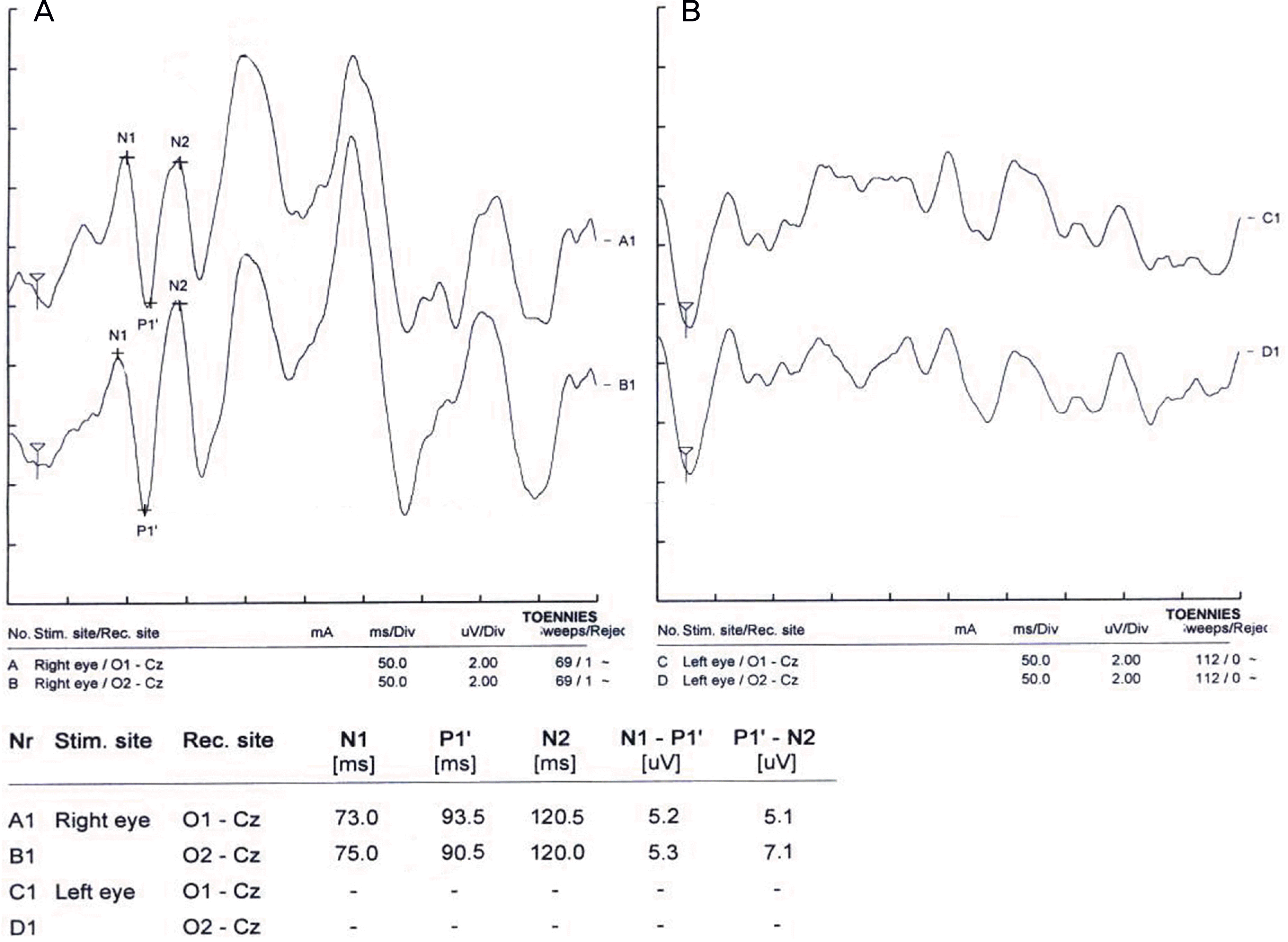
A 53-year-old man with a history of pituitary adenomectomy visited our clinic complaining of decreased vision of 7 weeks ducation. His best-corrected visual acuity was 1.2 in his right eye, and he had no light perception (NLP) in his left eye. Light reflex was absent in the left eye, and no consistent wave was detected in the left eye on flash visual evoked potential (FVEP). A pituitary adenoma 3×3.5 cm in diameter was found following magnetic resonance imaging (MRI), and was removed using a transsphenoidal approach and gamma knife radiosurgery. Six weeks postoperatively, his best corrected visual acuity improved to 1.0 in the left eye.
Go to : 
References
1. Asa SL, Kovacs K. Clinically non-functioning human pituitary adenomas. Can J Neurol Sci. 1992; 19:228–35.

2. Melen O. Neuro-opthalmologic features of pituitary tumors. Endocrinol Metab clin North Am. 1987; 16:585–608.
3. Jallu A, Kanaan I, Rahm B, Siqueira E. Suprasellar meningioma and blindness: a unique experience in Saudi Arabia. Surg Neurol. 1996; 45:320–3.

4. Kang SM, Kim JH, Cheong JH. Relationship between location and size of pituitary adenoma and visual field change. J Korean Ophthalmol Soc. 2005; 46:1690–6.
5. Elkington SG. Pituitary adenoma. Preoperative symptomatology in a series of 260 patients. Br J Ophthalmol. 1968; 52:322–8.

6. Dekkers OM, de Keizer RJ, Roelfsema F, et al. Progressive improvement of impaired visual acuity during the first year after transsphenoidal surgery for non-functioning pituitary macro-adenoma. Pituitary. 2007; 10:61–5.

7. Bampoe J, Ranalli P, Bernstein M. Postoperative reversal of complete (monocular) blindness in skull base meningioma: case report. Can J Neurol Sci. 2003; 30:72–4.

8. McGirt MJ, Cowan JA Jr, Gala V, et al. Surgical reversal of prolonged blindness from a metastatic neuroblastoma. Childs Nerv Syst. 2005; 21:583–6.

9. Suri A, Narang KS, Sharma BS, Mahapatra AK. Visual outcome after surgery in patients with suprasellar tumors and preoperative blindness. J Neurosurg. 2008; 108:19–25.

10. Clifford-Jones RE, Landon DN, McDonald WI. Remyelination during optic nerve compression. J Neurol Sci. 1980; 46:239–43.

11. Smith EJ, Blakemore WF, McDonald WI. Central remyelination restores secure conduction. Nature. 1979; 280:395–6.

12. Kerrison JB, Lynn MJ, Baer CA, et al. Stages of improvement in visual fields after pituitary tumor resection. Am J Ophthalmol. 2000; 130:813–20.

Go to : 
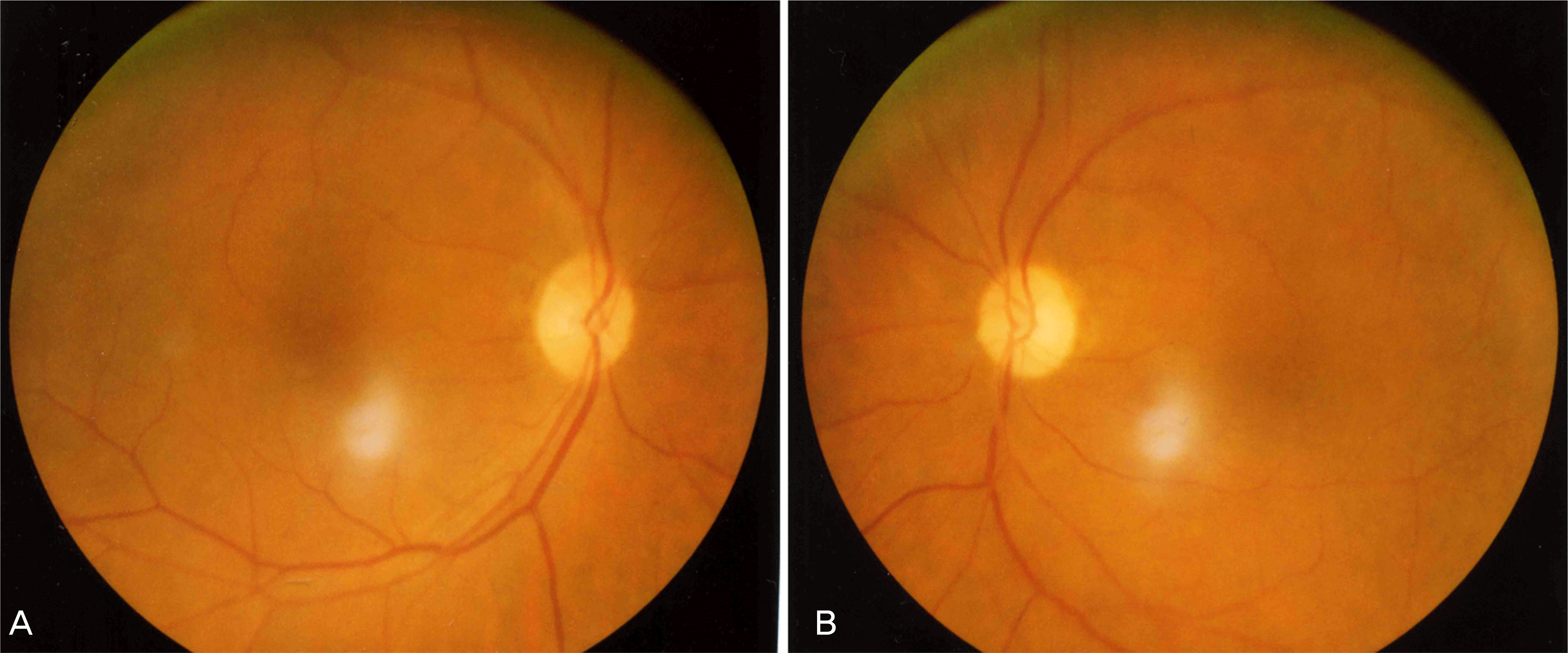
 | Figure 1.Preoperative fundus photographs show a relatively pale optic disc in the left eye. |
 | Figure 2.Preoperative flash visual evoked potential (FVEP) of the right (A) and the left eye (B). No consistent wave is detected in the left eye compared with normal latency seen in the right eye. |
 | Figure 3.Humphrey visual field test of the right eye (A) preoperatively and the left (B) and the right eye (C) 6 weeks after transsphenoidal pituitary adenoma resection. Postoperatively, generalized reduction in the sensitivity and constricted visual field is found in the left eye. |
 | Figure 4.Preoperative coronal (A) and sagittal (B) enhanced T1-weighted MR image showed peripheral enhancing sellar and suprasellar mass markedly compressing the optic chiasm (arrow). Postoperative coronal (C) and sagittal (D) enhanced T1-weighted MR image showed decreased degree of optic chiasm compression by the partial tumor resection. |


 PDF
PDF ePub
ePub Citation
 Print
Print


 XML Download