

 PDF
PDF ePub
ePub Citation
 Print
Print


Abstract
Case summary
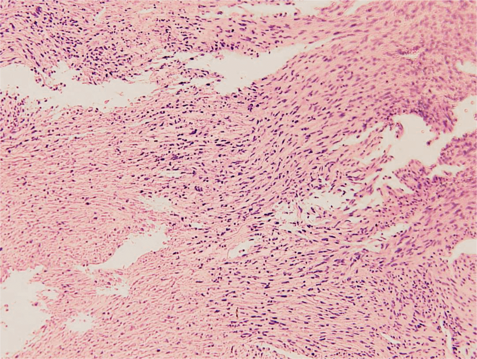
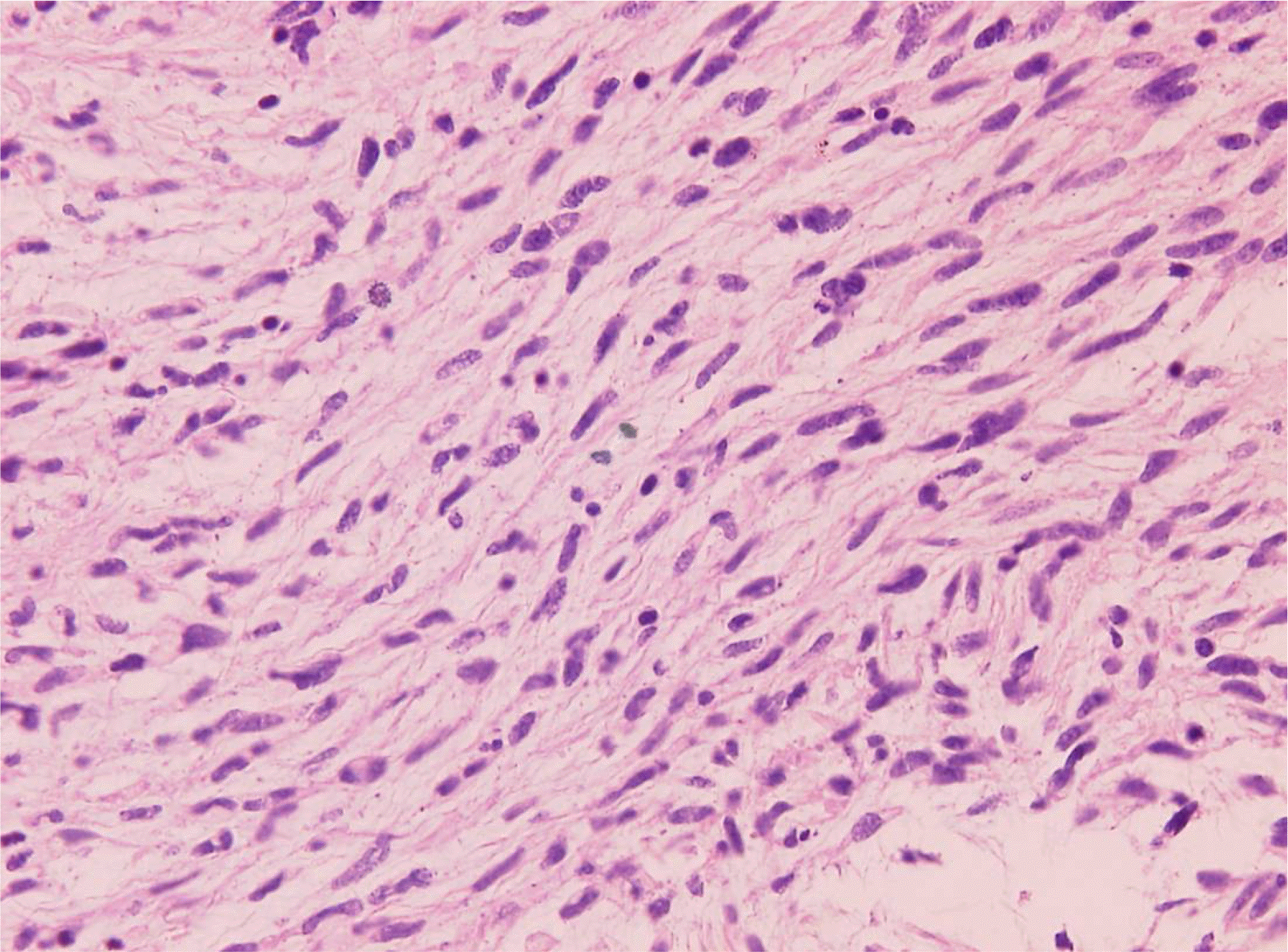
A 66-year-old man was evaluated for swelling of the left upper eyelid without pain that developed 2 months earlier. ACT scan and MRI of the orbit showed a large superior orbital mass with adjacent bony erosion, which had a central necrotic area. Incisional biopsy of the orbital mass was performed through a sub-brow incision. Histopathologic examination revealed a hypercellular tumor composed of spindle-shaped pleomorphic cells arranged in a fascicular pattern with necrotic foci. High-power magnification showed mitotic figures and nuclear pleomorphism. Subsequently, a malignant peripheral nerve sheath tumor was diagnosed.
References
1. Gupta TK, Brasfield RD. Solitary malignant schwannoma. Ann Surg. 1970; 171:419–28.
2. Jakobiec FA, Font RL, Zimmerman LE. Malignant peripheral nerve sheath tumours of the orbit: a clinicopathologic study of eight cases. Trans Am Ophthalmol Soc. 1985; 83:332–66.
3. Sack GH Jr. Malignant complications of neurofibromatosis. Clin Oncol. 1983; 9:17–23.
4. Enzinger FM, Weiss SW. Malignant tumors of the peripheral nerves. Soft Tissue Tumors. 1995; 32:889–928.
5. D'Agostino AN, Soule EH, Miller RH. Primary malignant neo-plasm of nerves (malignant neurilemomas) in patients without manifestations of multiple neurofibromatosis (von Recklinghau-sen's disease). Cancer. 1963; 16:1003–14.
6. Ducatman BS, Scheithauer BW. Post-irradiation neurofibrosar-coma. Cancer. 1983; 51:1028–33.

7. Ducatman BS, Scheithauer BW, Piepgras DG, et al. Malignant peripheral nerve sheath tumours. A clinicopathologic study of 120 cases. Cancer. 1986; 57:2006–21.
8. Wanebo JE, Malik JM, Vandenberg SR, et al. Malignant peripheral nerve sheath tumors. A clinicopathological study of 28 cases. Cancer. 1993; 71:1247–53.
9. Chitale AR, Dickersin GR. Electron microscopy in the diagnosis of malignant schwannomas. A report of six cases. Cancer. 1983; 51:1448–61.
10. Daimaru Y, Hashimoto H, Enjoji M. Malignant peripheral nerve-sheath tumors malignant schwannomas): An immunohisto-chemical study of 29 cases. Am J Surg Pathol. 1985; 9:434–44.
11. Stull MA, Moser RP Jr, Kransdorf MJ, et al. Magnetic resonance appearance of peripheral nerve sheath tumors. Skeletal Radiol. 1991; 20:9–14.
12. Colmero C, Rives T, Patron M, et al. Maxillofacial malignant peripheral nerve sheath tumours. J Craniomaxillofac Surg. 1991; 19:40–6.
13. Sordillo PP, Helson L, Hajdu SI, et al. Malignant schwannoma- clinical characteristics, survival, and response to therapy. Cancer. 1981; 47:2503–9.
14. Wanebo J, Malik J, Vanden Berg S, et al. Malignant peripheral nerve sheath tumors. Cancer. 1993; 71:1247–53.
15. Barbara SD, Bernd WS, David GP, et al. Malignant peripheral nerve sheath tumors. Cancer. 1986; 57:2006–21.
16. Greager JA, Reichard KW, Campana JP, et al. Malignant schwannoma of the head and neck. Am J Surg. 1992; 163:440–2.
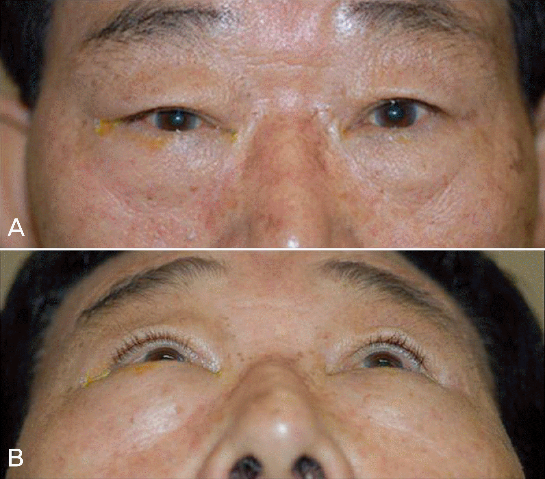
Figure 2.
(A) Axial CT reveals a large superior orbital mass with adjacent bony erosion. (B) Coronal CT depicts invasion of the superior muscle by the mass with destruction of the superior orbital wall.
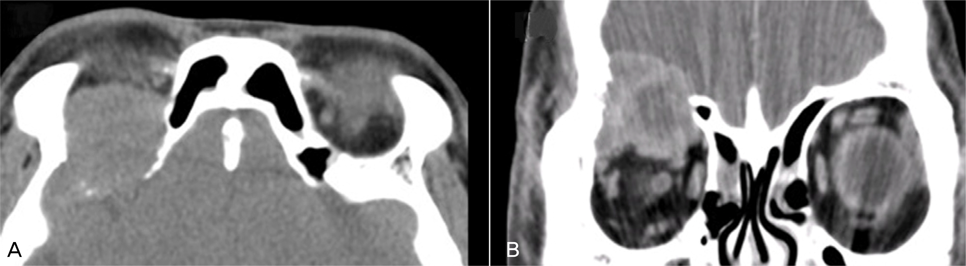
Figure 3.
(A) T1-weighted precontrast axial image demonstrating a well-defined mass in the right orbit with central necrosis. (B) T2-weighted precontrast axial image showing high signal intensity in the central necrotic area. (C, D, E) T1-weighted postcontrast images demonstrating significant homogenous peripheral enhancement and invasion into the dura mater without intracranial extension.

 XML Download