Abstract
Purpose
To compare anterior segment parameters in angle‐ closure glaucoma (ACG), primary open angle glaucoma (POAG), and normal subjects (N) using a Schiempflug camera.
Methods
Central corneal thickness (CCT), lens thickness (LT), axial length (AL), anterior chamber angle (ACA), anterior chamber depth (ACD), and anterior chamber volume (ACV) were measured in ACG (93 eyes of 92 patients), POAG (90 eyes of 87 patients), and normal (91 eyes of 88 subjects) with PentacamⓇ and A‐ scan. All of the results and measurements were then compared.
Results
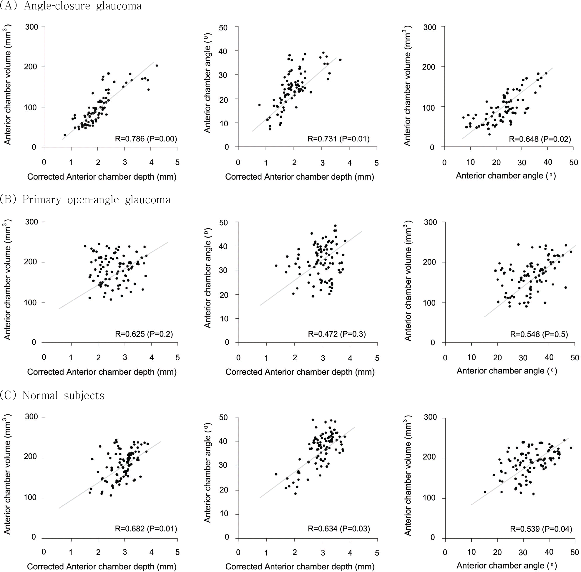
Compared to normal and POAG patients, ACG patients presented with significantly different measurements of CCT, LT, AL, and ACA, ACD, and ACV (p<0.05). Further, correlations were high between three measurements (ACA, ACD, ACV) in ACG, and the best correlations were found in acute angle‐ closure glaucoma (P<0.05).
Conclusions
By using a Schiempflug camera it was possible to assess the correlation between anterior segment parameters (ACA, ACD, ACV) in glaucoma patients. The best correlations were found in acute angle‐closure glaucoma, and thus anterior segment parameters can offer reciprocally complementary information.
Go to : 
References
1. Holladay JT. Standardizing constants for ultrasonic biometry, keratometry, and intraocular lens power calculations. J Cataract Refractive Surg. 1997; 23:1356–70.

2. Kim YY, Jung HR. Clarifying the nomenclature for primary angle closure glaucoma. Surv Ophthalmol. 1997; 42:125–36.
3. Ang MH, Baskaran M, Kumar RS. . National survey of ophthalmologists in Singapore for the assessment and management of asymptomatic angle closure. J Glaucoma. 2008; 17:1–4.

4. Lavanya R, Wong TY, Friedman DS. . Determinants of angle closure in older Singaporeans. Arch Ophthalmol. 2008; 126:686–91.

5. Koranyi G, Lydahl E, Norrby S, Taube M. Anterior chamber depth measurement: A‐ scan versus optical methods. J Cataract Refract Surg. 2002; 28:243–7.
6. Meinhardt B, Stachs O, Stave J. . Evaluation of biometric methods for measuring the anterior chamber depth in the non ‐contact mode. Graefes Arch Clin Exp Ophthalmol. 2006; 244:559–64.
7. Ryu HW, Kim KR, Chung SK. Comparison of A‐ scan, scheimpflug camera, and orbscan for measurement of anterior chamber depth. J Korea Ophthalmol Soc. 2006; 47:1287–91.
8. Giers U, Epple C. Comparison of A‐ scan device accuracy. J Cataract Refract Surg. 1990; 16:235–42.
9. Konstantopoulos A, Hossain P, Anderson DF. Recent advances in ophthalmic anterior segment imaging: a new era for ophthalmic diagnosis? Br J Ophthalmol. 2007; 91:551–7.
10. Swartz T, Marten L, Wang M. Measuring the cornea: the latest developments in corneal topography. Curr Opin Ophthalmol. 2007; 18:325–33.

11. Elbaz U, Barkana Y, Gerber Y. . Comparison of different techniques of anterior chamber depth and keratometric measurements. Am J Ophthalmol. 2007; 143:48–53.

12. Emre S, Doganay S, Yologlu S. Evaluation of anterior segment parameters in keratoconic eyes measured with the Pentacam system. J Cataract Refract Surg. 2007; 33:1708–12.

13. Friedman DS, Gazzard G, Foster P. . Ultrasonographic biomicroscopy, Scheimpflug photography, and novel provocative tests in contralateral eyes of Chinese patients initially seen with acute angle closure. Arch Ophthalmol. 2003; 121:633–42.

14. Bourne RR, Alsbirk PH. Anterior chamber depth measurement by optical pachymetry: systematic difference using the Haag‐ Streit attachments. Br J Ophthalmol. 2006; 90:142–5.
15. Foster PJ, Devereux JG, Alsbirk PH. Detection of gonioscopically occludable angles and primary angle closure glaucoma by estimation of limbal chamber depth in Asians: modified grading scheme. Br J Ophthalmol. 2000; 84:186–92.

16. Congdon NG, Spaeth GL, Augsburger J. . A proposed simple method for measurement in the anterior chamber angle: biometric gonioscopy. Ophthalmology. 1999; 106:2161–7.
17. Lowe R. Primary creeping angle‐ closure glaucoma. Br J Ophthalmol. 1964; 48:544–50.
18. Foster PJ, Buhrmann R, Quigley HA, Johson GJ. The definition and classification of glaucoma in prevalence surveys. Br J Ophthalmol. 2002; 86:238–42.

19. Shields MB, Ritch R. The secondary glaucomas. St Louis: CV Mosby;1982. p. 3–7.
20. Tomlision A, Leighton DA. Ocular dimensions in the heredity of angle closre glaucoma. Br J Ophthalmol. 1973; 57:475–86.
21. Dandona L, Dandona R, Mandal P. . Angle closure glaucoma in an urban population in South India. Ophthal-mology. 2000; 107:1710–6.
22. Lowe RF. Aetiology of the anatomical basis for primary angle closure glaucoma. Biometrical comparisons between normal eyes and eyes with primary angle closure glaucoma. Br J Ophthalmol. 1970; 54:161–9.
23. Sihota R, Lakshmaiah NC, Agarwal HC. . Ocular parameters in the subgroups of angle closure glaucoma. Clin Experiment Ophthalmol. 2000; 28:253–8.

24. Oka N, Otori Y, Okada M. . Clinical study of anterior ocular segment topography in angle‐ closure glaucoma using the three‐ dimensional anterior segment analyzer Pentacam. Nippon Ganka Gakkai Zasshi. 2006; 110:398–403.
25. Lee DA, Brubaker RF, Ilstrup DM. Anterior chamber dimensions in patients with narrow angles and angle‐ closure glaucoma. Arch Ophthalmol. 1984; 102:46–50.
26. Congdon N, Wang F, Tielsch JM. Issues in the epidemiology and population‐ based screening of primary angle‐ closure glaucoma. Surv Ophthalmol. 1992; 36:411–23.
27. Javitt J, Sommer A. A population‐ based evaluation of glaucoma screening: the Baltimore eye surgery. Am J Ophthalmol. 1991; 134:1102–10.
28. Shiose Y, Kitazawa Y, Tsukuhara S. . Epidemiology of glaucoma in Japan: a nationwide glaucoma survey. Jpn J Ophthalmol. 1991; 35:133–55.
29. Tomlinson A, Leighton DA. Ocular dimensions and the heredity of open‐ angle glaucoma. Br J Ophthalmol. 1974; 58:68–74.
30. Silver DM, Quigley HA. Aqueous flow through the iris‐ lens channel: estimates of differential pressure between the anterior and posterior chambers. J Glaucoma. 2004; 13:100–7.
31. Singh RP, Goldberg I, Graham . . Central corneal thickness, tonometry, and ocular dimensions in glaucoma and ocular hypertension. J Glaucoma. 2001; 10:206–10.

32. Sihota R, Dada T, Gupta R. . Ultrasound biomicroscopy in the subtypes of primary angle closure glaucoma. J Glaucoma. 2005; 14:387–91.

33. Lee DA, Brubaker RF, Ilstrup DM. Anterior chamber dimensions in patients with narrow angles and angle‐ closure glaucoma. Arch Ophthalmol. 1984; 102:46–50.
34. Mimiwati Z, Fathilah J. Ocular biometry in the subtypes of primary angle closure glaucoma in University Malaya Medical Centre. Med J Malaysia. 2001; 56:341–9.
35. Lee JY, Kim YY, Jung HR. Distribution and characteristics of peripheral anterior synechiae in primary angle‐ closure glaucoma. Korean J Ophthalmol. 2006; 20:104–8.
36. Buehl W, Stojanac D, Sacu S. . Comparison of three methods of measuring corneal thickness and anterior chamber depth. Am J Ophthalmol. 2006; 141:7–12.

37. Barkana Y, Gerber Y, Elbaz U. . Central corneal thickness measurement with the Pentacam Scheimpflug system, optical low‐ coherence reflectometry pachymeter, and ultrasound pachymetry. J Cataract Refract Surg. 2005; 31:1729–35.
38. O'Donnell C, Maldonado‐ Codina C. Agreement and repeat-ability of central thickness measurement in normal corneas using ultrasound pachymetry and the OCULUS Pentacam. Cornea. 2005; 24:920–4.
39. Lackner B, Schmidinger G, Skorpik C. Validity and repeat-ability of anterior chamber depth measurements with Pentacam and Orbscan. Optom Vis Sci. 2005; 82:858–61.
40. Lackner B, Schmidinger G, Pieh S. . Repeatability and reproducibility of central corneal thickness measurement with Pentacam, Orbscan, and ultrasound. Optom Vis Sci. 2005; 82:892–9.

41. Fujioka M, Nakamura M, Tatsumi Y. . Comparison of Pentacam Scheimpflug camera with ultrasound pachymetry and noncontact specular microscopy in measuring central corneal thickness. Curr Eye Res. 2007; 32:89–94.

42. Shin YJ, Kim NH, Kim DH. Comparison of pentacam with orbscan. J Korea Ophthalmol Soc. 2007; 48:637–41.
Go to : 
 | Figure 1.Correlation plots between each pair of three anterior segment parameters for angle‐closure glaucoma (A), primary open‐angle glaucoma (B), and normal subjects (C). (Pearson’s correlation coefficients (R)and p‐value were determined for each pair of parameters) |
Table 1.
Ocular parameters in the subtypes of angle closure‐glaucoma, primary open‐angle glaucoma, and normal subjects
| (Mean± SD) | Acute angle‐ closure glaucoma (n=31) | Angle closure glaucoma (n=93) Subacute angel closure glaucoma (n=30) | Chronic angle‐closure glaucoma (n=32) | Total | Primary open‐angle glaucoma (n=90) | Normal subjects (n=91) | p‐value∗ |
|---|---|---|---|---|---|---|---|
| Age | 51.74±9.87 | 57.48±8.54 | 53.18±8.12 | 54.13±9.54 | 56.79±6.67 | 54.39±7.41 | (‐) |
| Refractive error (D) | +1.87±0.91 | +1.54±0.84 | +1.76±0.79 | +1.72±0.87 | +1.32±1.02 | +1.67±0.65 | (‐) |
| Central pachymetry (µm) | ) 571.38±41.3 | 569.58±43.7 | 570.42±37.92 | 570.45±39.21 † | 547.26±36.8 b | 565.74±43.2 c | 0.00 |
| Corneal diameter (mm) | 10.53±0.31 | 10.89±0.28 | 10.69±0.34 | 10.71±0.32 a | 10.94±0.26 b | 11.03±0.32 b | 0.04 |
| Axial length (mm) | 22.36±1.43 | 22.42±1.51 | 22.38±1.62 | 22.40±1.54 a | 23.41±1.13 b | 23.45±1.76 b | 0.00 |
| Lens thickness (mm) | 4.87±0.18 | 4.62±0.20 | 4.68±0.23 | 4.72±0.22 a | 4.24±0.42 b | 4.12±0.36 b | 0.01 |
| cACD (mm) | 1.42±0.73 | 2.08±0.87 | 1.74±0.69 | 1.75±0.76 a | 2.42±0.83 b | 2.31±0.92 b | 0.00 |
| ACA (0) | 24.64±9.12 | 28.98±10.28 | 26.71±8.34 | 26.78±9.85 a | 36.21±12.05 b | 34.29±9.43 b | 0.00 |
| ACV (mm3) | 94.32±41.27 | 136.82±44.83 | 117.47±38.19 | 116.21±43.11 a | 163.23±47.13 b | 171.25±42.82 b | 0.00 |
| cACD/AL | 0.063±0.021 | 0.093±0.028 | 0.079±0.024 | 0.078±0.026 a | 0.102±0.031 b | 0.094±0.028 b | 0.03 |
| CD/AL | 0.471±0.018 | 0.486±0.021 | 0.477±0.023 | 0.479±0.019 | 0.467±0.024‡ | 0.471±0.027 | (‐) |
† Values followed by equal letters (a, b, c) in the same row do not differ among them according to the Tukey and Duncan post‐hoc analysis, p<0.05;
‡ In primary open‐angle glaucoma, CD/AL value was lower than in angle‐closure glaucoma and normal subjects. cACD=corrected anterior chamber depth (mm); ACA=anterior chamber angle; ACV=anterior chamber volume; AL=axial length; CD=corneal diameter; SD=standard deviation; D=diopter; ACG=angle‐closure glaucoma; POAG=primary open‐angle glaucoma.
Table 2.
Correlation analysis between anterior segment parameters in the subgroups of angle‐closure glaucoma
| Correlation between α and β | Correlation coefficient | p‐value∗ | |
|---|---|---|---|
| Acute angle‐closure glaucoma | α=cACD†, β=ACV§ | 0.847 | 0.00 |
| (n=31) | α=cACD, β=ACA‡ | 0.792 | 0.01 |
| α=ACA, β=ACV | 0.723 | 0.00 | |
| Subacute angle‐closure glaucoma (n=30) | α=cACD, β=ACV α=cACD, β=ACA | 0.682 0.615 | 0.02 0.01 |
| Chronic angle‐closure glaucoma (n=32) | α=ACA, β=ACV α=cACD, β=ACV α=cACD, β=ACA | 0.539 0.716 0.673 | 0.03 0.00 0.01 |
| α=ACA, β=ACV | 0.624 | 0.03 |


 PDF
PDF ePub
ePub Citation
 Print
Print


 XML Download