Abstract
Case summary:
A 68-year-old woman with a six-month history of using oral steroids complained of decreased vision in both eyes. Fundus examination revealed a circular area of macular elevation measuring approximately 1.5 disc diameter size in both eyes. Optical coherence tomography (OCT) showed serous retinal detachment, but pigment epithelial detachment was seen only on fluorescein angiography and indocyanine green angiography. The patient received a diagnosis of chronic central chorio-retinopathy with choroidal neovascularization. Photodynamic therapy (PDT) and intravitreal bevacizumab (IVB) injections were prescribed as treatment, but were ineffective. For a definitive diagnosis, we performed an electro-oculogram (EOG) and the result was abnormal with an Arden ratio below 1.5 in both eyes. A final diagnosis of Best’ s disease was established. Spectral domain OCT findings at the last visit showed a clearly visible RPE split and a low reflective space between the split RPE layers, as well as a high reflectivity corresponding to the subretinal material.
Go to : 
References
1. Best F. Uber eine hereditaere makulaaffection; Beitraege zurverer-bungslehre. Zschr Augenheilk. 1905; 13:199–212.
2. Marmorstein AD, Marmorstein LY, Rayborn M, et al. Bestrophin, the product of the Best vitelliform macular dystrophy gene (VMD2), localizes to the basolateral plasma membrane of the retinal pigment epithelium. Proc Natl Acad Sci U S A. 2000; 97:12758–63.

3. Deutman AF. Electro-oculography in families with vitelliform dystrophy of the fovea. Detection of the carrier state. Arch Ophthalmol. 1969; 81:305–16.
4. Deutman AF, Hoyng CB, van Lith-Verhoeven. Macular dys-trophies / vitelliform dystrophy. Ryan SJ, Schachat AP, editors. Retina. 4th ed.Baltimore: Elsevier Mosby;2006. 2:chap. 64.
5. Andrade RE, Farah ME, Costa RA. Photodynamic therapy with verteporfin for subfoveal choroidal neovascularization in best disease. Am J Ophthalmol. 2003; 136:1179–81.

6. Leu J, Schrage NF, Degenring RF. Choroidal neovascularisation secondary to Best's disease in a 13-year-old boy treated by intravitreal bevacizumab. Graefes Arch Clin Exp Ophthalmol. 2007; 245:1723–5.

7. Miyake Y, Shiroyama N, Horiguchi M, et al. Bull's-eye maculo-pathy and negative electroretinogram. Retina. 1989; 9:210–5.

8. Yannuzzi LA, Flower RW, Slakter JS. Central serous Chorio-retinopathy. Craven L, editor. Indocyanine green angiography. 1st ed.St. Louise: Mosby Co;1997. 1:chap. 22.
9. Chan WM, Lam DS, Lai TY, et al. Treatment of choroidal neovas-cularization in central serous chorioretinopathy by photodynamic therapy with verteporfin. Am J Ophthalmol. 2003; 136:836–45.

10. Chan WM, Lai TY, Liu DT, Lam DS. Intravitreal bevacizumab (avastin) for choroidal neovascularization secondary to central serous chorioretinopathy, secondary to punctate inner choroido-pathy, or of idiopathic origin. Am J Ophthalmol. 2007; 143:977–83.

11. Spaide RF, Noble K, Morgan A, Freund KB. Vitelliform macular dystrophy. Ophthalmology. 2006; 113:1392–1400.

12. O'Gorman S, Flaherty WA, Fishman GA, Berson EL. Histo-pathologic findings in Best's vitelliform macular dystrophy. Arch Ophthalmol. 1988; 106:1261–8.
Go to : 
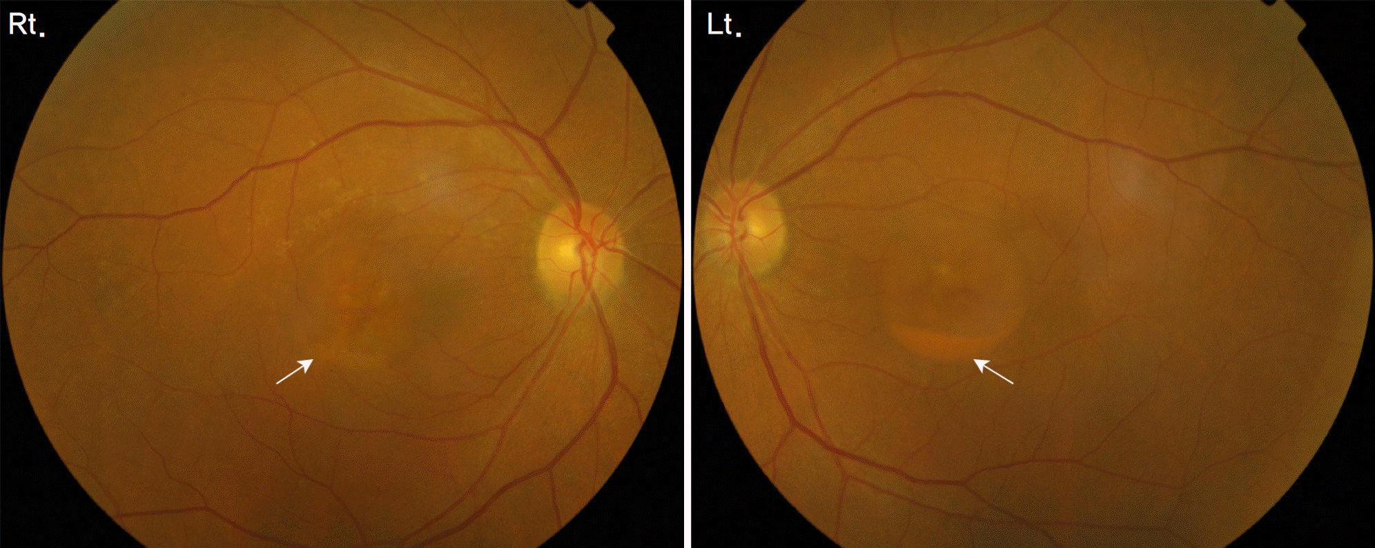 | Figure 1.Fundus findings show a well-circumscribed, circular area of macula elevation in both eyes. Subretinal exudates with fluid levels in the inferior part of the lesion are seen in both eyes, more marked in the left eye (arrows). |
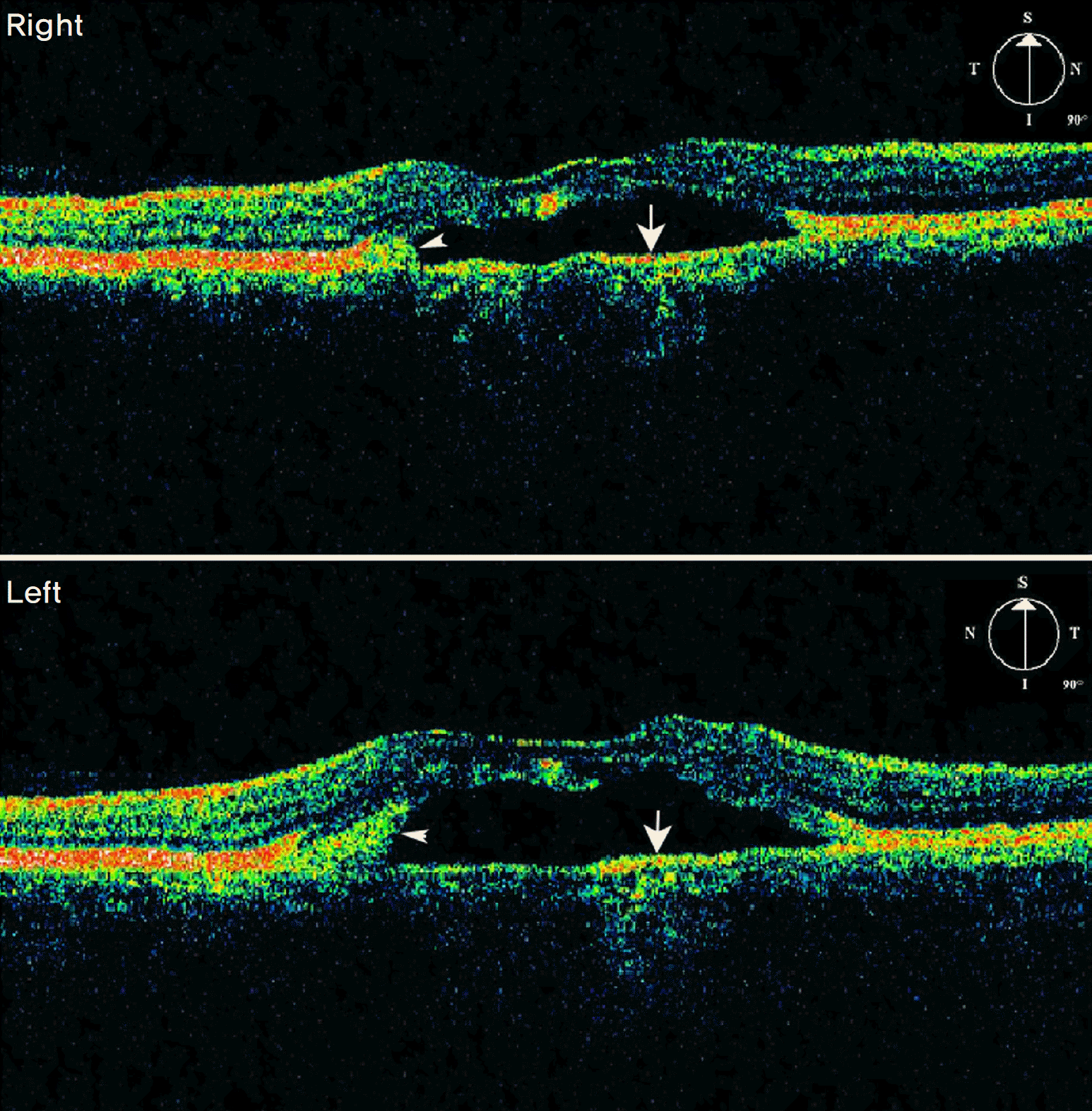 | Figure 2.Optical coherenece tomography findings of both eyes show serous retinal detachment. High reflectivity areas corresponding to the outer retinal pigment epithelium band (arrows) are visible under the low reflective space. High reflectivity areas corresponding to subretinal exudates are also seen (arrow heads). |
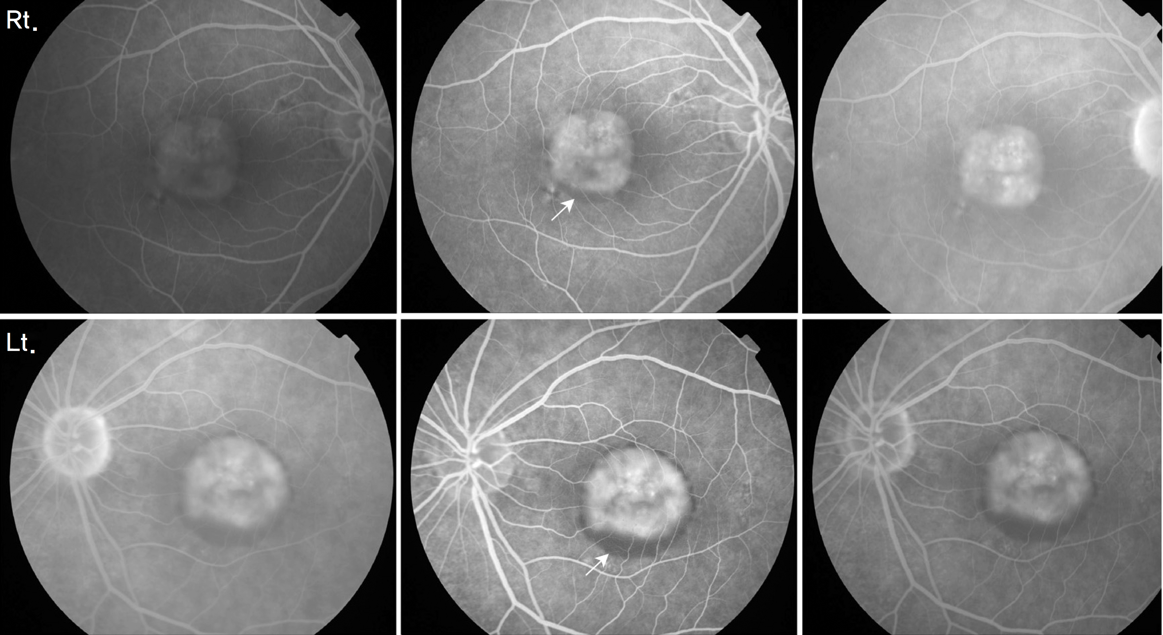 | Figure 3.Fluorescein angiography findings of both eyes reveal well-defined hyperfluorescence in the early phase and persistent hyperfluoresence in the late phase, corresponding to pigment epithelial detachment. Blocked fluorescence due to subretinal exudates are also seen in both eyes, especially in the left eye (arrows). |
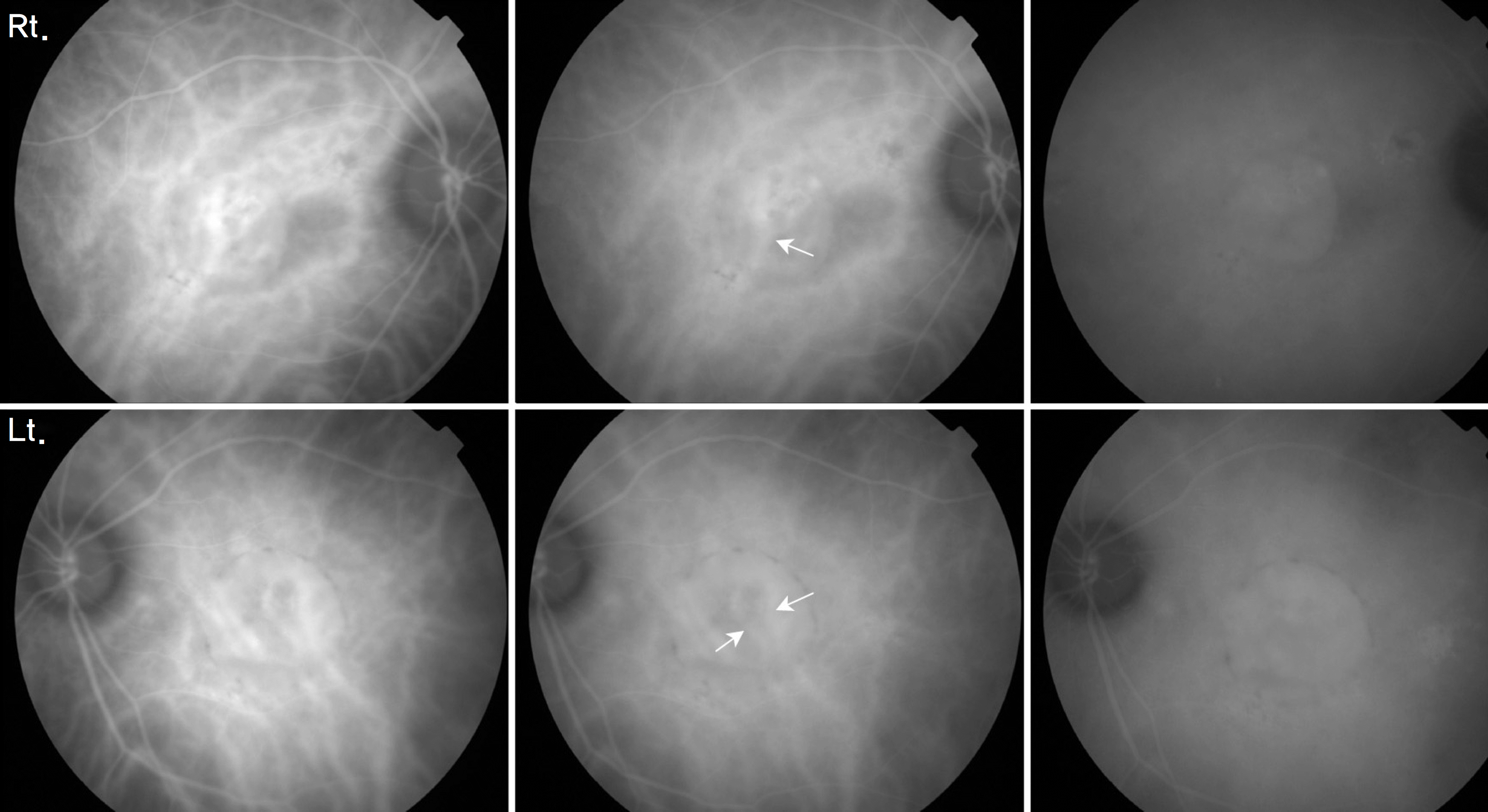 | Figure 4.Indocyanine green angiography findings of both eyes reveal well-demarcated early hyperfluorescence and persistent hyperfluorescence in the late phase, corresponding to pigment epithelial detachment. Large choroidal vessels (arrows) are visible under pigment epithelial detachment due to relatively clear fluid. |
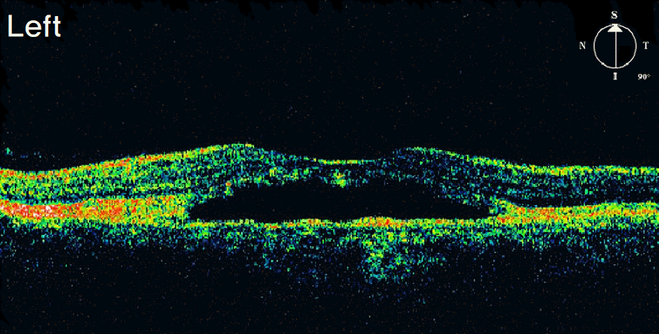 | Figure 5.Optical coherence tomography findings of the left eye at 1 month after photodynamic therapy plus intravitreal bevacizumab shows the same features as in the previous one. |
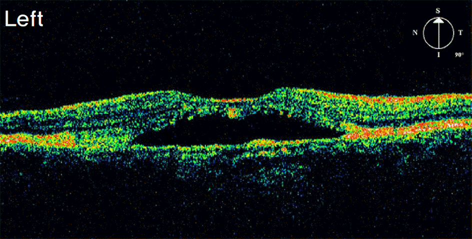 | Figure 6.Optical coherence tomography findings of the left eye at the last visit show the same features as in the previous ones. |
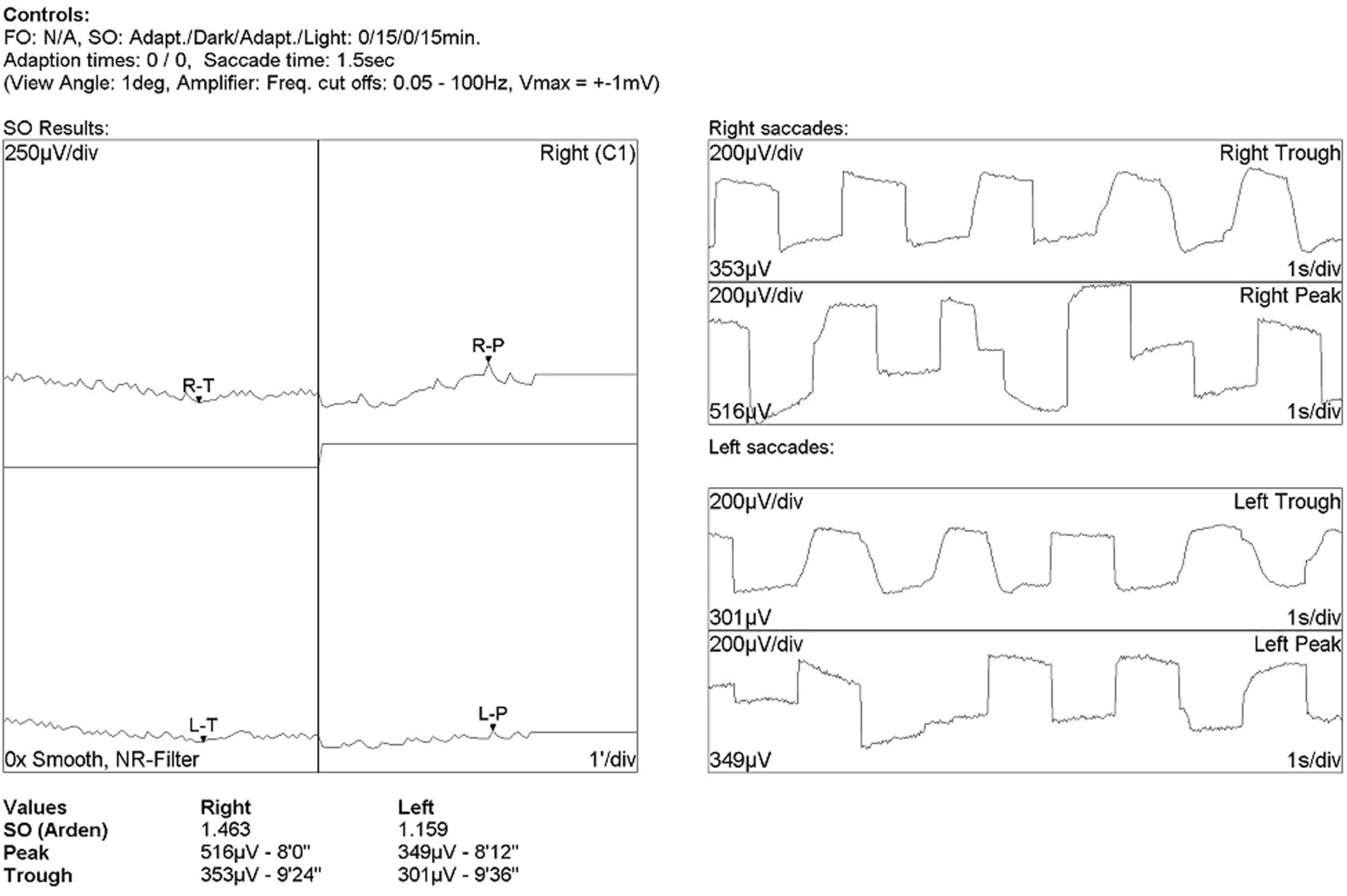 | Figure 7.An electro-oculogram is abnormal with an Arden ratio of 1.463, 1.159 in both eyes, respectively. A diagnosis of Best's disease is established. |
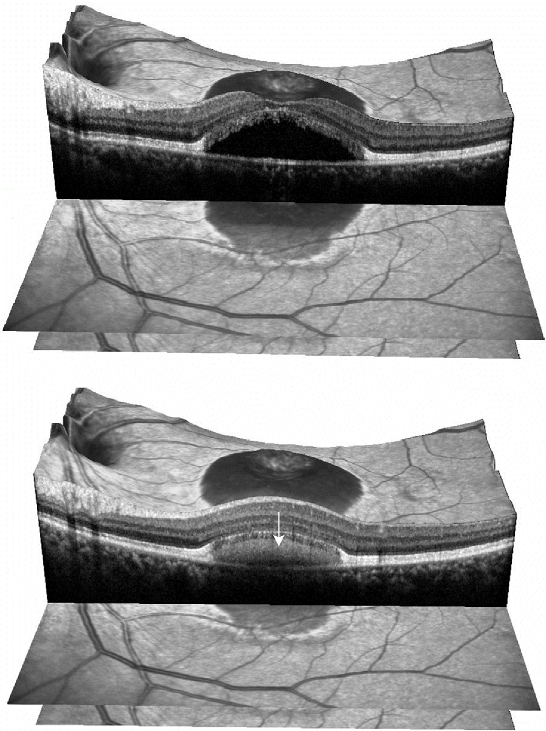 | Figure 8.Spectral domain optical coherence tomography findings of the left eye at the last visit show a clearly visible retinal pigment epithelium split and a low reflective space between split retinal pigment epithelium layers. High reflectivity can be seen between split retinal pigment epithelium layers, corresponding to subretinal exudates (arrows). |


 PDF
PDF ePub
ePub Citation
 Print
Print


 XML Download