Abstract
Purpose:
To evaluate the effects of oxidative stress and antioxidantson heme oxygenase-1 (HO-1) in cultured human retinal pigment epithelial (RPE) cells.
Methods:
Cultured RPE cells were challenged with different concentrations of hydrogen peroxide (H2 O2), and the HO-1 mRNA level was determined by RT-PCR after 24 hours and 48 hours of incubation independently. Additionally, the HO-1 mRNA level was measured after preincubating RPE cells with N-acetylcystein (NAC) as the antioxidant for 30 minutes and then challenging the cells with H2 O2.
Go to : 
References
1. Friedman DS, O'Colmain BJ, Muñoz B, et al. Eye Diseases Prevalence Research Group. Prevalence of Age-Related Macular Degeneration in the United States. Arch Ophthalmol. 2004; 122:564–72.
2. Klein R, Wang Q, Klein BE, et al. The relationship of age related maculopathy, cataract, and glaucoma to visual acuity. Invest Ophthalmol Vis Sci. 1995; 36:182–91.
3. Williams RA, Brody BL, Thomas RG, et al. The psychosocial impact of macular degeneration. Arch Ophthalmol. 1998; 116:514–20.

4. Young RW. Pathophysiology of age-related macular degeneration. Surv Ophthalmol. 1987; 31:291–306.

6. Beatty S, Koh H, Phil M, et al. The role of oxidative stress in the pathogenesis of age-related macular degeneration. Surv Ophthalmol. 2000; 45:115–34.

7. Winkler BS, Boulton ME, Gottsch JD, Sternberg P. Oxidative damage and age-related macular degeneration. Mol Vis. 1999; 5–32.
8. Jaattela M. Heat shock proteins as cellular lifeguards. Ann Med. 1999; 31:261–71.
9. David JT, Michael VM, David AN. Phagocytosis and H2 O2 induce catalase and metallothionein gene expression in human retinal pigment epithelial cells. Invest Ophthalmol Vis Sci. 1995; 36:1271–9.
10. Tenhunen R, Marver HS, Schmid R. The enzymatic conversion of heme to bilirubin by microsomal heme oxygenase. Proc Natl Acad Sci. 1968; 61:748–55.

11. Clark JE, Foresti R, Green CJ, Motterlini R. Dynamics of haem oxygenase-1 expression and bilirubin production in cellular protection against oxidative stress. Biochem J. 2000; 348:615–9.

12. Stocker R, Yamamoto Y, McDonagh AF, et al. Bilirubin is an antioxidant of possible physiological importance. Science. 1987; 235:1043–6.

13. Baranano DE, Wolosker H, Bae BI, et al. A mammalian iron ATPase induced by iron. J Biol Chem. 2000; 275:15166–73.
14. Peyton KJ, Reyna SV, Chapman GB, et al. Heme oxygenase-1− derived carbon monoxide is an autocrine inhibitor of vascular smooth muscle cell growth. Blood. 2002; 99:4443–8.
15. Otterbein LE, Bach FH, Alam J, et al. Carbon monoxide has anti-inflammatory effects involving the mitogen-activated protein kinase pathway. Nat Med. 2000; 6:422–8.

16. Otterbein LE, Zuckerbraun BS, Haga M, et al. Carbon monoxide suppresses arteriosclerotic lesions associated with chronic graft rejection and with balloon injury. Nat Med. 2003; 9:183–90.

18. Maines MD, Trakshel GM, Kutty RK. Characterization of two constitutive forms of rat liver microsomal heme oxygenase: only one molecular species of the enzyme is inducible. J Biol Chem. 1986; 261:411–9.

19. Maines MD. Heme oxygenase: function, multiplicity, regulatory mechanisms, and clinical applications. FASEB J. 1988; 2:2557–68.

20. McCoubrey WK Jr, Huang TJ, Maines MD. Isolation and charac-terization of a cDNA from the rat brain that encodes hemoprotein heme oxygenase− 3. Eur J Biochem. 1997; 247:725–32.
21. Ryter SW, Alam J, Choi AM. Heme oxygenase-1/carbon monoxide: From basic science to therapeutic applications. Physiol Rev. 2006; 86:583–650.

22. Kutty RK, Nagineni CN, Kutty G, et al. Increased expression of heme oxygenase-1 in human retinal pigment epithelial cells by transforming growth factor-beta. J Cell Physiol. 1994; 159:371–8.
23. Kuesap J, Li B, Satarug S, et al. Prostaglandin D2 induces heme oxygenase-1 in human retinal pigment epithelial cells. Biochem Biophys Res Commun. 2008; 367:413–9.

24. Udono-Fujimori R, Takahashi K, Takeda K, et al. Expression of heme oxygenase-1 is repressed by interferon-gamma and induced by hypoxia in human retinal pigment epithelial cells. Eur J Biochem. 2004; 271:3076–84.
25. Alizadeh M, Wada M, Gelfman CM, et al. Downregulation of Differentiation Specific Gene Expression by Oxidative Stress in ARPE-19 Cells. Invest Ophthalmol Vis Sci. 2001; 42:2706–713.
26. McCoubrey WK Jr, Maines MD. The structure, organization and differential expression of the gene encoding rat heme oxygenase-2. Gene. 1994; 139:155–61.

27. Hayashi S, Omata Y, Sakamoto H, et al. Characterization of rat heme oxygenase-3 gene: Implication of processed pseudogenes derived from heme oxygenase-2 gene. Gene. 2004; 336:241–50.

28. Age-Related Eye Disease Study Research Group . A randomized, placebo-controlled, clinical trial of high-dose supplementation with vitamins C and E, beta carotene, and zinc for age-related macular degeneration and vision loss: AREDS report No. 8. Arch Ophthalmol. 2001; 119:1417–36.
30. Hoekstra KA, Godin DV, Cheng KM. Protective role of heme oxygenase in the blood vessel wall during atherogenesis. Biochem Cell Biol. 2004; 82:351–9.

31. Nath KA. Heme oxygenase-1: A provenance for cytoprotective pathways in the kidney and other tissues. Kidney Int. 2006; 70:432–43.

32. Donoso LA, Kim D, Frost A, et al. The Role of Inflammation in the Pathogenesis of Age-related Macular Degeneration. Surv Ophthalmol. 2006; 51:137–52.

33. Hanneken A, Lin FF, Johnson J, Maher P. Flavonoids Protect Human Retinal Pigment Epithelial Cells from Oxidative-Stress– Induced Death. Invest Ophthalmol Vis Sci. 2006; 47:3164–77.
34. Frank RN, Amin RH, Puklin JE. Antioxidant enzymes in the macular retinal pigment epithelium of eyes with neovascular age-related macular degeneration. Am J Ophthalmol. 1999; 127:694–709.
35. Milbury PE, Graf B, Curran-Celentano JM, Blumberg JB. Bilberry (Vaccinium myrtillus) anthocyanins modulate heme oxygenase-1 and glutathione S-transferase-pi expression in ARPE− 19 cells. Invest Ophthamol Vis Sci. 2007; 48:2343–9.
Go to : 
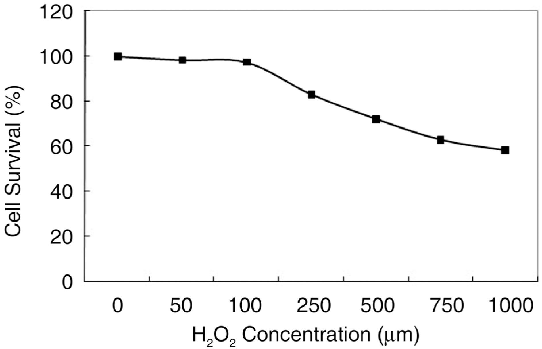
 | Figure 1.Human retinal pigment epithelial (RPE) cells were cultured for 24 hours at various concentrations of H2 O2. RPE cell survival showed progressive decrease with increasing concentrations of H2 O2. The cell viability was determined using MTT assay. The experiment was performed three times independently. |
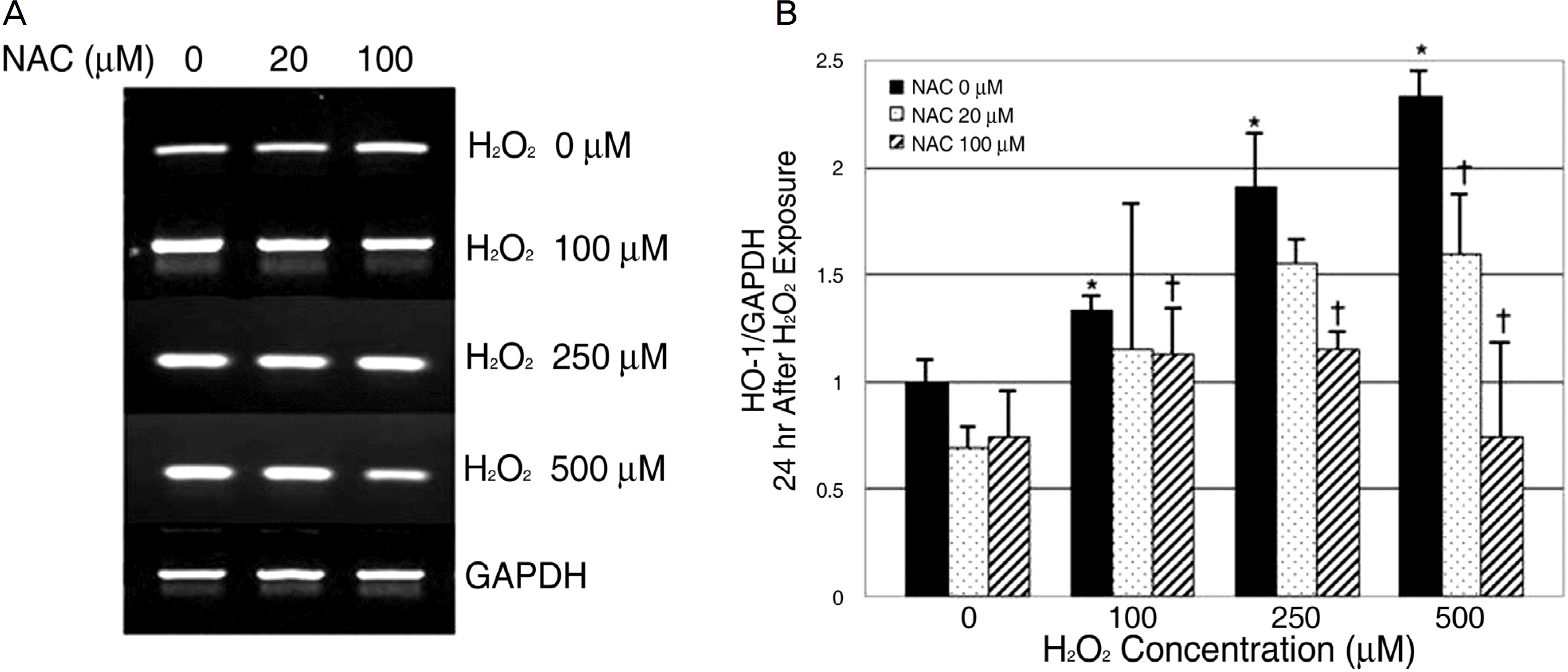
 | Figure 2.RT-PCR results of HO-1 mRNA expression in 24 hours culture. Human retinal pigment epithelial cells were cultured at various concentrations of H2 O2 with or without NAC for 24 hours. HO-1 mRNA expression increased with H2 O2 concentration increase. And after NAC treatment, HO-1 mRNA expression decreased in comparison to the cells without NAC treatment. The mRNA expression of HO-1 was normalized to GAPDH (house keeping gene). Data are shown as mean± SD of five experiments. ∗ p<0.05 versus control (H2 O2 0 μM, NAC 0 μM). † p<0.05 versus the cells treated with H2 O2 without NAC. |
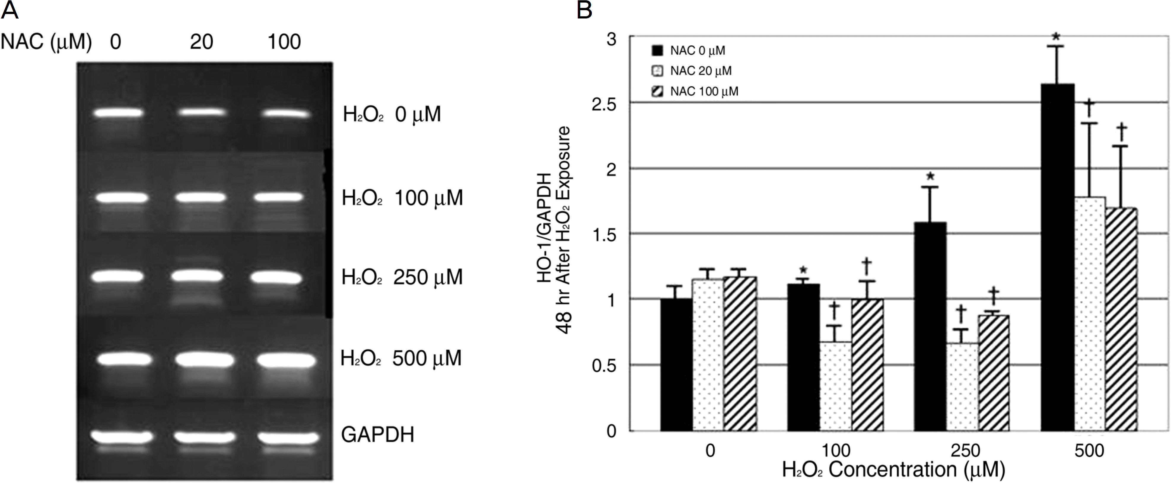
 | Figure 3.RT-PCR results of HO-1 mRNA expression in 48 hours culture. Human retinal pigment epithelial cells were cultured at various concentrations of H2 O2 with or without NAC for 48 hours. HO-1 mRNA expression increased with H2 O2 concentration increase. And after NAC treatment, HO-1 mRNA expression decreased in comparison to the cells without NAC treatment. The mRNA expression of HO-1 was normalized to GAPDH (house keeping gene). Data are shown as mean± SD of five experiments. ∗ p<0.05 versus control (H2 O2 0 μM, NAC 0 μM). † p<0.05 versus the cells treated with H2 O2 without NAC. |


 PDF
PDF ePub
ePub Citation
 Print
Print


 XML Download