Abstract
Purpose:
To evaluate of the range and relationship between intraocular pressure (IOP), and central corneal thickness (CCT) in premature infants.
Methods:
To investigate the correlation of IOP and CCT with gestational age and body weight, 58 premature infants 37 weeks-old or younger were examined. Under topical anesthesia, IOP was measured with Tono-PenⓇ XL (Medtronic Solan, Jacksonville, FL) and the CCT with pachymeter (SP-2000, TOMEYⓇ, Japan). The fundus was examined in infants with a risk of retinopathy of prematurity (ROP).
Results:
Average gestational age of the subjects was 33 weeks and 6 days and body weight was 1506±520 grams (mean±standard deviation). Forty-five subjects had oxygen therapy, and 10 patients were found to have any one of the stages of ROP. Average IOP was 15.14±4.64 mmHg in the right eye and 15.29±3.70 mmHg in the left eye. CCT was 594.72±74.87 μ m in the right eye and 599.78±74.17 μ m in the left eye. No statistically significant correlation was found between IOP or CCT and gestational age or body weight.
Conclusions:
Gestational age and body weight did not appear to affect IOP or CCT in the gestational age between 26 and 37 weeks. The maturing eye in the neonate is known for fast development in the first year after birth. There are, however, few reports in the literature regarding the changes in dimensions of ocular structures in the premature neonate. These normative values may aid ophthalmologists in assessing IOP and CCT in premature infants.
Go to : 
References
1. Tucker SM, Enzenauer RW, Levin AV, et al. Corneal diameter, axial length, and intraocular pressure in premature infants. Ophthalmology. 1992; 99:1296–300.

2. Kim WJ, Park SH, Shin H. The change of axial length according to age in the eyeball of premature infants by ultrasonic biometry. J Korean Ophthalmol Soc. 1993; 34:667–71.
3. Ha DW, Kim CK, Yoon YH. IOP & Corneal diameter in premature infants. J Korean Ophthalmol Soc. 2000; 41:1953–9.
4. Remón L, Cristóbal JA, Castillo J, et al. Central and peripheral corneal thickness in full-term newborns by ultrasonic pachymetry. Invest Ophthalmol Vis Sci. 1992; 33:3080–3.
5. Johnson M, Kass MA, Moses RA, Grodzki WJ. Increased corneal thickness simulating elevated intraocular pressure. Arch Opthal-mol. 1978; 96:664–5.

6. Magora F, Clooins VJ. The influence of general anesthetic agents on intraocular pressure in man. Arch Ophthalmol. 1961; 66:806–11.

7. Kornbleuth W, Abrahamov A, Aladjemoff L, et al. Intraocular pressure in the newborn measured under general anesthesia. Arch Ophthalmol. 1962; 67:750–2.
8. Radtke N, Cohan B. Intraocular pressure measurement in the newborn. Am J Ophthalmol. 1974; 78:501–4.

10. Muir KW, Jin J, Freedman SF. Central corneal thickness and its relationship to intraocular pressure in children. Ophthalmology. 2004; 111:2220–3.

11. Ehlers N, Sorensen T, Bramsen T, Poulsen EH. Central corneal thickness in newborns and children. Acta Ophthalmol. 1976; 54:285–90.

12. Autzen T, Bjørnstrøm L. Central corneal thickness in full-term newborns. Acta Ophthalmol. 1989; 67:719–20.

13. Autzen T, Bjørnstrøm L. Central corneal thickness in premature babies. Acta Ophthalmol. 1991; 69:251–2.

14. Kirwan C, O'Keefe M, Fitzsimon S. Central corneal thickness and corneal diameter in premature infants. Acta Ophthalmol Scand. 2005; 83:751–3.

15. Zheng Y, Ge J, Huang G, Zhang J, et al. Heritability of central corneal thickness in Chinese: The Guangzhou Twin Eye Study. Invest Ophthalmol Vis Sci. 2008; 49:4303–7.

Go to : 
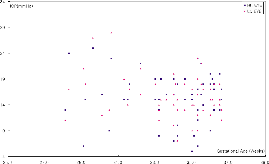 | Figure 1.Intraocular pressure (IOP) does not seem to be affected by gestational age between 26 and 37 weeks (Rt, r=−0.076, p=0.571/Lt, r=−0.220, p=0.097, Pearson correlation analysis). |
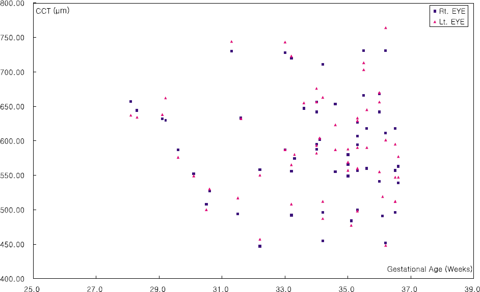 | Figure 2.Central corneal thickness (CCT) does not seem to be affected by gestational age between 26 and 37 weeks (Rt, r=−0.081, p=0.546/Lt, r=−0.050, p= 0.712, Pearson correlation analysis). |
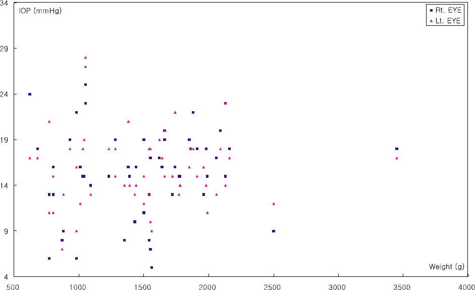 | Figure 3.Intraocular pressure (IOP) does not seem to be affected by body weight (Rt, r=0.113, p=0.397/Lt, r=0.024, p=0.858, Pearson correlation analysis). |
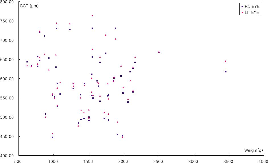 | Figure 4.Central corneal thickness (CCT) does not seem to be affected by body weight between 500 g and 4,000 g (Rt, r=−0.082, p=0.541/Lt, r=−0.021, p=0.877, Pearson correlation analysis). |
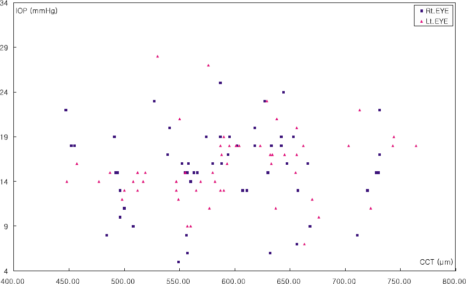 | Figure 5.Central corneal thickness (CCT) and intraocular pressure (IOP) has no correlation between each other (Rt, r=0.047, p=0.724/Lt, r=0.136, p=0.309, Pearson correlation analysis). |
Table 1.
Correlation of central corneal thickness, intraocular pressure, body weight and gestational age
|
CCT∗ (µm) |
IOP† (mm Hg) |
||||
|---|---|---|---|---|---|
| Rt | Lt | Rt | Lt | ||
| Body weight | Pearson correlation coefficient | -0.082 | -0.021 | 0.113 | 0.024 |
| p value | 0.541 | 0.877 | 0.397 | 0.858 | |
| Gestational age | e Pearson correlation coefficient | -0.081 | -0.050 | -0.076 | -0.220 |
| p value | 0.546 | 0.712 | 0.571 | 0.097 | |
Table 2.
Statistical analysis of clinical factors associated with intraocular pressure
| Factors |
IOP∗ (mm Hg) |
p value† |
|||
|---|---|---|---|---|---|
| Rt | Lt | Rt | Lt | ||
| Sex | Male | 14.4±4.9 | 15.0±4.5 | 0.060 | 0.222 |
| Female | 16.7±3.8 | 16.4±3.2 | |||
| Delivery | NSVD‡ | 14.9±4.2 | 15.4±4.4 | 0.414 | 0.709 |
| C/Sec§ | 15.9±4.3 | 15.8±3.6 | |||
| Oxygen treatment | (+) | 15.3±4.7 | 15.7±4.1 | 0.618 | 0.361 |
| (−) | 17.0±2.8 | 13.0±2.8 | |||
Table 3.
Statistical analysis of clinical factors associated with central corneal thickness
| Factors |
CCT∗ (µm) |
p value† |
|||
|---|---|---|---|---|---|
| Rt | Lt | Rt | Lt | ||
| Sex | Male | 574.57±80.01 | 581.42±78.02 | 0.027 | 0.038 |
| Female | 617.39±50.95 | 620.73±51.26 | |||
| Delivery | NSVD‡ | 590.46±73.07 | 593.21±71.39 | 0.901 | 0.655 |
| C/Sec§ | 592.88±73.39 | 601.69±71.32 | |||
| Oxygen treatment | (+) | 593.85±72.49 | 598.76±71.29 | 0.203 | 0.324 |
| (−) | 527.00±50.91 | 548.00±41.02 | |||


 PDF
PDF ePub
ePub Citation
 Print
Print


 XML Download