Abstract
Purpose:
To investigate the clinical characteristics of retinopathy associated with systemic lupus erythematosus (SLE) and its risk factors.
Methods:
Medical records of patients who were diagnosed with SLE were reviewed retrospectively. The presence of retinal hemorrhage, vasculitis and a cotton wool patch were regarded as lupus retinopathy, but concomitant diabetic retinopathy and hypertensive retinopathy were excluded from the study. The correlation between the development of lupus retinopathy and the presence of positive autoantibodies was also investigated.
Results:
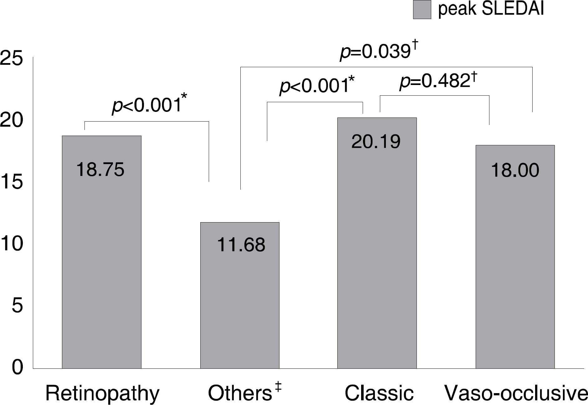
Ocular morbidity was found in 173 of 260 (66%) SLE patients. Retinopathy was detected in 52 eyes of 33 patients (12%), which included 36 eyes of 21 patients (63%) with classic retinopathy and 11 eyes of 10 patients (30%) with vaso-occlusive retinopathy. The presence of classic retinopathy coincided with the flare-up of lupus activity and completely resolved without visual impairment. However, vaso-occlusive retinopathy was not related with lupus activity, and resulted in significant visual impairments of 20/200 or less in six eyes of five patients. The disease activity of lupus assessed by the maximum SLE disease activity index was higher in patients with retinopathy (p<0.05), and the prevalence of antiphospholipid antibody was higher in patients with vaso-occlusive retinopathy than in patients with classic retinopathy (66.7% vs. 37.5%, p<0.05).
Go to : 
References
1. Wallace DJ, Hahn BH. Dubois' Lupus Erythematosus. 7th ed.Philadelphia: Lippincott Williams & Wilkins;2007. p. 65–86.
2. Kim IT, Chang SD. Papilledema and cerebral venous thrombosis in a patient with systemic lupus erythematosus. J Korean Ophthalmol Soc. 1999; 40:2015–9.
3. Hwang HS, Kim DH. Transient myopia with severe chemosis associated with systemic lupus erythematosus. J Korean Ophthalmol Soc. 2007; 48:1445–8.

4. Im CY, Kim SS, Kim HK. Bilateral optic neuritis as first manifestation of systemic lupus erythematosus. Korean J Ophthalmol. 2002; 16:52–8.

5. Shin SY, Lee JM. A case of multiple cranial nerve palsies as the initial ophthalmic presentation of antiphospholipid syndrome. Korean J Ophthalmol. 2006; 20:76–8.

6. Oh PC, Kim GH, Jin CH, Baek HJ. A case of systemic lupus erythematous associated with neuromyelitis optica (Devic's Syndrome). J Korean Rheum Assoc. 2007; 14:263–7.

7. Yun KA, Kim JG. A case of orbital myositis secondary to systemic lupus erythematosus. J Korean Rheum Assoc. 2006; 13:171–6.
8. Kim IT, Na SC, Lee KJ. Vascular occlusion associated with antiphospholipid antibodies in systemic lupus erythematosus. J Korean Ophthalmol Soc. 2000; 41:427–32.
9. Park DH, Koo HM, Chung SK. A case of optic neuritis and central retinal vein occlusion associated with systemic lupus erythematosus. J Korean Ophthalmol Soc. 1994; 35:116–21.
10. Jung NH, Kim SY. A case of severe retinal vaso-occlusive disease in systemic lupus erythematosus. J KoreanOphthalmol Soc. 1993; 34:1287–92.
11. Yawm SC, Uhm KB, Choe JK. A case of severe vaso-occlusive disease in systemic lupus erythematosus. J Korean Ophthalmol Soc. 1986; 27:681–6.
12. Jabs DA, Fine SL, Hochberg MC, et al. Severe retinal vaso− occlusive disease in systemic lupus erythematous. Arch Ophthalmol. 1986; 104:558–63.
13. Lanham JG, Barrie T, Kohner EM, Hughes GR. SLE retinopathy: evaluation by fluorescein angiography. Ann Rheum Dis. 1982; 41:473–8.

14. Stafford-Brady FJ, Urowitz MB, Gladman DD, Easterbrook M. Lupus retinopathy. Patterns, associations, and prognosis. Arthritis Rheum. 1988; 31:1105–10.
15. Bombardier C, Gladman DD, Urowitz MB, et al. Derivation of the SLEDAI. A disease activity index for lupus patients. The Committee on Prognosis Studies in SLE. Arthritis Rheum. 1992; 35:630–40.
16. Gold DH, Morris DA, Henkind P. Ocular findings in systemic lupus erythematosus. Br J Ophthalmol. 1972; 56:800–4.

17. Au A, O'Day J. Review of severe vaso-occlusive retinopathy in systemic lupus erythematosus and the antiphospholipid syndrome: associations, visual outcomes, complications and treatment. Clin Experiment Ophthalmol. 2004; 32:87–100.

18. Giorgi D, Pace F, Giorgi A, et al. Retinopathy in systemic lupus erythematosus: pathogenesis and approach to therapy. Hum Immunol. 1999; 60:688–96.

19. Klinkhoff AV, Beattie CW, Chalmers A. Retinopathy in systemic lupus erythematosus: relationship to disease activity. Arthritis Rheum. 1986; 29:1152–6.

20. Aronson AJ, Ordonez NG, Diddie KR, Ernest JT. Immune− complex deposition in the eye in systemic lupus erythematosus. Arch Intern Med. 1979; 139:1312–3.
21. Graham EM, Spalton DJ, Barnard RO, et al. Cerebral and retinal vascular changes in systemic lupus erythematosus. Ophthalmology. 1985; 92:444–8.

22. Ushiyama O, Ushiyama K, Koarada S, et al. Retinal disease in patients with systemic lupus erythematosus. Ann Rheum Dis. 2000; 59:705–8.

23. Kleiner RC, Najarian LV, Schatten S, et al. Vaso-occlusive retinopathy associated with antiphospholipid antibodies (lupus anticoagulant retinopathy). Ophthalmology. 1989; 96:896–904.

24. Montehermoso A, Cervera R, Font J, et al. Association of anti-phospholipid antibodies with retinal vascular disease in systemic lupus erythematosus. Semin Arthritis Rheum. 1999; 28:326–32.
25. Alarcon-Segovia D, Deleze M, Oria CV, et al. Antiphospholipid antibodies and the antiphospholipid syndrome in systemic lupus erythematosus. A prospective analysis of 500 consecutive patients. Medicine (Baltimore). 1989; 68:353–65.
27. Soo MP, Chow SK, Tan CT, et al. The spectrum of ocular involvement in patients with systemic lupus erythematosus without ocular symptoms. Lupus. 2000; 9:511–4.
Go to : 
 | Figure 1.Comparison of peak SLEDAI among patients with retinopathy, without retinopathy, and each subtype of retinopathy. ∗ttest; †Mann Whitney-U test; ‡ Patients without retinopathy. |
Table 1.
Ocular manifestations found in SLE
Table 2.
Difference of demographics between patients with retinopathy and without retinopathy
| No retinopathy (n=227) | Retinopathy (n=33) | p-value | |
|---|---|---|---|
| Age at ocular exam | 37.2±12.5 | 36.2±13.7 | 0.657∗ |
| Age at SLE diagnosis | 33.6±11.9 | 34.3±13.3 | 0.749∗ |
| M:F | 21 (9%): 206 (91%) | 5 (15%): 28 (85%) | 0.346† |
Table 3.
Funduscopic findings in each subtype of lupus etinopathy
Table 4.
Difference of systemic manifestations between patients with retinopathy and without retinopathy
| Systemic manifestations | No retinopathy N=148 (%) | Retinopathy N=30 | p-value∏ |
|---|---|---|---|
| Rash | 64 (43.2) | 19 (63.3) | 0.048# |
| Photosensitivity | 26 (17.6) | 4 (13.3) | 0.790 |
| Mucosal ulcer | 35 (23.6) | 6 (20.0) | 0.814 |
| Arthritis | 62 (41.9) | 13 (43.3) | 1.000 |
| Serositis | 26 (17.6) | 8 (26.7) | 0.307 |
| Renal involvement∗ | 63 (42.6) | 20 (66.7) | 0.026# |
| Neurological involvement† | 7 (4.7) | 2 (6.7) | 0.649 |
| Hematologic abnormality‡ | 103 (69.6) | 17 (56.7) | 0.201 |
| Immunologic abnormality§ | 120 (81.1) | 26 (86.7) | 0.606 |
Table 5.
Relationship between common autoantibodies found in SLE and lupus retinopathy
| Autoantibodies | No retinopathy N(%) | All retinopathies N (%) | Classic retinopathy N (%) | occlusive retinopathy N (%) | p∗ | p† | p‡ |
|---|---|---|---|---|---|---|---|
| APL§ | 53 (30.1) | 13 (48.1) | 6 (37.5) | 6 (66.7) | 0.078 | 0.576 | 0.031‡‡ |
| LE cell | 27 (44.3) | 4 (40.0) | 3 (42.9) | 1 (50.0) | 1.000 | 1.000 | 1.000 |
| Ro / La∏ | 67 (63.2) | 10 (58.8) | 7 (63.6) | 3 (60.0) | 0.790 | 1.000 | 1.000 |
| Sm# | 32 (27.8) | 6 (33.3) | 5 (45.5) | 1 (20.0) | 0.779 | 0.298 | 1.000 |
| RNP∗∗ | 30 (52.6) | 5 (50.0) | 5 (62.5) | 0 (0) | 1.000 | 0.716 | 0.237 |
| RF†† | 27 (19.3) | 4 (25.0) | 3 (21.4) | 1 (100.0) | 0.526 | 0.737 | 0.199 |


 PDF
PDF ePub
ePub Citation
 Print
Print


 XML Download