

 PDF
PDF ePub
ePub Citation
 Print
Print


Abstract
Purpose
To report the results of managing dislocated posterior chamber intraocular lenses (PC IOLs) by externalizing both haptics alternately through a single clear corneal incision.
Methods
The study included 10 eyes of 10 patients in which a dislocated PC IOL was managed with pars plana vitrectomy. With our modified method, both haptics were alternately externalized through a single clear corneal incision for suture knot placement and then reinserted.
Results
The dislocated PC IOLs were repositioned successfully in all 10 eyes, and the corrected visual acuity improved postoperatively in 9 eyes at a minimum follow-up of six months. Abnormal IOL position, IOL capture by the iris, or posterior synechia was not observed. Hypotony maculopathy occurred in one eye, but the macular folding disappeared and the visual acuity improved after adding one-bite suture.
Go to : 
References
1. Stark WJ Jr, Maumene AE, Datiles M, et al. Intraocular lenses: complications and visual results. Trans Am Ophthalmol Soc. 1983; 81:280–309.
2. Smith SG, Lindstrom RL. Malpositioned posterior chamber lenses: etiology, prevention, and management. J Am Intraocul Implant Soc. 1985; 11:584–91.

3. Mello MO Jr, Scott IU, Smiddy WE, et al. Surgical management and outcomes of dislocated intraocular lenses. Ophthalmology. 2000; 107:62–7.
4. Chan CK, Agarwal A, Agarwal S, Agarwal A. Management of dislocated intraocular implants. Ophthalmol Clin North Am. 2001; 14:681–93.
5. Smiddy WE, Ibanez GV, Alfonso E, Flynn HW Jr. Surgical management of dislocated intraocular lenses. J Cataract Refract Surg. 1995; 21:64–9.
6. Bloom SM, Wyszynski RE, Brucker AJ. Scleral fixation suture for dislocated posterior chamber intraocular lens. Ophthalmic Surg. 1990; 21:851–4.
7. Campo RV, Chung KD, Oyakawa RT. Pars plana vitrectomy in the management of dislocated posterior chamber lenses. Am J Ophthalmol. 1989; 108:529–34.
8. Akduman L. Transscleral fixation of a dislocated silicone plate haptic intraocular lens via the pars plana. Ophthalmic Surg Lasers. 1998; 29:519–21.
9. Azar DT, Wiley WF. Double-knot transscleral suture fixation technique for displaced intraocular lenses. Am J Ophthalmol. 1999; 128:644–6.
10. Koh HJ, Kim CY, Lim SJ, Kwon OW. Scleral fixation technique using 2 corneal tunnels for a dislocated intraocular lens. J Cataract Refract Surg. 2000; 26:1439–41.
11. Kwok AK, Cheng AC, Lam DS. Surgical technique for transcleral-fixation of a dislocated posterior chamber intraocular lens. Am J Ophthalmol. 2001; 132:406–8.
12. Maguire AM, Blumenkranz MS, Ward TG, Winkelman JZ. Scleral loop fixation for posteriorly dislocated intraocular lenses; operative technique and long-term results. Arch Ophthalmol. 1991; 109:1754–8.
13. Lawrence FC II, Hubbard WA. “Lens lasso” repositioning of dislocated posterior chamber intraocular lenses. Retina. 1994; 14:47–50.
14. Navia-Aray EA. A technique for knotting a suture around the loops of a dislocated intraocular lens, within the eye, for fixation in the ciliary sulcus. Ophthalmic Surg. 1993; 24:702–7.
15. Lee SC, Tseng SH, Cheng HC, Chen FK. Slipknot for scleral fixation of intraocular lenses. J Cataract Refract Surg. 2001; 27:662–4.
16. Virata SR, Holekamp NM, Meredith TA. “Luggage-tag” suture fixation of partially dislocated intraocular lenses. Ophthalmic Surg Lasers. 2001; 32:346–8.
17. Hanemoto T, Ideta H, Kawasaki T. Dislocated intraocular lens fixation using intraocular cowhitch knot. Am J Ophthalmol. 2001; 131:265–7.
18. Cho SH, Kang SW, Jung MS. Four cases of modification of scleral fixation using 30 G needle for posterior chamberintraocular lens dislocation. J Korean Ophthalmol Soc. 2002; 43:917–21.
19. Lee SJ, Choi KS, Park SH, Jung GY. A Reverse Ab Externo Scleral Fixation for Posterior Chamber Intraocular Lens Dislocation. J Korean Ophthalmol Soc. 2007; 48:1341–5.
20. Chan CK. An improved technique for management of dislocated posterior chamber implants. Ophthalmology. 1992; 99:51–7.
21. Kokame GT, Atebara NH, Bennett MD. Modified technique of haptic externalization for scleral fixation of dislocated posterior chamber lens implants. Am J Ophthalmol. 2001; 131:129–31.
22. Thach AB, Dugel PU, Sipperley JO, et al. Outcome of sulcus fixation of dislocated posterior chamber intraocular lenses using temporary externalization of the haptics. Ophthalmology. 2000; 107:480–4.
23. Kokame GT, Yamamoto I, Mandel H. Scleral fixation of dislocated posterior chamber intraocular lenses: Temporary haptic externalization through a clear corneal incision. J Cataract Refract Surg. 2004; 30:1049–56.
Go to :
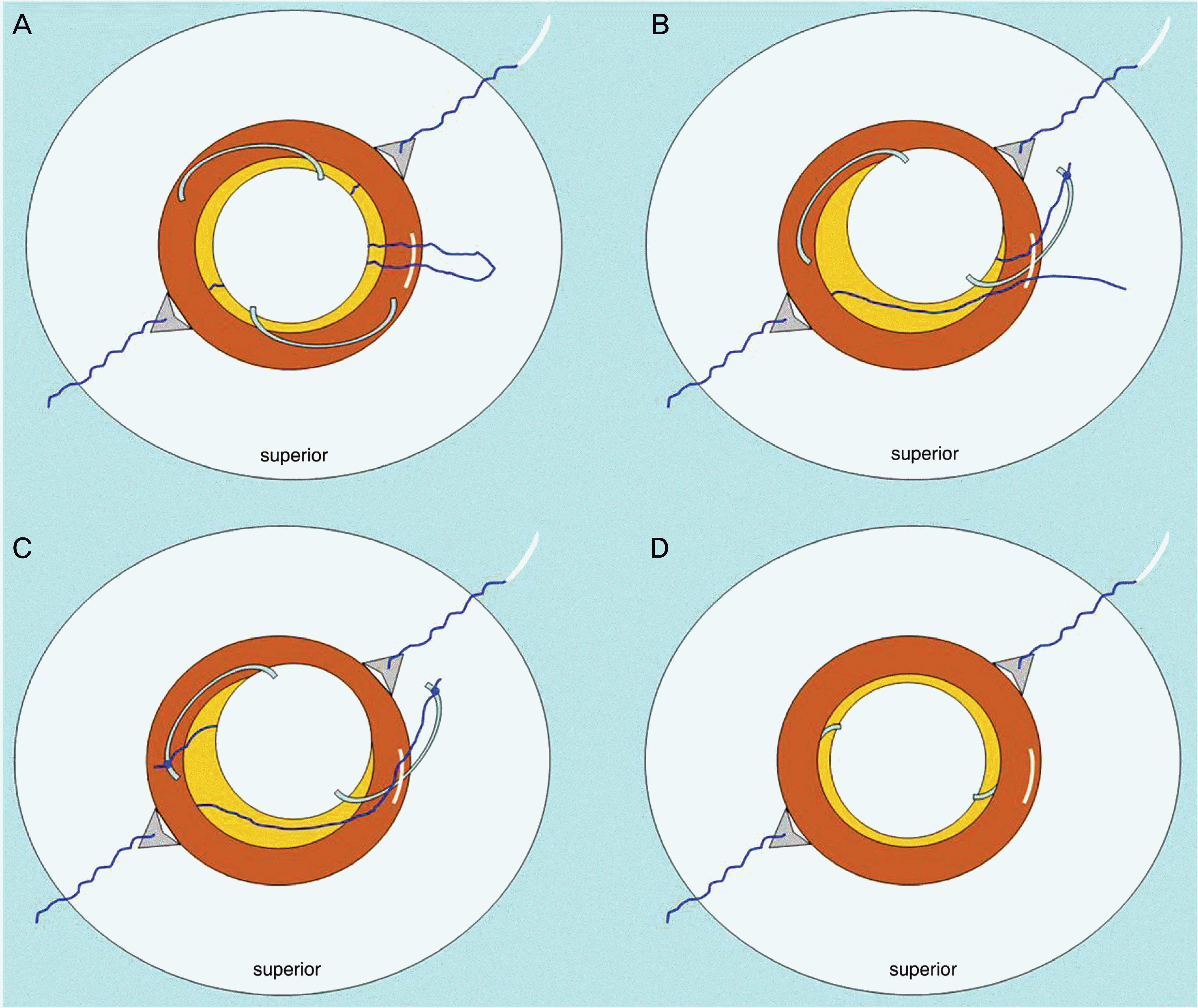 | Figure 1.(A) The PC IOL is placed in the anterior chamber through the pupil and over the iris. And a 10-0 Prolene is placed posterior to the iris through a sclerotomy under the scleral flap. Middle portion of the 10-0 Prolene is pulled out through a clear corneal incision. (B) One haptic is externalized through a clear corneal incision under direct visualization, and tied with the divided end of 10-0 Prolene. (C) The PC IOL is rotated in the anterior chamber and the other haptic is externalized through a clear corneal incision, and tied with the other end of 10–0 Prolene. (D) The PC IOL is reimplanted and the sutures attached to each haptic are pulled through the fixation sclerotomy under the scleral flap. |
Table 1.
Results
| No. | Age/Sex | Follow-up period (months) | Preop*. BCVA | Postop†. BCVAП (Sph/Cyl) | Complications |
|---|---|---|---|---|---|
| 1 | 54/M‡ | 18 | 1.0 | 0.5 (−1.0/-1.0 70°) | Macular edema |
| 2 | 70/M | 23 | 0.1 | 0.8 (+1.0/-2.0 90°) | N-S§ |
| 3 | 57/M | 27 | 1.0 | 1.2 (0/-0.75 40°) | N-S |
| 4 | 57/M | 12 | 0.3 | 0.4 (+1.0/-2.5 80°) | N-S |
| 5 | 68/M | 28 | 0.4 | 0.9 (−0.5/-1.0 130°) | N-S |
| 6 | 55/M | 26 | 0.2 | 1.0 (−1.0/-2.25 60°) | N-S |
| 7 | 52/M | 14 | 0.8 | 1.0 (+0.75/-2.0 110°) | N-S |
| 8 | 52/M | 9 | 0.03 | 1.0 (−1.0/-0.5 180°) | N-S |
| 9 | 17/F | 12 | 0.6 | 0.8 (−0.25/-1.5 30°) | N-S |
| 10 | 26/F | 12 | 0.8 | 1.0 (+0.75/-1.0 10°) | Hypotony maculopathy |
 XML Download