

 PDF
PDF ePub
ePub Citation
 Print
Print


Abstract
Purpose
The peripapillary retinal nerve fiber layer thickness (RNFL) was measured in normal children using optical coherence tomography (OCT), and the effect of various factors on the RNFL thickness was examined. Methods: From April 2006 to January 2007, the RNFL thickness of 74 normal children (148 eyes) between the ages of 4 and 17 years old was measured by OCT, and the effect of factors such as age, gender, refractive error, C/D ratios, cooperation, and laterality on the peripapillary RNFL thickness was analyzed.
Results
The mean age of the patients was 10.2 years (4∼17 years), and the mean peripapillary RNFL thickness was 106.3±12.8 µm. As to the thickness of the different peripapillary locations, the superior side was thickest (135.3±20.6 µm), followed in order by the inferior side (130.9±23.0 µm), the temporal side (86.3±18.9 µm), and the nasal side (71.9±20.8 µm). The refractive error was correlated positively with RNFL thickness (r=0.277, p=0.001), and age correlated negatively with RNFL thickness (r=-0.194, p=0.018).
Conclusions
RNFL thickness in normal children increases as the refractive error becomes hyperopic and decreases with age. The data about RNFL thickness of normal children obtained in this study may provide useful information for an early diagnosis of pediatric neuroophthalmologic disease and for monitoring its progression.
Go to : 
References
1. Quigley HA, Katz J, Derick RJ, et al. An evaluation of optic disc and nerve fiber examinations in monitoring progression of early glaucoma damage. Ophthalmology. 1992; 99:19–28.
2. Quigley HA, Addicks EM, Green WR. Optic nerve damage in human glaucoma. Arch Ophthalmol. 1982; 100:135–46.

3. Tuulonen A, Lehtola J, Airaksinen PJ. Nerve fiber layer defect with normal visual fields. Ophthalmology. 1993; 100:587–98.
4. Sommer A, Miller NR, Pollack I, et al. The nerve fiber layer in the diagnosis of glaucoma. Arch Ophthalmol. 1977; 95:2149–56.
5. Quigley HA, Dunkelberger GR, Green WR. Retinal ganglion cell atrophy correlated with automated perimetry in human eyes with glaucoma. Am J Ophthalmol. 1989; 107:452–64.
6. Sommer A, Katz J, Quigley HA, et al. Clinically detectable nerve fiber atrophy precedes the onset of glaucomatous field loss. Arch Ophthalmol. 1991; 109:77–83.
7. Quigley HA, Dunkelberger GR, Green WR. Chronic human glaucoma causing selectively greater loss of large optic nerve fibers. Ophthalmology. 1988; 95:357–63.
8. Huang D, Swanson EA, Lin CP, et al. Optical coherence tomography. Science. 1991; 254:1178–81.
9. Paunescu LA, Schuman JS, Price LL, et al. Reproducibility of nerve fiber thickness, macular thickness, and optic nerve head measurements using Stratus OCT. Invest Ophthalmol Vis Sci. 2004; 45:1716–24.
10. Hee MR, Izatt JA, Swanson EA, et al. Optical coherence tomography of the human retina. Arch Ophthalmol. 1995; 113:325–32.
11. Huynh SC, Wang XY, Rochtchina E, et al. Peripapillary retinal nerve fiber layer thickness in a population of 6-year-old children. Ophthalmology. 2006; 113:1583–92.
12. Varma R, Bazzaz S, Lai M. Optical tomography-measured retinal nerve fiber layer thickness in normal Latinos. Invest Ophthalmol Vis Sci. 2003; 44:3369–73.
13. Alamouti B, Funk J. Retinal thickness decreases with age: an OCT study. Br J Ophthalmol. 2003; 87:899–901.
14. Poinoosawmy D, Fontana L, Wu JX, et al. Variation of nerve fibre layer thickness measurements with age and ethnicity by scanning laser polarimetry. Br J Ophthalmol. 1997; 81:350–4.
15. Ahn HC, Son HW, Kim JS, et al. Quantitative analysis of retinal nerve fiber layer thickness of normal children and adolescents. Korean J Ophthalmol. 2005; 19:195–200.
16. Salchow DJ, Oleynikov YS, Chiang MF, et al. Retinal nerve fiber layer thickness in normal children measured with optical coherence tomography. Ophthalmology. 2006; 113:786–91.
17. Dichtl A, Jonas JB, Naumann GO. Retinal nerve fiber layer thickness in human eyes. Graefes Arch Clin Exp Ophthalmol. 1999; 237:474–9.
18. Mikelberg FS, Drance SM, Schulzer M, et al. The normal human optic nerve. Axon count and axon diameter distribution. Ophthalmology. 1989; 96:1325–8.
19. Repka MX, Quigley HA. The effect of age on normal human optic nerve fiber number and diameter. Ophthalmology. 1989; 96:26–32.
20. Hoh ST, Greenfield DS, Mistlberger A, et al. Optical coherence tomography and scanning laser polarimetry in normal, ocular hypertensive, and glaucomatous eyes. Am J Ophthalmol. 2000; 129:129–35.
21. Jonas JB, Dichtl A. Optic disc morphology in myopic primary open-angle glaucoma. Graefes Arch Clin Exp Ophthalmol. 1997; 253:627–33.
Go to :
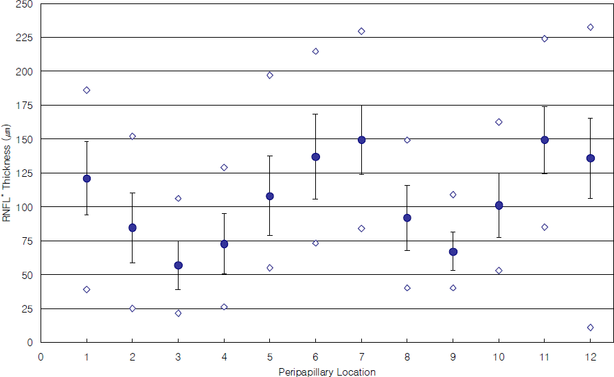 | Figure 1.Retinal nerve fiber layer thickness in normal children as peripapillary location. Mean (blue dot), standard deviation (whiskers), highest and lowest balues (white dot) are shown. * RNFL = retinal nerve fiber layer. |
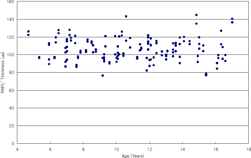 | Figure 2.Average retinal nerve fiber layer thickness as function of age (r=-0.194, p=0.018). * RNFL = retinal nerve fiber layer. |
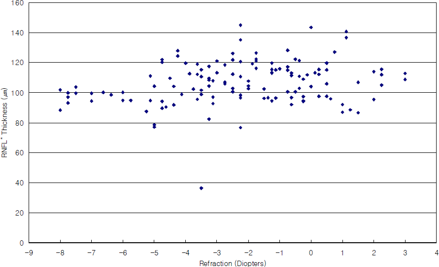 | Figure 3.Average retinal nerve fiber layer thickness as function of refraction (r=0.277, p=0.001). * RNFL = retinal nerve fiber layer. |
Table 1.
Distribution of age and sex
| Age (years) |
Sex |
Total | |
|---|---|---|---|
| Male | Female | ||
| 4∼6 | 5 | 4 | 9 |
| 7∼12 | 21 | 21 | 42 |
| 13∼15 | 9 | 8 | 17 |
| 16∼17 | 3 | 3 | 6 |
| Total | 38 | 36 | 74 |
Table 2.
Characteristics of study subjects
| Characteristics | Value |
|---|---|
| Age (years) | |
| Mean±SD* | 10.2±3.3 |
| Range | 4∼17 |
| Gender | |
| Male | 41 (55.4%) |
| Female | 33 (44.6%) |
| Refraction (diopters) | |
| Mean±SD | -2.4±3.2 |
| Range | -8.0∼+3.0 |
| Intraocular pressure (mmHg) | |
| Mean±SD | 13.5±1.4 |
Table 3.
Peripapillary retinal nerve fiber layer thickness by quadrants
| RNFL* Thickness (µm) | Superior | Nasal | Inferior | Temporal | Mean |
|---|---|---|---|---|---|
| Mean±SD† | 135.3±20.6 | 71.9±20.8 | 130.9±23.0 | 86.3±18.9 | 106.3±12.8 |
| Range | 80.5∼190 | 36∼117.5 | 37∼196 | 51∼131 | 76.6∼144.9 |
Table 4.
Comparison of studies reporting optical coherence tomography-measured retinal nerve fiber layer thickness in normal subjects
| Author | Size (eyes) | Range (year) |
RNFL* thickness (µm) |
||||
|---|---|---|---|---|---|---|---|
| Mean | Superior | Inferior | Nasal | Temporal | |||
| Salchow et al16 | 92 | 4∼17 | 108.0 | 135.4 | 136.9 | 83.0 | 72.5 |
| Ahn et al15 | 72 | 9∼18 | 105.5 | 134.0 | 132.0 | 70.0 | 87.8 |
| Huynh et al11 | 1369 | 6 | 103.7 | 129.5 | 127.8 | 81.7 | 75.7 |
| Cho et al (present) | 148 | 4∼17 | 106.3 | 135.3 | 130.9 | 71.9 | 86.3 |
 XML Download