Abstract
Purpose
To evaluate the safety of posterior subtenon’s injection of triamcinolone acetonide (PSTA) after intravitreal triamcinolone acetonide injection (IVTA).
Methods
We reviewed the charts of 34 patients who had previously treated with IVTA. Patients were categorized as steroid responder or non-responder. Responders were defined as having a relative intraocular pressure increase of 5 mmHg and absolute intraocular pressure greater than 24 mmHg. Relative risk of intraocular pressure was prospectively evaluated after PSTA.
Result
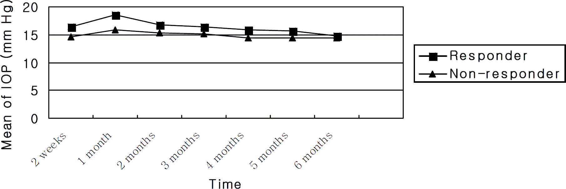
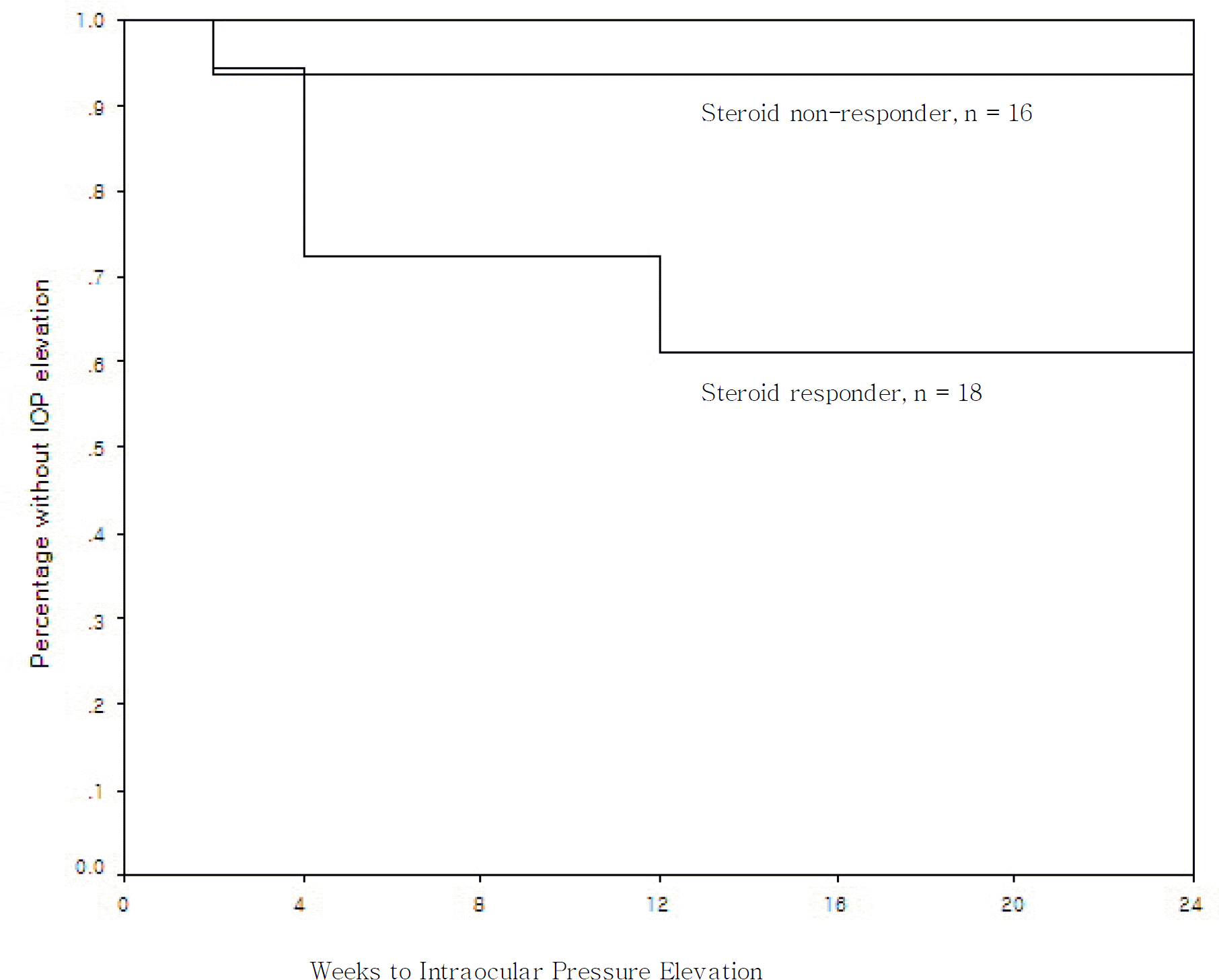
Eighteen eyes were categorized as steroid responders after IVTA injection and sixteen eyes were categorized as non-responders. For the actual amount of increase in the intraocular pressures, the steroid responder group (39%) was shown to be statistically higher than the non-responder group (6%) (P=0.044). However, the mean pressure values did not show a significant difference (P>0.05). Only one eye required the use of glaucoma medications and the intraocular pressure remained normal after treatment.
Go to : 
References
1. Danis RP, Ciulla TA, Pratt LM, et al. Intravitreal triamcinolone acetonide in exudative age related macula degeneration. Retina. 2000; 20:244–50.
2. Martidis A, Duker JS, Greenberg PB, et al. Intravitreal triamcinolone for refractory diabetic macular edema. Ophthal mology. 2002; 109:920–7.

3. Ip MS, Kumar KS. Intravitreous triamcinolone acetonide as treatment for macular edema from central retinal vein occlusion. Arch Ophthalmol. 2002; 120:1217–9.

4. Park HY, Yi K, Kim HK. Intraocular pressure elevation after intravitreal triamcinolone acetonide injection. Korean J Ophthalmol. 2005; 19:122–7.

5. Singh IP, Ahmad SI, Yeh D, et al. Early rapid rise in intraocular pressure after intravitreal triamcinolone acetonide injection. Am J Ophthalmol. 2004; 138:286–7.

6. McGhee CN, Dean S, Danesh-Meyer H. Locally administered ocular corticosteroids: benefits and risks. Drug Saf. 2002; 25:33–55.
7. Thach AB, Dugel PU, Flindall RJ, et al. A comparison of retrobulbar versus sub‐ Tenon’s corticosteroid therapy for cystoid macular edema refractory to topical medications. Ophthalmology. 1997; 104:2003–8.
8. Choi YJ, Oh IK, Oh JR, et al. Intravitreal versus posterior subtenon injection of triamcinolone acetonide for diabetic macular edema. Korean J Ophthalmol. 2006; 20:205–9.

9. Becker B. Diabetes mellitus and primary open‐ angle glaucoma. The XXVII Edward Jackson Memorial Lecture. Am J Ophthalmol. 1971; 1:1–16.
10. Podos SM, Becker B, Morton WR. High myopia and primary open‐ angle glaucoma. Am J Ophthalmol. 1966; 62:1038–43.
11. Gaston H, Absolon MJ, Thurtle OA, et al. Steroid responsiveness in connective tissue diseases. Br J Ophthalmol. 1983; 67:487–90.

12. Unlu N, Robinson JR. Scleral permeability to hydrocortisone and mannitol in the albino rabbit eye. J Ocul Pharmacol Ther. 1998; 14:273–81.

13. Jonas JB. Intraocular availibility of triamcinolone acetonide after intravitreal injection. Am J Ophthalmol. 2004; 137:560–2.
14. Forster DJ, Rao NA, Smith RE. Corticosteroids in the treatment of intermediate uveitis. Dev Ophthalmol. 1992; 23:163–70.

15. Weijtens O, Feron EJ, Schoemaker RC, et al. High concentration of dexamethasone in aqueous and vitreous after subconjunctival injection. Am J Ophthalmol. 1999; 128:192–7.

16. Freeman WR, Green RL, Smith RE. Echographic localization of corticosteroids after periocular injection. Am J Ophthalmol. 1987; 15:281–8.

17. Herschler J. Increased intraocular pressure induced by repository corticosteroids. Am J Ophthalmol. 1976; 82:90–3.

18. Akduman L, Kolker AE, Black DL, et al. Treatment of persistent glaucoma secondary to periocular corticosteroids. Am J Ophthalmol. 1996; 122:275–7.

19. Nozik RA. Periocular injection of steroids. Trans Am Acad Ophthalmol Otolaryngol. 1972; 76:695–705.
20. Mueller AJ, Jian G, Banker AS, et al. The effect of deep posterior subtenon injection of corticosteroids on intraocular pressure. Am J Ophthalmol. 1998; 125:158–63.

21. Lafranco Dafflon M, Tran VT, Guex‐ Crosier Y, et al. Posterior sub‐ Tenon’s steroid injections for the treatment of posterior ocular inflammation: indications, efficacy and side effects. Graefes Arch Clin Exp Ophthalmol. 1999; 237:289–95.
22. Kim YJ, Kang SW, Ahn BH, et al. The results of posterior subtenon steroid injection in uveitis patients. J Korean Ophthalmol Soc. 2003; 44:66–72.
23. Kalina RE. Increased intraocular pressure following subconjunctival corticosteroid administration. Arch Ophthalmol. 1969; 81:788–90.

24. Jea SY, Byon IS, Oum BS. Triamcinolone‐ induced intraocular pressure elevation: intravitreal injection for macular edema and posterior subtenon injection for uveitis. Korean J Ohthalmol. 2006; 20:99–103.
25. Thomas ER, Wang J, Ege E, et al. Intravitreal triamcinolone acetonide concentration after subtenon injection. Am J Ophthalmol. 2006; 142:860–1.

Go to : 
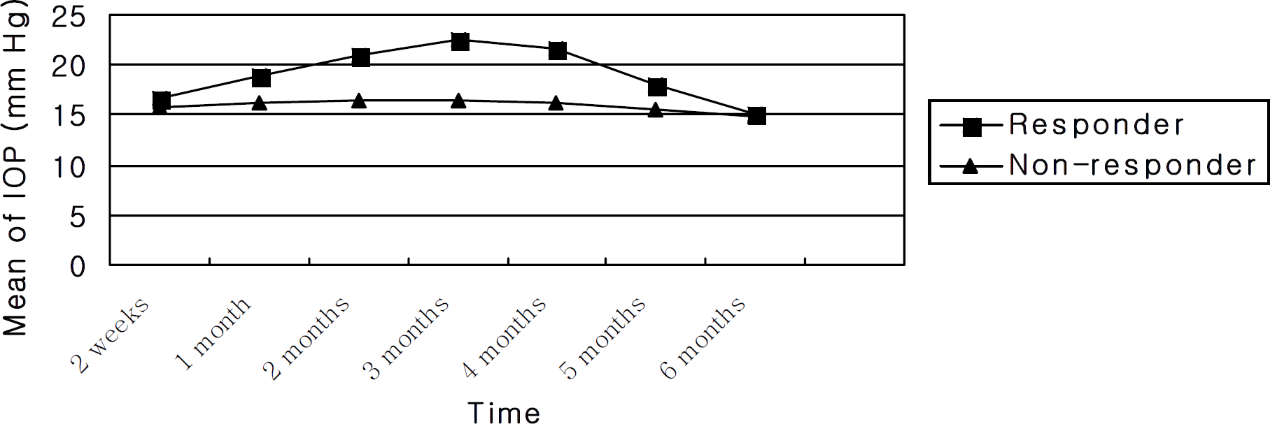
 | Figure 1.Comparison of mean intraocular pressure after intravitreal triamcinolone acetonide injection between responders and non-responders. |
 | Figure 2.Comparison of mean intraocular pressure after posterior subtenon’s injection of triamcinolone acetonide between responders and non-responders. |
 | Figure 3.Kaplan-Meier survival curves comparing probability of intraocular pressure elevation between steroid responders and non-responders after posterior subtenon’s injection of triamcinolone acetonide (P=0.034 Kaplan-Meier analysis). |
Table 1.
Comparison between responder and non-responder groups
| Non-responder | Responder | P-value* | |
|---|---|---|---|
| Mean age (years) | 60.87±10.74 | 61.11±8.87 | 0.945 |
| Baseline intraocular pressure | 15.56±1.93 mmHg | 15.44±1.42 mmHg | 0.839 |
| Male : Female | 8:8 | 11:7 | |
| RVO : CSME | 4:12 | 6:12 |
Table 2.
Mean intraocular pressure after IVTA (mmHg)
Table 3.
Mean intraocular pressure after PSTA (mmHg)
Table 4.
Characteristics of patients with intraocular pressure increase after posterior subtenon injection of triamcinolone acetonide


 PDF
PDF ePub
ePub Citation
 Print
Print


 XML Download